Sensitive and selective detection of Hg2+ based on an electrochemical platform of PDDA functionalized rGO and glutaraldehyde cross-linked chitosan composite film

Yongxiang Wang, Lihui Zhou, Sen Wang, Jinxia Li*, Jing Tang, Shaolei Wang and Ying Wang
School of Chemistry and Molecular Engineering, East-China University of Science and Technology, Shanghai, 200237, China. E-mail: lijinxia@ecust.edu.cn; Fax: +86-21-64252947; Tel: +86-21-64253847

Received 19th April 2016 , Accepted 16th July 2016

First published on 18th July 2016


Abstract

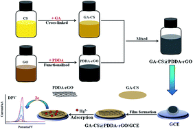
In this paper, a uniform PDDA-functionalized graphene composite film (GA–CS@PDDA-rGO) was utilized for detection of trace Hg2+ by using glutaraldehyde cross-linked chitosan (GA–CS) as a Hg2+-chelating adsorbent and film-forming agent. The results showed that a well-defined and high-sensitivity stripping peak at 0.06 V for Hg2+ was observed at the GA–CS@PDDA-rGO/GCE. Moreover, two important affecting factors of the content of PDDA-rGO and deposition potential were optimized on the GA–CS@PDDA-rGO/GCE modified electrode. Under the optimal conditions, the GA–CS@PDDA-rGO/GCE modified electrode showed a good linearity in the range of 0.03–5 μM between the concentration of the Hg2+ and stripping peak current. The detection limit was estimated to be 7.7 nM (S/N = 3). The interference and selectivity of other heavy metal ions were evaluated, showing no obvious interference on the Hg2+ detection. The results indicated that the GA–CS@PDDA-rGO composite film provided an efficient strategy and a new promising platform for detection of Hg2+.


1. Introduction

With the development of electrochemical techniques, it is very promising to conduct trace analysis of heavy metal ions by fabricating nanotechnology-based electrochemical platforms.1,2 Among all kinds of heavy metal ions, the mercury ion (Hg2+) is one of the most harmful ions to human health and environmental security, so it has become increasingly important to develop optimized electrochemical measurements for the detection of Hg2+.3,4 For ultrasensitive detection of heavy metals based on electrochemical analysis, the key is the design of modified electrodes with unique properties, which is expected to greatly improve the sensitivity, selectivity and stability of electrodes after modification with suitable materials.

Recently, graphene-based materials have been highly concerned as excellent electrode materials to construct electrochemical determination platforms.5,6 Graphene exhibits two-dimensional (2D) planar structure with high surface area, remarkable conductivity, good film-forming ability and easily chemical modification. Wang et al.7 reported that graphene enhanced electron transfer at aptamer modified electrode and its application in the detection of small molecule adenosine triphosphate (ATP) and Hg2+. The experimental results confirmed such electrochemical aptasensor possessed a good sensitivity and high selectivity for ATP and Hg2+. Li et al.8 established a new electrochemical platform based on the Nafion–graphene nanocomposite film for the determination of Cd2+ by anodic stripping voltammetry analysis. The nanocomposite film combined the advantages of graphene and the cationic exchange capacity of Nafion, which enhanced the sensitivity of Cd2+ assay. In most of reported graphene-based materials, graphene is usually prepared by chemical reduction of graphene oxide (GO), which is reduced graphene (rGO).9,10 However, rGO is hydrophobic and tends to agglomerate irreversibly through van der Waals interactions during reduction process. In order to avoid the agglomeration of rGO, it is necessary to carry out functionalization of graphene.

Poly(diallyldimethylammonium chloride) (PDDA) is not only an electronic conducting polymer but also a positively charged polyelectrolyte, which can non-covalently functionalize negatively charged GO and prevent the aggregation of rGO through electrostatic repulsion.11,12 During the reduction process of graphene, PDDA can act as a stabilizer and synergistic reducing agent, resulted in good dispersion of rGO. The corresponding electrochemical platforms based on PDDA-functionalized rGO have been fabricated and applied in the detection of biomolecules, such as angiogenin,13 uric acid,14 quinoline yellow,15 and so on. Wang et al.16 designed a sensitive electrochemical luteolin sensor based on PDDA-functionalized reduced graphene oxide (PDDA-rGO), which exhibited a wide detection range and low detection limit for luteolin. Peng et al.17 developed a novel electrochemical sensor based on PDDA functionalized graphene (PDDA-G) composite film for the detection of 4-nitropenol, which showed remarkably improved sensitivity and selectivity.

Chitosan (CS), a widely distributed natural polysaccharide with abundant hydroxyl and amino active functional groups, possessing excellent film-forming ability and high metal-chelating ability, which can act as effective adsorbent for capturing heavy-metal ions. It has been reported that nanocomposites containing CS can remarkably improve the adsorption capacity of Cu2+,18 Pb2+,19 especially for Hg2+.20–22 Deng et al.20 used thiol functionalized chitosan-multiwalled carbon nanotubes nanocomposite film electrode for the electrochemical determination of Hg2+ by square wave stripping voltammetry. Another chitosan derivative adsorbent [P-C-CTS-(Hg)] was prepared by employing the polyamination and Hg2+-imprinted technologies for Hg2+ removal from a low-concentration aqueous solution (CHg2+ ≤ 40 mg L−1).21 The adsorption capacity of P-C-CTS-(Hg) for Hg2+ reached 9.017 mg g−1, 2.2 times higher than that of chitosan.

Herein, a novel composite film of PDDA-functionalized graphene (PDDA-rGO) and glutaraldehyde cross-linked chitosan (GA–CS) was utilized to fabricate GA–CS@PDDA-rGO modified glass carbon electrode for the detection of trace Hg2+. It is worth noting that the as-prepared composite matrix in this work combines the advantages of the PDDA-rGO (such as high surface area, remarkable conductivity and good dispersion) and high Hg2+-chelating ability of GA–CS. PDDA-functionalized rGO (PDDA-rGO) was prepared by the reduction of GO using NaBH4 as reducing agent in the presence of PDDA. After being combined with glutaraldehyde to form cross-linked CS (GA–CS), the nanocomposite (GA–CS@PDDA-rGO) was used to fabricate GA–CS@PDDA-rGO modified glass carbon electrode (GA–CS@PDDA-rGO/GCE). After accumulating Hg2+, anodic stripping voltammetry (ASV) and differential pulse voltammetry (DPV) were applied to evaluate the electrochemical properties of GA–CS@PDDA-rGO/GCE modified electrode.

2. Experimental section

2.1. Reagent and apparatus

Natural graphite powder (purity of 99.9%), chitosan (CS, Mw = 130[thin space (1/6-em)]000 Da), glutaraldehyde (GA), HCl were purchased from Sinopharm Chemical Reagent Co., Ltd (China). PDDA was purchased from Shanghai Chemical Company, Shanghai (China). KMnO4, NaNO3 were purchased from Xi'an Chemical Company, Xi'an (China). H2O2, H2SO4, NaBH4, NaOH, CH3COOH were purchased from Shanghai Ling-Feng Chemical Reagent Co., Ltd (China). All chemicals used were of analytical grade and the solutions were prepared with doubly distilled water.

The field emission scanning electron microscopy (FESEM) analysis of GA–CS@PDDA-rGO/GCE was obtained on a Merlin Compact microscope (ZEISS, Germany). The transmission electron microscopy (TEM) images were recorded on a JEM-2100 microscope at an acceleration voltage of 200 kV (JEOL, Japan). X-ray diffraction (XRD) patterns were obtained on a D/max 2550 diffractometer (Rigaku, Japan) operating with Cu Kα radiation (λ = 0.154 nm, 40 kV, 100 mA, 2θ = 5–80°). The Raman spectroscope was carried on a Laser Raman Microscope (Renishaw, Iuvia reflerx). Fourier transform infrared (FTIR) spectra were collected with KBr pellet on a Nicolet 380 FTIR (THERMO, US).

2.2. Synthesis of PDDA-rGO

GO was synthesized from natural graphite powder using the modified Hummers method.23,24 In a typical procedure of chemical modification of GO to form PDDA-functionalized rGO, the prepared GO was exfoliated in water by ultrasonication to form 1.0 mg mL−1 GO dispersion. Then, 100 μL of PDDA (35 wt%) was dropped into 100 mL of 1.0 mg mL−1 GO dispersion. The mixture was continuously ultrasonicated for 20 min. Next, 200 mg of NaBH4 was added into the mixture. After stirring for 30 min, the mixture was transferred into an oil bath at 125 °C for 3 h of reflux condensation reaction. After cooling to room temperature, the mixture was washed with distilled water to neutral. Finally, the black PDDA-functionalized rGO (PDDA-rGO) produce was obtained after being dried at 40 °C under vacuum for 12 h.

2.3. Synthesis of GA–CS

CS was dissolved in 2% (v/v) aqueous acetic acid to get the 0.5 wt% CS solution. Glutaraldehyde cross-linked CS (GA–CS) was prepared by drop-by-drop addition of 150 μL glutaraldehyde (25% v/v solution) into 20 mL 0.5 wt% CS solution under magnetic stirring and then kept on stirring for 24 h at room temperature. Finally, the color of the mixing solution became darker from light yellow.

2.4. Electrode modification

Prior to preparation of modified electrode, GC electrode (GCE) of 3.0 mm in diameter was mechanically polished to a mirror finish with 0.05 μm alumina slurry, then ultrasonically washed with 1[thin space (1/6-em)]:[thin space (1/6-em)]1 HNO3 (v/v), NaOH (1.0 mol L−1), ethanol and redistilled water for 30 s, respectively.

2 mg mL−1 PDDA-rGO and 0.5 wt% GA–CS solutions were mixed with different volume ratio (volume ratio of PDDA-rGO[thin space (1/6-em)]:[thin space (1/6-em)]GA–CS = 1[thin space (1/6-em)]:[thin space (1/6-em)]4, 1[thin space (1/6-em)]:[thin space (1/6-em)]2, 1[thin space (1/6-em)]:[thin space (1/6-em)]1, 2[thin space (1/6-em)]:[thin space (1/6-em)]1, 3[thin space (1/6-em)]:[thin space (1/6-em)]1 and 4[thin space (1/6-em)]:[thin space (1/6-em)]1, corresponding to the PDDA-rGO content of 0.4 mg mL−1, 0.67 mg mL−1, 1 mg mL−1, 1.33 mg mL−1, 1.5 mg mL−1 and 1.6 mg mL−1, respectively) to get the homogeneous GA–CS@PDDA-rGO mixture. Then, 10 μL of the obtained GA–CS@PDDA-rGO mixture was coated on the fresh surface of GCE and naturally dried at ambient to form GA–CS@PDDA-rGO modified electrode, which was denoted as GA–CS@PDDA-rGO/GCE. Next, the GA–CS@PDDA-rGO/GCE was immersed into 1 mol L−1 NaOH solution for 3 min to neutralize the CH3COOH and rinsed with distilled water to remove physically adsorbed species before use. For comparison, GA–CS/GCE and PDDA-rGO/GCE electrodes were prepared through similar procedure.

In order to reveal the effect of rGO on the electrochemical response of Hg2+, the GA–CS@PDDA-GO/GCE electrode was also prepared. 200 μL of PDDA (35 wt%) was dropped into 100 mL of 2.0 mg mL−1 GO dispersion and stirred at room temperature for 3 h without the reduction of NaBH4. Then 2 mg mL−1 of PDDA-GO mixture was obtained, which was mixed with 0.5 wt% GA–CS solutions with the volume ratio of 2[thin space (1/6-em)]:[thin space (1/6-em)]1 to get the homogeneous GA–CS@PDDA-GO mixture. Next, GA–CS@PDDA-GO/GCE modified electrode was prepared through similar procedure.

2.5. Electrochemical measurements

Electrochemical detection of Hg2+ was carried out on a CHI 660 electrochemical workstation (Shanghai Chenhua, China) with a three electrode system: a modified GC electrode as the working electrode, a platinum wire electrode as the counter electrode and a saturated calomel electrode (SCE) as the reference electrode. The fabrication process of GA–CS@PDDA-rGO/GCE and determination process of target Hg2+ on the GA–CS@PDDA-rGO/GCE are illustrated in Scheme 1. In the adsorption step, the GA–CS@PDDA-rGO/GCE was immersed into 0.1 mol L−1 HCl solution containing target Hg2+ for 30 min at ice-bath under constant moderate magnetic stirring. Then the electrode was rapidly rinsed with water and transferred to a blank 0.1 mol L−1 HCl solution, followed by a potentiostatic deposition treatment at −0.3 V for 120 s. Next, a differential pulse voltammetric (DPV) scan response was recorded in a potential range from −0.3 V to 0.3 V at scan rate of 100 mV s−1 after 20 s.
image file: c6ra10075a-s1.tif
Scheme 1 Illustration of the fabrication process of GA–CS@PDDA-rGO/GCE and the determination of Hg2+.

3. Results and discussion

3.1. Characterization of electrode materials

PDDA-functionalized rGO (PDDA-rGO) was prepared by the reduction of GO using NaBH4 as reducing agent in the presence of PDDA. The obtained PDDA-rGO was systematically investigated by XRD, FTIR, Raman, SEM and TEM analysis. Fig. 1a shows the XRD patterns of GO and PDDA-rGO. Compared with GO, PDDA-rGO displays a broad peak at 2θ of 25.4°, corresponding to (002) plane of amorphous carbon with d-spacing of 0.35 nm, which is smaller than d-spacing (0.94 nm) of GO at 2θ of 9.4°. This change suggested that GO was successfully reduced by NaBH4. However, from the XRD patterns of PDDA-rGO, the characteristic peak of PDDA can't be found due to the low content of PDDA in PDDA-rGO. In order to further analyze the surface functionalization structure of PDDA-rGO, the FTIR spectra of GO and PDDA-rGO nanosheets were obtained (Fig. 1b). As well known, GO synthesized by Hummers method not only contains hydroxyl, carboxyl, epoxy and other oxidation groups, but also remains part of unoxidized graphite structure. As expected, the FTIR spectrum of GO shows the characteristic absorption peaks from oxygen-containing functional groups at 3417 cm−1, 1734 cm−1, 1380 cm−1, 1223 cm−1 and 1047 cm−1. The absorption bands at 3417 and 1380 cm−1 can be assigned to the stretching vibration and deformation vibration of O–H, respectively. The bands at 1734 cm−1, 1223 cm−1 and 1047 cm−1 belong to the C[double bond, length as m-dash]O stretching of –COOH groups and stretching vibration of C–O (epoxy) and C–O (alkoxy), respectively.15,16 Besides, the band at 1627 cm−1 should be assigned to the C[double bond, length as m-dash]C skeletal vibration of unoxidized sp2 graphitic domain. In the spectrum of PDDA-rGO, after NaBH4 reduction process, the C[double bond, length as m-dash]C skeletal vibration of sp2 graphitic domain still remains and relatively shifts to 1571 cm−1 while the intensities of oxygen-containing functional groups significantly decrease or even vanish, suggesting that GO has been successfully reduced. Moreover, the FTIR spectrum of PDDA-rGO also exhibits two new bands at 1466 cm−1 (–CH2) and 1113 cm−1 (C–N), belonging to the characteristic bands of PDDA.25–27 The results indicate that the rGO surface has been successfully functionalized with PDDA.
image file: c6ra10075a-f1.tif
Fig. 1 (a) The XRD patterns, (b) FTIR spectra, (c) Raman spectra of GO and PDDA-rGO.

The Raman spectra of GO and PDDA-rGO are shown in Fig. 1c. It can be clearly seen that the two samples exhibit a similar Raman spectrum with a distinct pair of broad absorption bands at around 1354 cm−1 and 1590 cm−1, corresponded to the two main characteristic absorptions of D (∼1350 cm−1) band and G (∼1580 cm−1) band for carbon materials. The D band is attributed to the disordered structures of carbon, reflecting the disorder of graphene sheets. The G band corresponds to the first order scattering of the E2g phonon of sp2 carbon atoms,28 reflecting the symmetry and crystallinity. The ratio of the integral peak area of two bands (ID/IG) can be used as an indicator for disorder degree of carbon materials. From Fig. 1c, the ID/IG ratio of PDDA-rGO is determined to be 1.66, which is higher than that of GO (1.43). It is suggested that a decrease of sp2 domain induced by the NaBH4 reduction, which is in agreement with the results of reported literatures.16,29,30

The obtained PDDA-rGO powder was ultrasonicated in deionized water to get a suspension. There are no obvious precipitation was found in the bottom of the bottle after one month, indicating that the PDDA-rGO dispersion is still uniform and stable (as shown in Fig. 2a), which is favorable to further electrochemical applications. Fig. 2b shows the TEM image of PDDA-rGO nanosheet, which clearly appears the flaky morphology with rolled edges and wrinkles. The selected area electron diffraction (SAED) pattern (Fig. 2b inset) reveals the well-crystallized nature and the hexagonal structure characteristics of a single exfoliated graphene nanosheet.30,31


image file: c6ra10075a-f2.tif
Fig. 2 (a) Photograph and (b) TEM image of PDDA-rGO, inset: SAED pattern of PDDA-rGO.

Because of film-forming ability and high metal-chelating ability, the appropriate proportion of GA–CS solution was added into the aforementioned PDDA-rGO dispersion. After the GA–CS@PDDA-rGO mixture was cast on the surface of GCE and dried in room temperature, a film visible to the naked eye was obtained. Using a special detachable electrode head, the surface morphology and structure of GA–CS@PDDA-rGO composite film on the GC electrode were further characterized via FE-SEM. Fig. 3a showed that a flat and uniform coating with GA–CS@PDDA-rGO layer exists on the surface of GC electrode. The zoomed-in SEM image (Fig. 3b) clearly exhibits the numerous wrinkles of graphene, indicating the layer-folding structure of GA–CS@PDDA-rGO.


image file: c6ra10075a-f3.tif
Fig. 3 SEM micrographs of GA–CS@PDDA-rGO/GCE with (a) low magnification and (b) high magnification.

What's more, the stability of GA–CS@PDDA-rGO film on the electrode surface was investigated by immersing in the HCl solution, which is a key influencing factor in electrochemical applications. No obvious film falling off was found from prolonged immersion. The results indicated that the GA–CS@PDDA-rGO film may be promising as a potential material for the determination of Hg2+.

3.2. Electrochemical determination of trace Hg2+ at different modified electrodes

The electrochemical determination of Hg2+ with different modified electrodes was investigated by DPV analysis (as shown in Fig. 4). Before the detection, all of the different electrodes were immersed into 0.1 mol L−1 HCl solution containing 1 μM Hg2+ for the adsorption of Hg2+ for 30 min. No obvious stripping peak was observed at the bare GC electrode, which can be attributed to almost no adsorption Hg2+ on the surface of bare GC electrode (curve a). Under comparable conditions, a small stripping peak can be observed at the PDDA-rGO/GCE (curve b), GA–CS/GCE (curve c) and GA–CS@PDDA-GO/GCE (curve d). However, the corresponding peak intensity and peak symmetry was not satisfactory. Whereas, a well-defined and high-sensitivity stripping peak at 0.06 V for Hg2+ was observed at the GA–CS@PDDA-rGO/GCE (curve e). It was suggested that the integration of GA–CS and PDDA-rGO provided a remarkable synergistic promotion for the DPV electrochemistry signals of Hg2+. Such an electrochemical platform combined the advantage of the good conductivity of PDDA functionalized rGO and easy film formation and strong Hg2+-chelating capacity of GA–CS, resulting in highly enhanced electron conductive nanostructure film for Hg2+ determination.
image file: c6ra10075a-f4.tif
Fig. 4 DPV voltammograms in a blank 0.1 mol L−1 HCl solution recorded at (a) bare GCE, (b) PDDA-rGO/GCE, (c) GA–CS/GCE, (d) GA–CS@PDDA-GO/GCE and (e) GA–CS@PDDA-rGO/GCE with a scan rate of 100 mV s−1. Pre-accumulation was conducted by immersing the bare/modified electrode in 0.1 mol L−1 HCl solution containing 1 μM Hg2+ for 30 min, followed by potentiostatic deposition at −0.3 V for 120 s in blank 0.1 mol L−1 HCl solution.

3.3. Optimization of determination conditions on GA–CS@PDDA-rGO/GCE

In order to obtain the optimum experimental conditions, two important affecting factors that PDDA-rGO content and deposition potential were studied on the GA–CS@PDDA-rGO/GCE. Fig. 5a showed the effect of PDDA-rGO content in GA–CS@PDDA-rGO on the determination of trace Hg2+. 2 mg mL−1 PDDA-rGO solution was mixed with GA–CS (0.5 wt%) solution (volume ratio of PDDA-rGO[thin space (1/6-em)]:[thin space (1/6-em)]GA–CS = 1[thin space (1/6-em)]:[thin space (1/6-em)]4, 1[thin space (1/6-em)]:[thin space (1/6-em)]2, 1[thin space (1/6-em)]:[thin space (1/6-em)]1, 2[thin space (1/6-em)]:[thin space (1/6-em)]1, 3[thin space (1/6-em)]:[thin space (1/6-em)]1 and 4[thin space (1/6-em)]:[thin space (1/6-em)]1, respectively). As shown in Fig. 5a, the highest stripping peak current occurred as the volume ratio of PDDA-rGO and GA–CS is 2[thin space (1/6-em)]:[thin space (1/6-em)]1, corresponding to PDDA-rGO content of 1.33 mg mL−1. The results indicated that the synergistic effect of PDDA-rGO and GA–CS is best as the ratio is 2[thin space (1/6-em)]:[thin space (1/6-em)]1.
image file: c6ra10075a-f5.tif
Fig. 5 The effect of (a) PDDA-rGO content (volume ratio of PDDA-rGO[thin space (1/6-em)]:[thin space (1/6-em)]GA–CS = 1[thin space (1/6-em)]:[thin space (1/6-em)]4, 1[thin space (1/6-em)]:[thin space (1/6-em)]2, 1[thin space (1/6-em)]:[thin space (1/6-em)]1, 2[thin space (1/6-em)]:[thin space (1/6-em)]1, 3[thin space (1/6-em)]:[thin space (1/6-em)]1 and 4[thin space (1/6-em)]:[thin space (1/6-em)]1, respectively) and (b) deposition potential on the stripping responses of Hg2+ at GA–CS@PDDA-rGO/GCE in 0.1 mol L−1 HCl solution. Other experimental conditions are the same as in Fig. 4.

Fig. 5b showed the stripping peak value recorded at different deposition potentials from −0.1 V to −0.6 V. The stripping peak current of Hg2+ increased as the deposition potential shift to a negative direction in the range −0.1 V to −0.3 V. However, further negative shift of the deposition potential can decrease the peak current. Therefore, −0.3 V was chosen as the optimum deposition potential to reduce the accumulated Hg2+ on GA–CS@PDDA-rGO/GCE before DPV detection.

In conclusion, we can determine the optimum experimental conditions. The modified GA–CS@PDDA-rGO/GCE was prepared as the volume ratio of PDDA-rGO and GA–CS is 2[thin space (1/6-em)]:[thin space (1/6-em)]1 and the deposition potential of −0.3 V was selected for the subsequent research work.

3.4. Analytical performance of GA–CS@PDDA-rGO/GCE for detection of Hg2+

Under the optimum experimental conditions, the GA–CS@PDDA-rGO/GCE was immersed into 0.1 mol L−1 HCl solution containing various Hg2+ concentration in the range from 0.03 μM to 5 μM. The DPV responses of Hg2+ were recorded in a potential range from −0.3 V to 0.3 V at scan rate of 100 mV s−1, as shown in Fig. 6a. The peak current was used for the quantitative analysis of Hg2+. The inset was an amplification of the stripping response with low concentrations of Hg2+ from 0.03 μM to 0.5 μM. As can be seen, the stripping peak currents increased linearly with the increasing concentration of Hg2+ (Fig. 6b). The regression equation is ipc = 11.23c − 1.16 (ipc in μA, c in μM) (R2 = 0.998). The limit of detection for Hg2+ (S/N = 3) was estimated to be 7.7 nM, which is lower than most of the reported values obtained on other modified electrodes, such as GO/DTT/Nafion modified electrode,1 MTTZ-MSU-2-CPE electrode,32 N-BDMP/CPEs,33 CAL-GCE34 and so on. The compared results are listed in Table 1.
image file: c6ra10075a-f6.tif
Fig. 6 (a) DPV responses and (b) regression curve for determination of Hg2+ in the concentration range from 0.03 μM to 5 μM on the GA–CS@PDDA-rGO/GCE.
Table 1 Comparison of linear ranges and detection limits of different modified electrodes for detection of Hg2+ by electrochemical method
Modified electrodes Linear range (nM) Detection limit (nM) Reference
GO/DTT/Nafion 5–50[thin space (1/6-em)]000 13 1
CS-SH-MWCNTs/GCE 10–140 3 20
MTTZ-MSU-2-CPE 25–1000 23 32
N-BDMP/CPEs 50–10[thin space (1/6-em)]000 41 33
CAL-GCE 125–1500 25 34
GA–CS@PDDA-rGO/GCE 30–5000 7.7 This work


3.5. Anti-interference, repeatability and stability of GA–CS@PDDA-rGO/GCE

To study the interference of some general metal ions on the detection of Hg2+, 50-fold of Cd2+, Ba2+, Pb2+, Mg2+, Ni2+, Zn2+, Cu2+ and Ca2+ were chosen to add into a preconcentration solution containing 0.5 μM Hg2+. Under the optimized experimental conditions, the detection of Hg2+ was individually carried out by DPV in the presence of different interfering ions. The results indicated that there was no obvious influence on the electrochemical stripping signals for Hg2+ (peak current changes <±5%), as shown in Fig. 7.
image file: c6ra10075a-f7.tif
Fig. 7 DPV responses for the determination of Hg2+ (0.5 μM) at GA–CS@PDDA-rGO/GCE in the presence of 50-fold different interfering ions.

Further assessment of repeatability of the GA–CS@PDDA-rGO/GCE was conducted through DPV. Eight parallel electrodes were fabricated to detect Hg2+ and the relative standard deviation (RSD) was around 3.1%. In addition, it was observed that the peak current of Hg2+ remained 97% of its initial value after the GA–CS@PDDA-rGO/GCE was stored in a refrigerator of 4 °C for a week. These results showed the GA–CS@PDDA-rGO/GCE had good repeatability and stability for electrochemical analysis, which could be employed to determine some real water samples. Herein, the recovery rates were performed for the detection of Hg2+ at GA–CS@PDDA-rGO/GCE by adding 0.2 μM, 0.3 μM and 0.4 μM into tap water containing 0.3 μM Hg2+ and applying the standard addition method, which led to a recovery of 92%, 98% and 101%, respectively. These results are relatively small and acceptable, indicating that the method is quite reproducible and accurate.

4. Conclusions

In this work, a novel GA–CS@PDDA-rGO composite material for easy film formation on the surface of GC electrode was successfully synthesized and employed to fabricate an advanced electrochemical platform for high sensitive detection of Hg2+ from 0.03 μM to 5 μM. The GA–CS@PDDA-rGO/GCE modified electrode exhibited excellent adsorption selectivity for Hg2+, low limit of detection (7.7 nM) and good reproducibility due to the synergistic promotion of PDDA-rGO and GA–CS. The GA–CS@PDDA-rGO composite material was supposed to be applied to the determination of low-concentration Hg2+ in waste waters.

Acknowledgements

This work was supported by the National Natural Science Foundation of China (Project No. 21305046).

References

  1. S. M. Choi, D. M. Kim, O. S. Jung and Y. B. Shim, Anal. Chim. Acta, 2015, 892, 77–84 CrossRef CAS PubMed.
  2. Y. Q. Yang, M. M. Kang, S. M. Fang, M. H. Wang, L. H. He, J. H. Zhao, H. Z. Zhang and Z. H. Zhang, Sens. Actuators, B, 2015, 214, 63–69 CrossRef CAS.
  3. J. M. Gong, T. Zhou, D. D. Song and L. Z. Zhang, Sens. Actuators, B, 2010, 150, 491–497 CrossRef CAS.
  4. Z. Q. Zhu, Y. Y. Su, J. Li, D. Li, J. Zhang, S. P. Song, Y. Zhao, G. X. Li and C. H. Fan, Anal. Chem., 2009, 81, 7660–7666 CrossRef CAS PubMed.
  5. L. H. Tang, H. B. Feng, J. S. Cheng and J. H. Li, Chem. Commun., 2010, 46, 5882–5884 RSC.
  6. L. Y. Chen, Y. H. Tang, K. Wang, C. B. Liu and S. L. Luo, Electrochem. Commun., 2011, 13, 133–137 CrossRef CAS.
  7. L. Wang, M. Xu, L. Han, M. Zhou, C. Z. Zhu and S. J. Dong, Anal. Chem., 2012, 84, 7301–7307 CrossRef CAS PubMed.
  8. J. Li, S. J. Guo, Y. M. Zhai and E. K. Wang, Electrochem. Commun., 2009, 11, 1085–1088 CrossRef CAS.
  9. N. I. Kovtyukhova, B. R. Ollivier, P. J. Martin, T. E. Mallouk, S. A. Chizhik, E. V. Buzaneva and A. D. Gorchinskiy, Chem. Mater., 1999, 11, 771–778 CrossRef CAS.
  10. J. T. Robinson, F. K. Perkins, E. S. Snow, Z. Q. Wei and P. E. Sheehan, Nano Lett., 2008, 8, 3137–3140 CrossRef CAS PubMed.
  11. F. D. Cai, Q. Zhu, K. Zhao, A. P. Deng and J. G. Li, Environ. Sci. Technol., 2015, 49, 5013–5020 CrossRef CAS PubMed.
  12. Y. Y. Zhang, C. Zhang, D. Zhang, M. Ma, W. Z. Wang and Q. Chen, Mater. Sci. Eng., C, 2016, 58, 1246–1254 CrossRef CAS PubMed.
  13. Z. B. Chen, C. M. Zhang, X. X. Li, H. Ma, C. Q. Wan, K. Li and Y. Q. Lin, Biosens. Bioelectron., 2015, 65, 232–237 CrossRef CAS PubMed.
  14. Y. Y. Yu, Z. G. Chen, B. B. Zhang, X. C. Li and J. B. Pan, Talanta, 2013, 112, 31–36 CrossRef CAS PubMed.
  15. L. Fu, Y. H. Zheng, A. W. Wang, W. Cai and H. T. Lin, Food Chem., 2015, 181, 127–132 CrossRef CAS PubMed.
  16. L. Fu, Y. H. Zheng, A. W. Wang, W. Cai and H. T. Lin, Int. J. Electrochem. Sci., 2015, 10, 3518–3529 CAS.
  17. D. Peng, J. X. Zhang, D. G. Qin, J. Chen, D. L. Shan and X. Q. Lu, J. Electroanal. Chem., 2014, 734, 1–6 CrossRef CAS.
  18. L. Z. Li, Z. Wang, P. M. Ma, H. Y. Bai, W. F. Dong and M. Q. Chen, J. Polym. Res., 2015, 22, 150–159 CrossRef.
  19. A. H. Gedam and R. S. Dongre, RSC Adv., 2015, 5, 54188–54201 RSC.
  20. W. F. Deng, Y. M. Tan, Y. Y. Li, Y. Q. Wen, Z. H. Su, Z. Huang, S. Q. Huang, Y. Meng, Q. J. Xie, Y. P. Luo and S. Z. Yao, Microchim. Acta, 2010, 169, 367–373 CrossRef CAS.
  21. X. J. Tang, D. Niu, C. L. Bi and B. X. Shen, Ind. Eng. Chem. Res., 2013, 52, 13120–13127 CrossRef CAS.
  22. C. H. Xiong, L. L. Pi, X. Y. Chen, L. Q. Yang, C. A. Ma and X. M. Zheng, Carbohydr. Polym., 2013, 98, 1222–1228 CrossRef CAS PubMed.
  23. J. Chen, Y. R. Li, L. Huang, C. Li and G. Q. Shi, Carbon, 2015, 81, 826–834 CrossRef CAS.
  24. S. Wang, Y. Wang, L. H. Zhou, J. X. Li, S. L. Wang and H. L. Liu, Electrochim. Acta, 2014, 132, 7–14 CrossRef CAS.
  25. L. Ding, Y. P. Liu, S. X. Guo, J. P. Zhai, A. M. Bond and J. Zhang, J. Electroanal. Chem., 2014, 727, 69–77 CrossRef CAS.
  26. D. Q. Yang, J. F. Rochette and E. Sacher, J. Phys. Chem. B, 2005, 109, 4481–4484 CrossRef CAS PubMed.
  27. X. L. Ye, Y. L. Du, D. B. Lu and C. M. Wang, Anal. Chim. Acta, 2013, 779, 22–34 CrossRef CAS PubMed.
  28. A. Jänes, H. Kurig and E. Lust, Carbon, 2007, 45, 1226–1233 CrossRef.
  29. S. Stankovich, D. A. Dikin, R. D. Piner, K. A. Kohlhaas, A. Kleinhammes, Y. Y. Jia, Y. Wu, S. B. T. Nguyen and R. S. Ruoff, Carbon, 2007, 45, 1558–1565 CrossRef CAS.
  30. G. X. Wang, J. Yang, J. Park, X. L. Gou, B. Wang, H. Liu and J. Yao, J. Phys. Chem. C, 2008, 112, 8192–8195 CAS.
  31. J. Shen, Y. Hu, M. Shi, X. Lu, C. Qin, C. Li and M. X. Ye, Chem. Mater., 2009, 21, 3514–3520 CrossRef CAS.
  32. S. Alfredo, M. Z. Sonia, P. Q. Damian, S. Isabel and H. Isabel del, Sens. Actuators, B, 2012, 163, 38–43 CrossRef.
  33. A. Afkhami, T. Madrakian, S. J. Sabounchei, M. Rezaei, S. Samiee and M. Pourshahbaz, Sens. Actuators, B, 2012, 161, 542–548 CrossRef CAS.
  34. J. Q. Liu, X. W. He, X. H. Zeng, Q. J. Wan and Z. Z. Zhang, Talanta, 2003, 59, 553–560 CrossRef.

This journal is © The Royal Society of Chemistry 2016
Click here to see how this site uses Cookies. View our privacy policy here.