Exploration of selective decoration of Janus silica particles within polymeric patterned pore arrays

Pinghui Yangab, Junjie Huangab, Wei Sun*ab, Yujie Weiab, Yuwei Liuab, Lingyun Dingab, Jinbiao Baoab and Zhong-Ren Chenab
aDepartment of Polymer Science and Engineering, Faculty of Materials Science and Chemical Engineering, Ningbo University, Ningbo, 315211, China. E-mail: sunwei@nbu.edu.cn
bKey Laboratory of Specialty Polymers, Faculty of Materials Science and Chemical Engineering, Ningbo University, Ningbo, 315211, China

Received 18th April 2016 , Accepted 6th June 2016

First published on 6th June 2016


Abstract

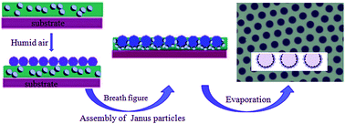
Janus particles, especially amphiphilic ones, are known to be highly active when involved in interfacial self-assembly processes. However, experimental reports on employing Janus particles as an interfacial stabilizer to establish functional structures are rather limited. In this paper, amphiphilic Janus silica particles were prepared by selective chemical treatment on the particle “colloidosomes”. Different ways (decoration of Au nanoparticles, interfacial adsorption test) were used to confirm the acquirement of the Janus character of the particles. Upon successful synthesis of amphiphilic Janus particles, they were employed in the breath figures method for the first time. It turned out that by using the Janus particles, a regularly arranged pore array could be easily obtained. Characterization of the morphology of the patterned structure shows that Janus particles were densely located on the interior wall of the entire holes, forming finer particle arrays compared to the case of isotropic particles. The result indicates that Janus particles show optimized behavior when they are involved in the interfacial self-assembly processes. With further work, by choosing Janus particles with functional features, selective functionalization of porous films could be achieved with high efficiency and great ease using this method.


1. Introduction

Janus particles are particles with two or more parts presenting different chemical or physical properties on a single particle's surface. This particular kind of particle was named “Janus particles” by de Gennes in 1991.1 According to de Gennes, Janus particles, especially with amphiphilic chemical features, would present unique behavior if they were put on oil–water interfaces. They would behave in ways which are quite different from molecular amphiphiles. Due to their anisometric characteristics, Janus particles could find novel applications in many fields, such as stabilization of emulsions and foams,1–4 optical and electronic devices.5,6 In the past few decades, the main research focus has been put on the synthesis of Janus particles. Many techniques, including surface coating by vapor deposition of metal thin films,7–9 masking of a photoresist layer,10 self-assembly of polymer,11,12 photo-polymerization in a micro-fluidic channel,13 and Pickering emulsion method,14 were established to fulfill the synthesis.

While in recent years, besides the effort on the synthesis techniques, the research on the self-assembly process of the Janus particles has attracted increasing attentions. The highly active behavior the Janus particles would show on the interfaces makes them interesting candidates to form unusual assembly structures. Nikoubashman investigated the self-assembly of Janus particles under shear flow by employing hybrid molecular dynamics simulations that explicitly consider the hydrodynamic interactions.15 Rickman and co-workers study the impact of anisotropy and interaction range on the self-assembly of Janus colloidal particles using Monte Carlo simulation.16 Not until very recently, Janus particles were used as building blocks to fabricate complex materials. Sun and co-workers have studied the influence of shape of Janus particles on their orientation and surface activity at fluid–fluid interfaces via molecular dynamics simulations.17 Harting also developed theoretical models describing the behavior of magnetic Janus particles adsorbed at fluid–fluid interfaces interacting with an external magnetic field.18

However, all the researches mentioned above concerning the self-assembly of Janus particles at fluid–fluid interfaces are studied via models or simulations, while the experimental reports are rather limited. What we tend to demonstrate here is a novel way to have the Janus particles experimentally involved in a self-assembly process, in which arrays of oil/water interfaces are presented. To be specific, amphiphilic silica Janus particles are, for the first time, employed in breath figures (BF) method as stabilizers. “Breath figure”, studied as early as 1911 by Lord Rayleigh, was originally referred to the organized arrays of water droplets that form when humid air comes in contact with cold solid or liquid surfaces.19,20 BF method is now emerging as a representative bottom-up technique for fabricating patterned surface of porous films. For BF process, polymers dissolved in a volatile solvent are cast under humid atmosphere. With the rapid evaporation of the solvent, water droplets condense onto the upper surface of the solution from humid environment. Hexagonal arrays of the water droplets formed minutes after the casting of the solution and act as templates for the structuring of the polymer. After the complete evaporation of the solvent and water droplets, the polymeric porous film with “imprints” of the arrays of the droplets will be obtained. Those porous films have potential applications in sensors,21 optoelectronic devices,22,23 biomaterials,24–26 separation membranes,27,28 catalysis29 and so forth.

In order to achieve preservation of the ordered arrays of water droplets during the BF process, certain materials, mostly polymers, must be used to prevent the coalescence of water droplets. Sun and co-workers utilized polymeric and silica particles to assist in preparing BF films.20,30 Particles were proven to be effective in serving as stabilizers for making fine BF arrays. Based on our previous work of the particle-assisted BF method, we aimed to evaluate the interfacial property of particles with chemical anisotropy in this work. A simple and repeatable way to synthesis amphiphilic Janus particles is presented. And the interfacial assembly behavior of amphiphlic Janus particles was evaluated in the BF method to assist in the fabrication of highly ordered hybrid films on the micrometer scale.

2. Experimental section

2.1 Materials

Paraffin wax (mp 58–62 °C), polystyrene (PS, Mw = 192[thin space (1/6-em)]000 g mol−1), (3-aminopropyl)trimethoxysilane (APTS) and dodecylethyldimethylammonium bromide (DDAB) were purchased from Sigma-Aldrich. Gold(III) chloride was purchased from Alfa. Silica particle (size of 1000 nm) was purchased from Nanjing Dongjian Biological Technology Co., Ltd. Octadecyl acrylate was obtained from Tokyo Chemical Industry co., Ltd. Other chemicals, including chloroform, methylbenzene, anhydrous ethanol, dimethylformamide (DMF) and tetraethyl orthosilicate (TEOS) were purchased from Shanghai Hushi Laboratorial Equipment Co., Ltd and were of analytical grade with purity ≧ 99%. And trisodium citrate dehydrate was obtained from Sinopharm Chemical Reagent Co., Ltd (China) and was of analytical grade with purity ≧ 99%.

2.2 Preparation of silica particles

Silica particles (size of 550 nm) were prepared according to the method developed by Stöber.31 TEOS (6 mL) and ethanol (36 mL) were firstly mixed in a beaker to make a solution. And ammonium hydroxide (9.5 mL) and ethanol (50 mL) were added to a flask. The TEOS solution was siphoned into a 60 mL syringe equipped with a long metal needle, which was inserted through a rubber stopper into the flask. Using a syringe pump, 35 mL of the TEOS solution were added at a rate of 0.05 mL min−1, and the contents of the flasks were continuously stirred at the speed of approximately 950 rpm. After the addition of TEOS solution was done (over a period of about 12 hours), the contents of the flask were left for stirring for an additional 12 hours to complete the reaction. The silica particles were centrifuged (9000 rpm, 10 min), washed with ethanol for three times to remove any excess TEOS and ammonia. At last, particles were dried at 80 °C under vacuum.

Subsequently, we modified the whole silica particles (size of 550 nm) with APTS. The silica particles were added into methanol solution (5 mL) mixing with APTS (5 mL) and reacted at 100 rpm and 25 °C for 12 h. Subsequently, the resulting particles were centrifuged (9000 rpm, 10 min), washed with anhydrous ethanol (three times), and then dried at 30 °C under vacuum for 24 h.

2.3 Fabrication of Janus particles

Janus particles were prepared utilizing oil/water interface from emulsified mix of water and melted wax based on the procedure proposed by Granick.32 To synthesize colloidosomes, 1 g of paraffin wax was melted and further mixed with SiO2 (550 nm) particles and APTS modified SiO2 (size of 550 nm and 1000 nm) respectively under the temperature of 75 °C, which is above the melting point of the wax, to make a oil-in-water emulsion. After the silica particles being fully dispersed, 10 mL aqueous solution of DDAB (75 °C, 60 mg L−1) was added into the mix. The emulsions were produced by vigorous stirring at 1500 rpm for 40 min and then cooled to room temperature. By applying plain filtration, the solids (colloidosomes) were collected from the aqueous suspension and washed by deionized water for several times to remove particles in the aqueous solution as well as weakly attached particles.

After the fabrication of colloidosomes, we chemically modified the particles wrapping the wax to prepare amphiphilic Janus particles. To graft hydrophobic alkyl chains onto the resulting exposed surfaces of the paraffin wax, the colloidosomes (550 nm-sized APTS-SiO2@wax) were put into reaction in a DMF solution containing octadecyl acrylate and then incubated at 45 °C for 24 h. The reaction solution was centrifuged (9000 rpm, 10 min) and then chloroform was added to dissolve the wax to release these modified silica particles. Then the obtaind particles were washed by centrifuging with anhydrous ethanol for 3 times and dried under vacuum for the subsequent experiments.

2.4 Adsorption test of Au nanoparticles on the Janus particles

Gold nanoparticles were prepared by the method described by Yang et al.33 To synthesize 40 nm-sized gold nanoparticles, 25 mL aqueous solution of HAuCl4 (0.01 wt%) was added to a 3-neck flask under vigorous stirring, and 0.15 mL of aqueous sodium citrate solution (1 wt%) was added to the stirred, boiling gold(III) chloride solution. After the solution turned to be in deep, blood red color and was no longer changed, the solution was stirred for 15 minutes and then stored at 4 °C.

Au nanoparticles were adsorbed onto Janus particles by dispersing the dried Janus particles in colloidal gold suspension. As Pastoriza-Santos reported, 3.3 mg of Janus particles was dispersed in 6.2 mL of aqueous suspension of Au nanoparticles via sonication and kept in incubation for 15 minutes.34 Lastly, Janus particles adsorbed with Au nanoparticles were collected by centrifugation (9000 rpm, 10 minutes), washed for three times in water and dried under vacuum.

2.5 Preparation of honeycomb-structured porous film

The casting solution was prepared by mixing PS/chloroform solution with particles suspensions. Firstly, PS was weighed and dissolved in chloroform to prepare polymer solution with the concentration of 15 mg mL−1. Janus particles were dispersed in chloroform (as-prepared hydrophilic SiO2 particles were dispersed in ethanol) ultrasonically. The casting solution was prepared by mixing PS chloroform solution with Janus particles (or hydrophilic SiO2 particles) suspensions via ultrasonic processing. Subsequently, the casting solution was transferred onto a clean glass substrate with a microsyringe. The environmental humidity was controlled to be around 70–80%. After fully evaporation of the solvent, film sample was obtained, and further dried in the ambient environment.

2.6 Characterizations

The surfaces and cross sections of the honeycomb-structured films were imaged using a scanning electron microscope (SU-70, SEM) at an accelerating voltage of 5 kV. All samples, except particles absorbed with Au nanoparticles, were coated with a gold layer in a vacuum chamber before SEM observation.

3. Results and discussion

3.1 Synthesizing technique of the amphiphilic Janus particles

A simple, inexpensive method to synthesize Janus colloidal particles in large quantity is well reported by Granick.32 For liquid–liquid interface, particles whose surface energy is between that of the two liquid can easily adsorb onto the interface and hence lower the total free energy.35 And such behavior of interfacial adsorption is well utilized in Granick's work. Unmodified particles were allowed to adsorb onto the water/molten wax interface and were later frozen there after the molten wax solidifies. The particle-covered wax referred as colloidosome was further subject to chemical modification. The exposed sides of the immobilized particles are treated utilizing chemical method in order to render the particles with chemical Janus character. However, it's very difficult to repeatedly obtain powder-like samples of the colloidosomes (with sphere-like shapes and regular sizes) following Granick's method. The greatest challenge encountered was how to properly control the solidification of silica particle-covered wax structures to prevent them from sticking onto the flask walls during the cooling of the emulsion. If the product had to be scraped off the walls of the flask, it would result in irregular, damaged colloidosomes (Fig. S1), which makes them not suitable for further treatment. To optimize the fabrication result of the colloidosomes, several measures were adopted to make the whole procedure be more reliable.

Plain silica particles were chemically modified before put into water/wax emulsions so that the particles could be better dispersed both at the water phase and at the interfaces. Firstly, the silica nanoparticles with APTS modification (550 nm sized SiO2–NH2) were applied to fabricate colloidosomes. As we can see in the Fig. 1, very little product stuck to the flask walls and fine powdered colloidosomes were obtained. Overall, colloidosomes were small and regularly shaped. As was reported in previous works, silica particles spontaneously adsorbed onto the wax–water interface to form colloidosomes.36,37 And such assembly behavior results in the morphology revealed by the SEM images, showing the silica particles closely packed to form monolayer on the surfaces of the wax droplets. And it facilitates further obtaining of the amphiphilic Janus character. Octadecyl acrylate was chemically bonded onto the exposed surface of the silica particles which was not embedded in the wax. After releasing the embedded particles, amphiphilic Janus character which is composed of hydrophobic alkane bond and hydrophilic amination bond was obtained.


image file: c6ra10035j-f1.tif
Fig. 1 (a) Digital photograph and (b–d) SEM images of colloidosomes made by the silica particles (550 nm SiO2–NH2). The scale bars are 200 μm (b), 2 μm (c) and 1 μm (d).

The cooling strategy is another experimental detail which has great influences on the formation of colloidosomes with fine structure. Instead of conventional self-cooling way, which performs the cooling from outside the emulsion to the inside, we adopted a reverse strategy delivering the cooling “inside out”. To be specific, once the heating was turned off to initiate the cooling, the “inside out” cooling method was pursued in which progressively cooler water was added to the emulsion in two steps. Firstly, 3 mL of 50 °C water was added to begin steady cooling and to provide a larger volume of liquid in which the emulsion droplets and, eventually, colloidosomes could be dispersed. Then, when the emulsion was cooled down to 60 °C, 3 mL of room temperature water was added. Subsequently the flask was promptly removed form the oil bath and the product was filtered. Such staged “inside out” cooling process resulted in better stabilization of the colloidosomes during the whole process comparing to the conventional self-cooling way. By following the “inside out” cooling procedure, one could collect the product of colloidosomes with finer particulate appearance (Fig. S2c and d). While in the most cases of which “outside in” strategy was employed, waxy colloidosomes tended to stick upon the inner wall of the flask, resulting in products with lumpy texture (Fig. S2a and b).

As Pieranski described, the spontaneous allocation of spherical particles towards the oil/water interface was determined by the decrease of the total free energy.38 For the given system, the energy gain is smaller and hence the interfacial self-assembly is less stable for particles with smaller size. According to that, larger particles with diameter of 1000 nm were utilized for fabrication of the colloidosomes. As shown in Fig. 2, regularly shaped colloidosomes were produced. Silica particles packed closely in monolayer on the surface of wax.


image file: c6ra10035j-f2.tif
Fig. 2 SEM images of colloidosomes. (a and b) Spherical colloidosomes, (c) enlarged view of SiO2–NH2 particle monolayer covering the surface of wax. The scale bars are 200 μm (a), and 5 μm (b and c).

Beside all the factors mentioned above, more experimental conditions have been optimized after repeated trial. For example, the choice of the emulsion generator is proved to have significant influence on the outcome of making waxy colloidosomes. We have chosen a high-shearing homogenizer to make emulsions so that the emulsion droplets could be more evenly distributed and more stable during the process in which the particles migrate onto the surface of the droplets. It provides promising support for securing the successful acquiring of the colloidosomes.

3.2 Confirmation of the Janus character

To confirm the anisometric chemical characteristics of the obtained Janus silica particles, we carried out two proof tests to reveal the anisotropy. In the first test, micrometer- and submicrometer-sized Janus particles with one side bonded with alkane and the other side grafted with amine group were incubated in the suspension of colloidal gold with the size of 40 nm. After the adsorption of the Au nanoparticles (NPs) took its path, the particles were separated and put under SEM to be characterized. Fig. 3 reveals the selective decoration of Au nanoparticles onto a fraction of the whole surface of each Janus particles. There are clear boundaries separating the Au-decorated region from that are Au-free. One can see that Au NPs coverages of most Janus particles are more than 50%, indicating that most of the surface of the Janus particles was rendered hydrophilic. And it reflected the fact that most of the silica particles' bulk was embedded in the wax after the colloidosomes were collected (Fig. 1d). Such result shows that the partial chemical treatment on the silica particles has been carried out successfully.
image file: c6ra10035j-f3.tif
Fig. 3 SEM images of Janus particles whose amine area was labeled with Au nanoparticles. Particles diameter are 550 nm (a and b) and 1000 nm (c and d). The scale bars are 500 nm (a), and 250 nm (b–d).

For the second test, we performed a liquid–liquid interfacial adsorption test to justify the special interfacial activity of Janus particles. Along with their hydrophilic and hydrophobic counterparts, Janus particles were added into a mixture of oil (chloroform) and water. As shown in Fig. 4, hydrophilic silica particles remained to be dispersed in the water phase, while homogenously hydrophobic silica particles partitioned into the chloroform phase. Amphiphilic Janus particles, which were originally placed in one phase, immediately migrated onto the water/oil interfacial plane right after they were added into the mixture. One could even visually spot the movement path particles took to achieve the migration. It has been extensively reported that solid particles would show adsorption preference towards the oil/water interface if they are of suitable size and wettability.39 While in our test, both hydrophilic and hydrophobic particles showed no obviously seen migration tendency towards the interfacial section. It should be due to the extreme hydrophilicity or hydrophobicity of the particles which kept them either in water phase or oil phase. The test shows the unusual activity of Janus particles upon interfaces. More importantly, it provides evidence for the successful synthesis of amphiphilic Janus particles in some measure.


image file: c6ra10035j-f4.tif
Fig. 4 (a) Unmodified particles dispersed in the water phase (left), homogeneously hydrophobic particles in the chloroform phase (right), and amphiphilic Janus particles adsorbed at the water–chloroform interface. (b) Magnified image of Janus particles adsorbed at the fluid interface. The diameter of the particle is 550 nm.

3.3 Janus-particle-assisted fabrication of the porous films

The BF patterning technique is known as a simple, robust method for producing micro-structured honeycomb-patterned porous films, which may have great potential in applications like size-selective separation,27 biosensing,21 electronics,40 superhydrophobic surfaces41 and assemblies of nanoparticles particles.42 As Ji and co-workers reported, particles can self-assemble into ordered arrays during the BF process by spontaneous adsorption onto the interfaces between the templating water droplets and the solvent, resulting in controlled micro- and nano-scaled hierarchical structures.20 Ji has showed that isotropic particles employed in the BFs method were able to assist in the formation of ordered porous arrays. Furthermore, it could result in hierarchically patterned surface with ordered arrays of silica nanoparticles decorating the microscale honeycomb-structured polymeric matrix. However, with higher concentration of silica particles being applied, the regularity of the BF pore arrays was considerably compromised while the decorating density of the particles within the pores increased accordingly. For isotropic particles, one can not achieve fine BF micro-arrays and high density of particle decoration at the same time. For addressing this limitation, we fabricated highly ordered films via the Janus-particle-assisted BF method, based on the fact that amphiphilic Janus particles would show highly effective adsorption behavior on the liquid–liquid interface and possess a maximum of 3-fold increase in interfacial activity than homogeneous particles.43 As shown in the Fig. 5, homogeneous (hydrophilic) particle-assisted BF has resulted in irregular array structures and alteration of pore shape with increasing amount of particles, while regularly ordered pore arrays were achieved with high concentrations of Janus particles suspension being employed. In general, the adsorption of particles onto the templating water droplets would slow down the evaporation of the droplets, while the prolonged evaporation makes the growing water droplets harder to be stabilized, resulting in poorer regularity. However, our result shows that amphiphilic Janus particles could assist in alleviating such conflict. We assumed that when Janus particles adsorbed onto the templating water droplets, strong stabilization effect, which originates both from the Pickering emulsion effect of the particle and the interfacial stabilization effect of the amphiphilic property, could be provided. Hence the uniformity and regularity of the BF pore arrays could be preserved with large amount of Janus particles addition.
image file: c6ra10035j-f5.tif
Fig. 5 SEM images of porous films fabricated from solutions prepared by adding (a–d) Janus particles and (e–h) hydrophilic particles suspension with concentration of 10 mg mL−1 into 1 mL PS chloroform solution with concentration of 15 g L−1. The amount of Janus particles (or hydrophilic SiO2 particles) suspension is 10 (a and e), 15 (b and f), 20 (c and g), 25 (d and h) μL, respectively. The scale bars are 4 μm.

More details of particle assembly were revealed by characterizing the cross-sectional morphology of particle-decorated pores under SEM. Fig. 6a and b shows that the Janus particles filled the entire pore up, whereas the homogeneous silica particles were shown to preferentially assemble onto the openings of the pores near the three-phase contact line (Fig. 6c and d). It can be explained that homogeneous silica particles were carried to the three phase contact lines via capillary flow induced by the evaporation of the solvent,20 whereas the Janus particles adhered strongly onto the water–chloroform interface, avoiding such evaporative influences. One can see that with the same amount of particle addition, Janus particles show much higher assembling preference towards the interface, later to be the interior walls of the pores, than the homogeneous counterparts, featuring their chemical anisotropy.


image file: c6ra10035j-f6.tif
Fig. 6 Cross-sectional SEM images of porous films prepared by adding (a, b and e) Janus particles and (c and d) homogeneous particles alcoholic suspension with the concentration of 10 mg mL−1 into 1 mL PS chloroform solution with concentration of 15 g L−1. The amounts of Janus particles (or hydrophilic SiO2 particles) suspension are both 30 μL. The scale bars are 3 μm.

It is also worth mentioning that the porous films prepared form adding Janus particles occasionally possessed a multilayered pore structure, whereas films prepared from adding homogenous particles all exhibit mono-layered pore structure. It also confirmed the fact that Janus-assisted BF process could result in longer growth of the templating water droplets, so the second layer of droplets could be able to form.44 As shown in the Fig. 6e, almost all Janus particles assembled into the lower layer of the pores, indicating that once the Janus particles adsorbed onto the water droplets, it is very hard for them to de-adsorb and re-assemble to the second layer of water droplets.

4. Conclusions

In conclusion, we have successfully fabricated amphiphilic Janus particles in a feasible way and studied particle-assisted surface patterning technique based on the combination of Pickering emulsions and BF method. Moreover, amphiphilic Janus particles enriching the patterned pores present unique assembling characteristics which are much different from previously reported cases with homogeneous particles. The increase in application quantity of the Janus particles does not compromise the regular BF morphology like the case of homogeneous particles. It opens an attractive route to produce functional hierarchically patterned materials by, for example, loading the biological Janus particles for single-cell cultures or catalytic Janus particles for micro-chemical reactors.

Acknowledgements

Financial support from the National Natural Science Foundation of China (No. 21104036), Program for Zhejiang Leading S&T Innovation Team (No. 2011R50001-04), the Natural Science Foundation of Ningbo (No. 2015A610057), Scientific Research Funding of Ningbo University (No. xkl11050 and No. xyl14014) and K. C. Wong Magna Fund in Ningbo University is gratefully acknowledged.

References

  1. P. G. de Gennes, Rev. Mod. Phys., 1992, 64, 645–648 CrossRef.
  2. Z. Meng, C. Xue, L. Lu, Y. Bo, H. Y. Xue, X. Kai and D. J. Xu, J. Colloid Interface Sci., 2011, 356, 429–433 CrossRef CAS PubMed.
  3. S. Jiang, Q. Chen, M. Tripathy, E. Luijten, K. S. Schweizer and S. Granick, Adv. Mater., 2010, 22, 1060–1071 CrossRef CAS PubMed.
  4. B. P. Binks, Curr. Opin. Colloid Interface Sci., 2002, 7, 21–41 CrossRef CAS.
  5. P. F. Damasceno, M. Engel and S. C. Glotzer, Science, 2012, 337, 453–457 CrossRef CAS PubMed.
  6. S. C. Glotzer and A. M. J. Solomon, Nat. Mater., 2007, 6, 557–562 CrossRef PubMed.
  7. D. Suzuki and H. Kawaguchi, Colloid Polym. Sci., 2006, 284, 1471–1476 CAS.
  8. O. Cayre, V. N. Paunov and O. D. Velev, J. Mater. Chem., 2003, 13, 2445–2450 RSC.
  9. Y. Lu, H. Xiong, X. C. Jiang, Y. N. Xia, M. Prentiss and G. M. Whitesides, J. Am. Chem. Soc., 2003, 125, 12724–12725 CrossRef CAS PubMed.
  10. Z. N. Bao, L. Chen, M. Weldon, E. Chandross, O. Cherniavskaya, Y. Dai and J. B. H. Tok, Chem. Mater., 2002, 14, 24–26 CrossRef CAS.
  11. R. Erhardt, M. F. Zhang, A. Boker, H. Zettl, C. Abetz, P. Frederik, G. Krausch, V. Abetz and A. H. E. Muller, J. Am. Chem. Soc., 2003, 125, 3260–3267 CrossRef CAS PubMed.
  12. I. K. Voets, R. Fokkink, T. Hellweg, S. M. King, P. de Waard, A. de Keizer and M. A. C. Stuart, Soft Matter, 2009, 5, 999–1005 RSC.
  13. D. Dendukuri and P. S. Doyle, Adv. Mater., 2009, 21, 4071–4086 CrossRef CAS.
  14. Y. Wang, B. H. Guo, X. Wan, J. Xu, X. Wang and Y. P. Zhang, Polymer, 2009, 50, 3361–3369 CrossRef CAS.
  15. E. Bianchi, A. Z. Panagiotopoulos and A. Nikoubashman, Soft Matter, 2015, 11, 3767–3771 RSC.
  16. D. P. Ruth, J. D. Gunton and J. M. Rickman, J. Chem. Phys., 2014, 141, 214903–214909 CrossRef CAS PubMed.
  17. H. M. Gao, Z. Y. Lu, H. Liu and Z. Y. Sun, J. Chem. Phys., 2014, 141, 134907–134915 CrossRef PubMed.
  18. Q. G. Xie, G. B. Davies, F. Günther and J. Harting, Soft Matter, 2015, 11, 3581–3588 RSC.
  19. M. Hernández-Guerrero and M. Stenzel, Polym. Chem., 2012, 3, 563–577 RSC.
  20. W. Sun, Z. Shao and J. Ji, Polymer, 2010, 51, 4169–4175 CrossRef CAS.
  21. P. C. Chen, L. S. Wan, B. B. Ke and Z. K. Xu, Langmuir, 2011, 27, 12597–12605 CrossRef CAS PubMed.
  22. J. E. Wijnhoven and W. L. Vos, Science, 1998, 281, 802–804 CrossRef CAS.
  23. M. Imada, S. Noda, A. Chutinan, T. Tokuda, M. Murata and G. Sasaki, Appl. Phys. Lett., 1999, 75, 316–318 CrossRef CAS.
  24. D. Beattie, K. H. Wong, C. Williams, L. A. Poole-Warren, T. P. Davis, C. Barner-Kowollik and M. H. Stenzel, Biomacromolecules, 2006, 7, 1072–1082 CrossRef CAS PubMed.
  25. Y. Fukuhira, M. Ito, H. Kaneko, Y. Sumi, M. Tanaka, S. Yamamoto and M. Shimomura, J. Biomed. Mater. Res., Part B, 2008, 86, 353–359 CrossRef PubMed.
  26. B. B. Ke, L. S. Wan and Z. K. Xu, Langmuir, 2010, 26, 8946–8952 CrossRef CAS PubMed.
  27. L. S. Wan, J. W. Li, B. B. Ke and Z. K. Xu, J. Am. Chem. Soc., 2012, 134, 95–98 CrossRef CAS PubMed.
  28. H. Cong, J. Wang, B. Yu and J. Tang, Soft Matter, 2012, 8, 8835–8839 RSC.
  29. A. Böker, Y. Lin, K. Chiapperini, R. Horowitz, M. Thompson, V. Carreon, T. Xu, C. Abetz, H. Skaff and A. Dinsmore, Nat. Mater., 2004, 3, 302–306 CrossRef PubMed.
  30. W. Sun, Y. C. Zhou and Z. R. Chen, Macromol. Res., 2013, 21, 414–418 CrossRef CAS.
  31. W. Stöber and A. Fink, J. Colloid Interface Sci., 1968, 26, 62–69 CrossRef.
  32. L. Hong, S. Jiang and S. Granick, Langmuir, 2006, 22, 9495–9499 CrossRef CAS PubMed.
  33. W. Y. Yang, Y. L. Cui and L. J. Dai, Natural Science Edition, 2003, 31, 71–73 CAS.
  34. I. Pastoriza-Santos, D. Gomez, J. Pérez-Juste, L. M. Liz-Marzán and P. Mulvaney, Phys. Chem. Chem. Phys., 2004, 6, 5056–5060 RSC.
  35. R. Aveyard, B. P. Binks and J. H. Clint, Adv. Colloid Interface Sci., 2003, 100, 503–546 CrossRef.
  36. P. F. Noble, O. J. Cayre, R. G. Alargova, O. D. Velev and V. N. Paunov, J. Am. Chem. Soc., 2004, 126, 8092–8093 CrossRef CAS PubMed.
  37. A. D. Dinsmore, M. H. Ming, M. G. Nikolaides, M. Marquez, A. R. Bausch and D. A. Weitz, Science, 2002, 298, 1006–1009 CrossRef CAS PubMed.
  38. P. Pieranski, Phys. Rev. Lett., 1980, 45, 569–572 CrossRef CAS.
  39. B. P. Binks, Curr. Opin. Colloid Interface Sci., 2002, 7, 21–24 CrossRef CAS.
  40. O. Karthaus, C. Adachi, S. Kurimura and T. Oyamada, Appl. Phys. Lett., 2004, 84, 4696–4698 CrossRef CAS.
  41. H. Yabu and M. Shimomura, Chem. Mater., 2005, 17, 5231–5234 CrossRef CAS.
  42. B. B. Ke, L. S. Wan, P. C. Chen, L. Y. Zhang and Z. K. Xu, Langmuir, 2010, 26, 15982–15988 CrossRef CAS PubMed.
  43. B. P. Binks and P. D. I. Fletcher, Langmuir, 2001, 17, 4708–4710 CrossRef CAS.
  44. M. Srinivasarao, D. Collings, A. Philips and S. Patel, Science, 2001, 292, 79–83 CrossRef CAS PubMed.

Footnote

Electronic supplementary information (ESI) available. See DOI: 10.1039/c6ra10035j

This journal is © The Royal Society of Chemistry 2016
Click here to see how this site uses Cookies. View our privacy policy here.