Sorption discrimination between secondary alcohol enantiomers by chiral alkyl-dicarboxylate MOFs

Yulia A. Satskaa, Elena A. Mikhalyovaa, Zhanna V. Chernenkoa, Sergey V. Kolotilov*a, Matthias Zellerbc, Igor V. Komarovd, Andriy V. Tymtsunikd, Andrey Tolmachevef, Konstantin S. Gavrilenkoef and Anthony W. Addison*g
aL. V. Pisarzhevskii Institute of Physical Chemistry of the National Academy of Sciences of the Ukraine, Prospect Nauki 31, Kiev 03028, Ukraine. E-mail: svk001@mail.ru; Fax: +380-44-525-62-16
bYoungstown State University, Department of Chemistry, One University Plaza, Youngstown, Ohio 44555-3663, USA
cPurdue University, Department of Chemistry, 560 Oval Drive, West Lafayette, IN 47907-2084, USA
dInstitute of High Technologies, Taras Shevchenko National University of Kyiv, 64/13, Volodymyrska Street, Kiev 01601, Ukraine
eChemBioCenter, National Taras Shevchenko University of Kyiv, Chervonotkatska str., 61, 03022, Kiev, Ukraine
fEnamine Ltd, A. Matrosova str. 23, Kiev 01103, Ukraine
gDepartment of Chemistry, Drexel University, Philadelphia, PA 19104-2816, USA. E-mail: AddisonA@drexel.edu; Fax: +215-895-1265

Received 11th April 2016 , Accepted 20th September 2016

First published on 20th September 2016


Abstract

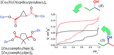
The 3D coordination polymer [Co2(H2O)(cpda)2(py)4·py]n (cpdaH2 is trans-(S,S)-1,2-cyclopropane dicarboxylic acid, py = pyridine) crystallizes from pyridine as 1·5py (one py is not coordinated) and was characterized by X-ray single crystal diffraction. Desolvation of 1·5py was accomplished with decoordination of pyridine and transformation of the CoII octahedral coordination into tetrahedral, as confirmed by electronic spectroscopy. Sorption of individual optical isomers – (S)-2-butanol and (R)-2-butanol – from the gas phase at 303 K by desolvated 1 was studied, and for comparison sorption of these substrates by the chiral MOFs [Zn2(camph)2(bipy)]n (2) and [Zn2(camph)2(dpe)]n (3) was examined (camphH2 is (1R,3S)-camphoric acid, bipy is 4,4′-bipyridine, dpe is trans-1,2-di(4-pyridyl)ethylene). Chiral sites in 1–3 contain only one polar group (carboxylate) in close proximity to the asymmetric C atom, while the other groups contain only C–H or C–C bonds. In the cases of 1 or 2 the absorption isotherms grew abruptly at certain pressure values P, and these values were different for the (R) or (S) isomers' sorptions. Such differential growth can be accounted for through the polymeric framework's rearrangement induced by interaction with 2-butanol, the difference in P values for (R) and (S) isomers being an indication of different interaction energies for these isomers with the MOF. There was no significant difference between the values of total sorption capacity of 1 for the two enantiomers of 2-butanol at pressures close to the saturation vapor pressure. In contrast, the sorption capacity of 3 was higher for (R)-2-butanol than for (S)-2-butanol over the whole pressure range.


Introduction

Porous coordination polymers, or metal–organic frameworks (MOFs), have been attracting attention due to their possible use in sorption and separation processes,1 catalysis,2 creation of new magnetic1a,b,3 or luminescent1a,b,4 materials, materials for non-linear optics,1a,5 etc. Fairly intensive studies of chiral MOFs were started in the previous decade, and a number of chiral materials for adsorption6 or chromatographic7 separation of enantiomers or chiral catalysts8 were developed on the basis of such compounds. In particular, MOFs possessing chiral lactate,6b α-aminocarboxylates or their derivatives,9,7a 2,2′-binaphthol10 or tartrate derivatives11 showed selectivity in adsorption or chromatographic separation of enantiomers of organic molecules. The efficiency of MOF's in such processes involving enantiomers is believed to depend on the structure and availability of a chiral center in the sorbent. Different enantiomers of the same substrate may adopt different orientations in the pores of a chiral MOF, associated with different energies of their interaction with the sorbent.6c However, the details of such interactions are not clear. In the majority of cases, the chiral center of the MOF is associated with a polar group, such as an hydroxo- or amino-group, which is able to form H-bonds or participate in dipole–dipole interactions. These groups provide the highest energy contribution to the binding, so that the lesser contribution of the chirality itself in the cavity cannot readily be quantified. On the other hand, it was shown that the match between size and shape of the cavity and guest molecule could be more important for recognition of optical isomers than the chirality of the coordination polymer.12

In comparison with the substantial number of extant publications on chiral MOFs, we describe here a rare case wherein the chiral centers in the ligands are constructed from three non-polar fragments (containing C–H or C–C bonds) and only one polar group (carboxyl). Here, we have examined sorption of pure enantiomers of 2-butanol by the new CoII complex [Co2(H2O)(cpda)2(py)4·py]n (1·5py) and by two previously reported13 ZnII complexes [Zn2(camph)2(bipy)·3DMF·2H2O]n (2·3DMF·2H2O) and [Zn2(camph)2(dpe)·5DMF·H2O]n (3·5DMF·H2O) (cpdaH2 is trans-(S,S)-1,2-cyclopropane dicarboxylic acid, camphH2 is (1R,3S)-camphoric acid, bipy is 4,4′-bipyridine, dpe is trans-1,2-di(4-pyridyl)ethylene; the chiral ligands are shown in Fig. 1). The specific feature of these chiral ligands (cpda2− and camph2−) is the presence of only one polar group in close proximity to each asymmetric C atom, in contrast to lactate, tartrate or any α-aminocarboxylate. Both cpda2− and camph2− contain two polar, hydrophilic carboxy groups, separated by a non-polar hydrophobic carbocyclic moiety, but they differ by the mutual orientation of these groups: trans- in the case of cpda2− and cis- in the case of camph2−. The carboxy groups in camph2− are more likely able to interact via a two-point binding with the same substrate molecule, while cpda2− is less likely to interact with guests having two closely spaced polar groups. It can be supposed that MOFs, containing this cpda2−, can show different interactions with chiral substrates due to chirality of the whole cavity rather than by chirality of a particular, localised site (such as a set of polar groups, etc.) for which the contribution to total substrate binding energy is the highest.


image file: c6ra09353a-f1.tif
Fig. 1 Structural formulae for trans-(S,S)-1,2-cyclopropane dicarboxylic acid (cpdaH2) and (1R,3S)-camphoric acid (camphH2).

The aim of this work was to study the influence of mutual orientation of polar hydrophilic carboxy groups in chiral dicarboxylates in MOFs on their ability to differentiate optical isomers of 2-butanol.

The new porous coordination polymer [Co2(H2O)(cpda)2(py)4·py]n (hereinafter abbreviated as 1·5py) was prepared and its X-ray structure was determined. The sorption of the two pure optical isomers of 2-butanol from the gas phase at 303 K by desolvated compounds 1–3 (the desolvated compounds are hereinafter referred to as 1′–3′, respectively) was studied and a dependence on the structure of MOF of the sorption capacity and of the shapes of the (R)- and (S)-isomers' sorption isotherms was investigated.

Results and discussion

Compound 1·5py was prepared by interaction of CoII nitrate and cpdaH2 in solution in the presence of pyridine, employed for deprotonation of cpdaH2 and avoidance of any necessity for additional counter-anions in the structures.

The crystal structure of 1·5py was determined by single-crystal X-ray diffraction. Compound 1 crystallizes in the tetragonal system (space group P41212), forming a 3D polymeric framework. Potential voids in the crystal lattice are filled by non-coordinated, solvating pyridine. The asymmetric unit of 1·5py contains one CoII ion, one cpda2− anion, two coordinated pyridine molecules, half a bridging water molecule (coordinated to CoII) and half a non-coordinated pyridine. The CoII ions possess cis-N2O4 donor sets, where the N atoms belong to coordinated pyridine and one O atom in each set belongs to the bridging water molecule, while the other oxygen donors are O-atoms of coordinated cpda2− anions (Fig. 2). Angular distortion of the CoN2O4 chromophore from an ideal octahedron D can be estimated by modification of a previously proposed procedure14 based on the deviation of the twelve D–Co–D cis-angles from 90° (see ESI for details). For CoII in 1, the D value is 3.2%, which is a little larger than in the more symmetric CoO6 chromophores in hexaaquacobalt(II) with nitrate15 (D = 1.6%), monoprotonated o-phthalate16 (D = 1.7%) or 1,1-cyclopropanedicarboxylate17 (D = 0.4%) as counter-ions. The bridging water Co–O(1)–Co angle in 1, at 113.07(9)°, is higher than similar angles in many reported aqua- or diaqua-bridged CoII complexes (Table S1, ESI).18 The cpda2− anions link three CoII ions, two of which are connected through one carboxylate group, while the second carboxylate group of each cpda2− is bound to only one CoII ion through one oxygen atom. Thus, dinuclear fragments Co2(μ-H2O)(–CO2)2 can be distinguished in the framework (where –CO2 signifies one carboxylate group of a cpda2− ligand). Each CoII links three cpda2− units, and each cpda2− anion links three CoII ions (two of which belong to the above-mentioned Co2(μ-H2O)(–CO2)2 block).


image file: c6ra09353a-f2.tif
Fig. 2 Co2 unit in coordination polymer 1·5py. Hydrogen atoms omitted for clarity of presentation. Symmetry operations: Co1′: ′−1 + y, 1 + x, 1 − z′; Co1′′: ′0.5 − x, −0.5 + y, 1.25 − z′; Co1′′′: ′0.5 − x, 0.5 + y, 1.25 − z′.

The 3D polymeric network in 1·5py can be represented as 2D layers, built and connected through cpda2− anions (Fig. 3 and S1, ESI). Three cpda2− anions linked to each Co2 unit participate in 2D layer formation, while the fourth cpda2− anion from each Co2 unit acts as a linker between adjacent 2D layers. Rings containing six Co2 units and six cpda2− anions can be distinguished in the 2D layers (Fig. 3).


image file: c6ra09353a-f3.tif
Fig. 3 2D layer in the polymeric network of 1·5py. cpda2− residues located on different faces of each 2D layer are marked by red and green. Coordinated and non-coordinated pyridine molecules, as well as all hydrogen atoms are omitted for clarity of presentation.

The putative cavities in which the non-coordinated pyridine molecules are found are not interconnected and do not form channels. Estimation of solvent-accessible voids for the lattice using the PLATON software19 applied to a lattice model from which just the solvating (non-coordinated) pyridines have been removed shows that there are no pores accessible for probe molecules with r equal to or greater than 2.02 Å (Fig. S2; the dynamic radius of pyridine is expected to be about 2.7–2.9 Å, similar to r of benzene.20 The apparent inconsistency between the size of solvent-accessible void estimated by PLATON and the dynamic radius of pyridine is a consequence of the non-spherical shape of the pore; the linear dimensions of the pyridine molecule are about 6.65 × 3.34 × 6.48 Å (ref. 21) so that it can fit into a “slot” not accessible to a spherical probe molecule. The total volume occupied by coordinated and non-coordinated pyridine in the lattice is 65.7% for a probe molecule with r = 1.4 Å, or 48.2% for a probe molecule with r = 2.8 Å (the latter value is equivalent to 0.54 cm3 g−1, which means the volume occupied by pyridine, but not the volume of voids which actually remain open after desolvation of 1·5py). Captured pyridine molecules are located inside a cavity surrounded by coordinated pyridines and cpda2− anions. The closest contact from a C atom of solvating pyridine to a cpda2− chiral carbon center is 3.934 Å (C17–C2), which serves as indirect evidence that the chiral center is potentially accessible for guest molecules.

Single crystals of 1·5py quickly lose/exchange the pyridine molecules when exposed to air and capture water, which leads to complete rearrangement of the crystal structure, as evidenced by comparison of X-ray powder diffraction patterns for an air-dried sample with a putative composition of 1·3.5py·2.5H2O with the pattern calculated for 1·5py (Fig. 4). Such collapse of the lattice is not surprising, taking into account that movement of captured pyridine from within the lattice cannot be mechanically trivial. Though we do not have enough data to conclude if the framework of 1 is flexible (i.e. can undergo reversible changes)22 or just friable, its ability to be changed upon guests' removal or exchange is critically important for discussion of its sorption properties, vide infra.


image file: c6ra09353a-f4.tif
Fig. 4 Powder X-ray diffraction patterns for air-dried sample 1·3.5py·2.5H2O (lower) and calculated for single crystal 1·5py (λ = 1.5406 Å, upper).

Complete desolvation of 1·3.5py·2.5H2O (formation of 1′), achieved by heating 1 in vacuum at 120 °C, is associated with a color change from pink to purple (Fig. S3, ESI) and complete loss of crystallinity, as can be concluded from the absence of peaks in the X-ray diffraction pattern. The λmax in its reflectance spectrum shifted from 495 to 560 nm (Fig. S3, band assignments provided in ESI). Such a change in the spectrum is explicable by decrease in coordination number of the initially hexacoordinate Co(II).23 Comparison of band positions in electronic spectra of CoII coordination polymers with different chromophores indicates that compounds with a tetrahedral donor set generally have bands with longer λmax than their octahedral analogues (Table S2, ESI), but the spectra do not rule out formation of trigonal-bipyramidal CoII (ref. 23) (Table S3, ESI). Desolvation of 1·3.5py·2.5H2O thus leads to a decrease of coordination number for at least some of the CoII ions, which in 1′ can then bind donor substrates. A tetrahedral donor set could be formed by μ-OH2 and three carboxylate oxygen atoms (O2, O5, O4′ for Co1), or μ-OH2 water could be eliminated and the coordination sphere of Co1 completed by carboxylate atom O3 (i.e., total composition O2, O3, O5, O4′ for Co1). A pentacoordinate form could, for example, result from elimination of two pyridine molecules and the above mentioned coordination of O3. However, an attempt to resolvate 1′ by 2-butanol did not lead to restoration of the initial electronic spectrum (Fig. S3), so it can be concluded that 2-butanol does not coordinate to CoII. This observation agrees with the observed 2-butanol sorption capacity, which was only 0.55 molecules per mole of CoII ions at the highest pressure, vide infra. It might have been expected that coordination of 2-butanol would facilitate its sorption to the level of one or two butanol molecules per one metal ion (for filling one or two vacancies in the cobalt coordination spheres, similarly to reported cases24); however, it seems that potential coordinatively-unsaturated sites do not play a significant role for sorption of 2-butanol. Also, no reflections were observed in the X-ray diffraction pattern of a sample of 1′ resolvated by 2-butanol (liquid) or water (vapor), evidencing that resolvation did not result in crystal lattice ordering. This observation agrees with the results of the electronic spectra measurement – resolvation by these compounds did not cause significant rearrangement of the crystal lattice of 1′.

Sorption of pure optical isomers of 2-butanol by compounds 1′–3′ (desolvated forms of 1–3, respectively) was measured in two separate experiments at T = 303 K. Although an increase in temperature led to a decrease of the enantiomeric excess in optical isomer adsorption by MOF from the racemate,10 we performed measurements at temperature above ambient in order to increase the pressure measurement accuracy (PS of 2-butanol at 303 K is 2.5 times higher than at room temperature). In neither case was the shape of sorption isotherms typical of adsorption (i.e. concentration of substrate on the interface25) in sorbents with rigid pores, but resembled absorption (i.e. penetration of substrate into the interior of the sorbent25) by compounds with flexible polymeric frameworks.26 The adjustable nature of the framework of 1′ is consistent with rearrangement of its crystal lattice upon desolvation (as shown above). We note that 1′ appeared to be non-porous, based on the results of N2 adsorption measurements (only surface adsorption of N2 was found). Similarly, it was shown that the crystal lattices of 2 and 3 collapsed upon solvent removal.13

The sorption behavior of 1′ and 2′ were quite similar, while the properties of 3′ were significantly different from those of these two MOFs'. Isotherms of both isomers' absorption by 1′ and 2′ were quite similar at low pressures. The difference between absorption of the two isomers occurred in a region of P/PS > 0.2 for 1′ and at P/PS > 0.4 for 2′ (Fig. 5a and b; P and PS are the pressure of 2-butanol and its saturation vapor pressure, respectively). At pressures higher than these values compounds 1′ and 2′ absorbed one of the isomers better than the other ((R)-2-butanol compared to (S)-2-butanol for 1′ and vice versa for 2′). It can be assumed that abrupt growth of the isotherm for one of the isomers sorption is associated with structural rearrangement – expansion of the voids – due to interaction of butanol with the structural elements of the coordination polymer. In the case of (S)-2-butanol sorption by 1′, similar growth of the isotherm was observed at higher P/PS (about 0.4). The positions of the maxima on the absorption isotherms derivatives vs. pressure (dV/dP) can serve as an additional metric to characterize the difference between sorption of the two isomers27 (Fig. S4, ESI). In the P/PS range from 0.1 to 0.9, the dV/dP maximum for (R)-2-butanol absorption by 1′ is located at P/PS = 0.2, while the maximum for (S)-2-butanol is observed at P/PS = 0.4. This difference between (R)- and (S)-isomers can be associated with different energies of their interaction with the structural elements of 1′, because the energy consumed for the structural rearrangement must be compensated by the energy obtained upon substrate absorption.


image file: c6ra09353a-f5.tif
Fig. 5 Isotherms for 2-butanol enantiomer sorption by 1′ (a), 2′ (b) and 3′ (c) from the gas phase at 303 K. image file: c6ra09353a-u1.tif – (S)-2-butanol, image file: c6ra09353a-u2.tif – (R)-2-butanol. Inset shows the same isotherms as on the main graph, on a coarser scale.

The volume of one of the absorbed isomers was greater than the volume of the other isomer with a maximum of ca. 1.35 times in the case of 1′ (at P/PS = 0.37) and ca. 1.55 times in the case of 2′ (at P/PS = 0.85). A pressure increase to P/PS > 0.9 led to significant growth of sorption capacity for both 2-butanol enantiomers, for both 1′ and 2′. Though there can be some contribution of inter-particle condensation of the alcohol at high P/PS, further expansion of the polymeric framework cannot be excluded.

In the case of 1′ and 2′, the difference between the isomers' sorptions occurred only in the pressure range associated with pore expansion. For example, one might posit that the energy of (R)-2-butanol sorption by 1′Gsorpt.) is slightly greater in magnitude compared to the energy of (S)-2-butanol sorption, but the difference can be seen only when this sorption energy is directly compared with the energy of structural rearrangement (ΔGrearrang.) – the isotherm grows only when |ΔGsorpt.| > |ΔGrearrang.|, the abrupt growth of the isotherm can serve as a kind of benchmark to evidence that the desired energy profit from sorption is already achieved.

In contrast to 1′ and 2′, the sorption capacity of 3′ for (R)-2-butanol exceeded the capacity for the (S)-isomer over the whole range of pressures, and the difference was 2.5-fold at P/PS = 0.9 (Fig. 5c). Some “steps” on the sorption isotherms might indicate incidents of structural rearrangement, and the (R)-isomer seems to induce more significant framework expansion than the (S)-isomer, as can be concluded from the relative increments of the isotherms on these “steps”. Surprisingly, compound 3′ absorbed (R)-2-butanol better, while 2′ was a better absorbant for (S)-2-butanol, despite similar structures of these sorbents. This difference can be considered as further evidence for host–guest interactions between 2-butanol and MOF rather than “classical” pore-filling by substrate where 2-butanol interacts only with chiral centers of certain camphorates, without participation of other functional groups from neighboring molecules or fragments. It should be noted, that as implied above, close inspection of the crystal structure of 1·5py leads to the conclusion that 2-butanol cannot enter the voids freely without framework rearrangement. The size of channels in 2 (5 × 7 Å for solvated single crystal) or 3 (5 × 10 Å)13 can allow for “free” 2-butanol capture, but only if the crystal lattice does not collapse upon desolvation, which is not the case. Diffusion of 2-butanol with some energy barrier associated with framework rearrangement seems to be the more probable option. It was shown that the sorption capacity of another chiral MOF, [Zn(Lact)(bdc)]n (LactH2 is (S)-lactic acid, and bdcH2 is 1,4-benzenedicarboxylic acid), with respect to (R)-2-butanol was significantly higher than for (S)-2-butanol in the whole pressure range, but the pores in this MOF are not sufficiently large for “unhindered” penetration of 2-butanol molecules.28 Thus, accessibility of the pores in the lattice “as-is” (i.e., before possible rearrangement) is not the critical factor which controls the recognition of optical isomers, and it probably should be considered as just one of the factors, along with contributions from the framework flexibility (rearrangement energy), host–guest interaction energy, etc. To support this idea, it can be mentioned, that MOFs based on camphorate (including 229e) allowed chromatographic separation of optical isomers of various compounds, molecules of which were apparently larger than the pore size.29 Though separation of isomers in these cases could be enabled mainly by surface interactions, absorption of substrates by MOFs also can make a contribution to differentiation of such compounds.

The difference between isotherms of pure isomers' sorption can be quantitatively evaluated as the ratio between the quantities of absorbed isomers at a given pressure, or enantioselectivity (es) (similarly to the definition proposed by Liu et al.30). Judging from the es criterion, the performance of compound 3′ exceeds that of other chiral MOFs for which sorption of pure isomers of alcohols has been measured (Table 1).

Table 1 Enantioselectivity of chiral PCPs in pure isomers' sorption from gas phase
Compounda Substrate esb Conditions Ref.
a H2Lact is (S)-lactic acid, H2bdc is 1,4-benzenedicarboxylic acid; H2asp is (R)-aspartic acid, bipy is 4,4′-bipyridine; H3TMTA is trimesoyltri(S-alanine); 1,2-pd is 1,2-propanediol, 3-pic = 3-picoline, H3btc = 1,3,5-benzenetricarboxylic acid; H2camph is (1R,3S)-camphoric acid; dabco is 1,4-diazabicyclo(2.2.2)octane.b es is ratio of quantities of isomers, letter indicates the better-absorbed isomer.c Ratio of VDR of (R)-ethyl-3-hydroxybutyrate, absorbed by sorbent containing (S)-1,2-propanediol, to VDR of (R)-ethyl-3-hydroxybutyrate, absorbed by sorbent containing (R)-1,2-propanediol, where VDR is the Dubinin–Radushkevich micropore volume.
1′ (R) and (S) isomers of 2-butanol 1.3(R) P/PS = 0.4, T = 303 K This work
2′ 1.7(S) P/PS = 0.9, T = 303 K This work
3′ 2.4(R) This work
[Zn(Lact)(bdc)]nb 2.1(R) 28
[Ni2(asp)2(bipy)]nb 1.4(R) 28
[Co3(TMTA)2(bipy)4]nb 1.6(S) 28
[Ni3(btc)2(3-pic)6(1,2-pd)3]nb (R)-Ethyl-3-hydroxybutyrate 1.06c T = 300 K 6a
[Zn2(camph)2(dabco)]nb (2R,5R)-2,5-He-xanediol/(2S,5S)-2,5-he-xanediol 1.5(2R,5R) Saturated vapor in N2, room temp. 30


Evacuation of 1′–3′ after butanol absorption did not lead to complete desorption of either isomer, significant absorption/desorption hysteresis being observed, with some part of the 2-butanol being absorbed irreversibly. The quantities of irreversibly-absorbed isomers were similar in the cases of 1′ and 3′ (noticeable desorption of (R)-isomer from sample 3′ occurred upon pressure decrease, and the quantity of (R)-isomer remaining was similar to the level of remaining (S)-isomer); irreversibility of sorption is consistent with the supposition about polymeric framework expansion upon inclusion of guest molecules. The volume occupied by irreversibly-absorbed 2-butanol can correspond to one of the meta-stable conformations, adopted by the polymeric framework as a consequence of its reorganisation.

The quantity of 2-butanol remaining after its partial pressure is reduced to zero in 1′ or 2′ corresponds to a volume much lower than that of the volumes occupied by coordinated and non-coordinated pyridine (vide supra) in the solvated single crystal of 1·5py (about 0.54 cm3 g−1) or solvent-accessible volume in 2′ (about 0.5 cm3 g−1, as estimated from structural data13). This estimation is consistent with some flexibility of the frameworks of these complexes and can be accounted for by a scenario in which desolvation causes crystal lattice collapse, while interaction with 2-butanol induces pore “expansion”, the level of which is governed by the host–guest interaction energy.

Potential coordination of 2-butanol to CoII ions in 1′ is likely not an important driving force for its absorption, because the sorption capacity expressed in moles of 2-butanol per mole of CoII at P/PS < 0.7, for example, is only ca. 0.2.

The ability of chiral guest and host's chiral recognition sites to participate in multi-center interactions (simultaneous interaction of several groups or atoms) is considered to be the key factor which controls the ability of a chiral sorbent to discriminate enantiomers.6c Such interactions can include formation of H-bonds and dipole–dipole interactions, as well as weaker interactions such as π-stacking, etc., and of course the configuration of the binding groups or atoms should be asymmetric. It seems reasonable to presume, that specificity of sorption (efficiency of discrimination) should be higher, if host–guest interactions are stronger, for example, selectivity of diols' adsorption or chromatographic separation by chiral MOFs is usually higher than selectivity achieved for monofunctional alcohols (or their derivatives),31 while sorption separation of isomers of aromatic alcohols is better than for aliphatic ones.32 From this viewpoint, cpda2− and camph2− would seem to be disadvantageous for enantiomer recognition compared to, for example, lactate, derivatives of α-aminoacids or β-cyclodextrins. All these ligands contain several polar carboxy groups close to asymmetric C atoms, which favor interaction of chiral guest with chiral host, while in cpda2− or camph2− only one carboxyl group is located close to the asymmetric C atom, and other groups (C–H) can probably participate in very weak dipole–dipole interactions at best. On the other hand, assembly of several cpda2− or camph2− anions by one metal ion can form a binding site containing multiple chiral centers in close proximity to polar groups. The distance between C-atoms of carboxyl groups belonging to two different cpda2− anions bound to one CoII ion in 1 is ca. 3.97 Å, which is comparable to the distance between C-atoms of carboxyl groups in one cpda2− anion (ca. 3.86 Å). Similarly, the distance between C-atoms of carboxyl groups belonging to two different camph2− anions bound to one ZnII ion in 2, is 3.63 Å – significantly shorter than the distance between C-atoms of carboxyl groups in one camph2− anion (ca. 5.07 Å). Thus, binding of several chiral ligands by metal ions in 1–3 (i.e. location of asymmetric centers of the different neighboring ligands in close proximity to each other) seems to play a more important role than the structure of one ligand.

Conclusions

Binding of CoII ions by trans-S,S-cyclopropane dicarboxylate allowed the preparation of a new chiral 3D coordination polymer, 1·5py. The crystal lattice of this compound collapsed upon removal of pyridine, but the desolvated sample 1′ was able to absorb 2-butanol. Though the isotherms of (S)-2-butanol and (R)-2-butanol sorption were quite similar at the low and high pressure extremes, a difference was found in the range of P/PS from 0.2 to 0.9, where dV/dP is itself large. Abrupt growth of the isotherms in this region can be associated with expansion of the porous framework of the sorbate due to lattice rearrangements.

The shapes of the isotherms for the 2-butanol enantiomers' absorption by 1′ and 2 were different, which also demonstrates the differences due to chirality and can be accounted for by these isomers exerting selectivity in inducing rearrangements of the frameworks of these MOFs. Notably, only examination of the full sorption isotherm allowed the discovery of the difference between sorption of the enantiomers by 1′, since sorption of both isomers of 2-butanol at either very low or high pressure, as well as residual sorption capacity, were the same. In any case, a single carboxy group near the asymmetric carbon atom in cpda2− was sufficient to generate a difference between sorption of the (R) and (S) isomers of 2-butanol. Along with this, the role of the metal ion as linker amongst several chiral ligands (yet not as a potential coordination site) seems to be critically important for generation of an efficient recognition site. Nevertheless, the efficiency of 1′ in the process of chiral molecule discrimination is less than for analogues with lactate, α-aminocarboxylate or similar ligands containing several polar groups near the asymmetric carbon, and this lower efficiency is consistent with the structure of sorbent (only one polar group near the asymmetric C atoms). Thus, pore accessibility is hardly the sole factor governing the recognition of optical isomers, and should be considered as one of several factors, along with contribution of the framework flexibility (rearrangement energy), host–guest interaction energy, etc.

The outcomes of this work may be helpful in understanding the mechanism for interaction of chiral compounds with porous coordination polymers, especially with sorbents possessing flexible frameworks.

Experimental

General

Reagents and solvents were commercially available (Aldrich, ABCR) and were used without further purification. trans-S,S-Cyclopropane-1,2-dicarboxylic acid was synthesized as previously described.33

Single crystal X-ray diffraction

Data were collected using Bruker ApexII CCD diffractometer with Mo-Kα radiation (λ = 0.71073 Å) with a fine focus sealed tube X-ray source with graphite monochromator. A single crystal was mounted on a Mitegen micromesh mount using a trace of mineral oil and cooled in situ to 100(2) K for data collection. Frames were collected, reflections were indexed and processed, and the files scaled and corrected for absorption using APEX2.34 The space group was assigned and the structure solved by direct methods using XPREP within the SHELXTL suite of programs35 and refined by full matrix least squares against F2 with all reflections using Shelxl2014.36 H atoms attached to carbon atoms were positioned geometrically and constrained to ride on their parent atoms, with carbon hydrogen bond distances of 0.95 Å for pyridine C–H, 1.00 and 0.99 for aliphatic C–H and CH2. Water H atoms were located in a difference density electron map and were refined with an O–H distance restraint of 0.84(2) Å. Uiso(H) values were set to a multiple of Ueq(O/C) with 1.5 for OH and 1.2 for C–H and CH2 units, respectively. The uncoordinated pyridine molecule is disordered around a twofold axis. It was constrained to resemble an ideal hexagon with C–C and C–N bonds of 1.39 Å. The Uij components of the ADPs of disordered atoms were restrained to be similar if closer than 1.7 Å to each other. Complete crystallographic data, in CIF format, have been deposited with the Cambridge Crystallographic Data Centre. CCDC 1454802 contains the supplementary crystallographic data for this paper.

Powder X-ray diffraction experiments were performed on a Bruker D8 instrument with Cu radiation (λ = 1.54 Å) in pellets or in a 0.1 mm capillary. Nitrogen sorption at 77.5 K was estimated using a Sorptomatic-1990 instrument; prior to measurement, the sample of 1′ was additionally activated in vacuum at 120 °C. Reflectance spectra were measured using a Specord 210 spectrometer (Analytik Jena AG) in BaSO4 pellets. Sorption of (S)- or (R)-isomers of 2-butanol was studied gravimetrically using a quartz microbalance at 303 K. Elongation of the springs was measured with an optical cathetometer (elongation of the springs during measurements reached ca. 10 cm, the accuracy of measurement being ±0.3 mm). The pressure in the system was measured with an Hg manometer, the Hg-level being measured with the same cathetometer as above. The changes in Hg level in the experiment ranged up to 29.3(±0.3) mm, which corresponded to the PS (in mm Hg) of 2-butanol at 303 K. Each point on the absorption and desorption isotherms corresponds to equilibrium conditions (i.e. no change of sample weight at the given P/PS, where PS is the saturation vapour pressure of the compound at 303 K). Similarly, measurements of the samples after complete evacuation (the last point on the desorption branch of the isotherm) were performed under equilibrium conditions. Prior to sorption measurements the samples were thermally activated at 120 °C in vacuo at 10−2 Torr.

The volume of the pores was estimated from the quantity of absorbed butanol by using the 303 K liquid density.

Synthesis of compound 1

Co(NO3)2·6H2O (0.029 g, 0.1 mmol) and cpdaH2 (0.013 g, 0.1 mmol) were dissolved in 3 mL of DMF, then 3 mL of MeOH were added. Diffusion of pyridine vapor led to formation of crystals of 1·5py after several weeks (composition of single crystals determined by X-ray diffraction). These crystals were collected by filtration, washed with MeOH and air-dried. Anal. Calcd for C27.5H32.5N3.5O11.5Co2 (air-dried/equilibrated sample – composition 1·3.5py·2.5H2O): C, 46.2; H, 4.55; N, 6.86. Found: C, 46.2; H, 4.51; N, 6.88.

References

  1. (a) C. Wang, D. Liu and W. Lin, J. Am. Chem. Soc., 2013, 135, 13222–13234 CrossRef CAS PubMed; (b) R. J. Kuppler, D. J. Timmons, Q.-R. Fang, J.-R. Li, T. A. Makal, M. D. Young, D. Yuan, D. Zhao, W. Zhuang and H.-C. Zhou, Coord. Chem. Rev., 2009, 253, 3042–3066 CrossRef CAS; (c) J.-R. Li, J. Sculley and H.-C. Zhou, Chem. Rev., 2012, 112, 869–932 CrossRef CAS PubMed; (d) S. Qiu and G. Zhu, Coord. Chem. Rev., 2009, 253, 2891–2911 CrossRef CAS; (e) B. Chen, C. Liang, J. Yang, D. S. Contreras, Y. L. Clancy, E. B. Lobkovsky, O. M. Yaghi and S. Dai, Angew. Chem., Int. Ed., 2006, 45, 1390–1393 CrossRef CAS PubMed; (f) Z.-Y. Gu, C.-X. Yang, N. Chang and X.-P. Yan, Acc. Chem. Res., 2012, 45, 734–745 CrossRef CAS PubMed.
  2. (a) A. Dhakshinamoorthy, M. Opanasenko, J. Cejka and H. Garcia, Catal. Sci. Technol., 2013, 3, 2509–2540 RSC; (b) J. Lee, O. K. Farha, J. Roberts, K. A. Scheidt, S. T. Nguyen and J. T. Hupp, Chem. Soc. Rev., 2009, 38, 1450–1459 RSC; (c) P. K. Thallapally, C. A. Fernandez, R. K. Motkuri, S. K. Nune, J. Liu and C. H. F. Peden, Dalton Trans., 2010, 39, 1692–1694 RSC.
  3. M. Kurmoo, Chem. Soc. Rev., 2009, 38, 1353–1379 RSC.
  4. (a) Y. Cui, Y. Yue, G. Qian and B. Chen, Chem. Rev., 2012, 112, 1126–1162 CrossRef CAS PubMed; (b) Z. Hu, B. J. Deibert and J. Li, Chem. Soc. Rev., 2014, 43, 5815–5840 RSC.
  5. C. Wang, T. Zhang and W. Lin, Chem. Rev., 2012, 112, 1084–1104 CrossRef CAS PubMed.
  6. (a) D. Bradshaw, T. J. Prior, E. J. Cussen, J. B. Claridge and M. J. Rosseinsky, J. Am. Chem. Soc., 2004, 126, 6106–6114 CrossRef CAS PubMed; (b) D. N. Dybtsev, A. L. Nuzhdin, H. Chun, K. P. Bryliakov, E. P. Talsi, V. P. Fedin and K. Kim, Angew. Chem., Int. Ed., 2006, 45, 916–920 CrossRef CAS PubMed; (c) D. N. Dybtsev, M. P. Yutkin, D. G. Samsonenko, V. P. Fedin, A. L. Nuzhdin, A. A. Bezrukov, K. P. Bryliakov, E. P. Talsi, R. V. Belosludov, H. Mizuseki, Y. Kawazoe, O. S. Subbotin and V. R. Belosludov, Chem.–Eur. J., 2010, 16, 10348–10356 CrossRef CAS PubMed.
  7. (a) S.-M. Xie, Z.-J. Zhang, Z.-Y. Wang and L.-M. Yuan, J. Am. Chem. Soc., 2011, 133, 11892–11895 CrossRef CAS PubMed; (b) X. Kuang, Y. Ma, H. Su, J. Zhang, Y.-B. Dong and B. Tang, Anal. Chem., 2014, 86, 1277–1281 CrossRef CAS PubMed; (c) P. Peluso, V. Mamane and S. Cossu, J. Chromatogr. A, 2014, 1363, 11–26 CrossRef CAS PubMed.
  8. (a) M. Yoon, R. Srirambalaji and K. Kim, Chem. Rev., 2012, 112, 1196–1231 CrossRef CAS PubMed; (b) M. J. Ingleson, J. P. Barrio, J. Bacsa, C. Dickinson, H. Park and M. J. Rosseinsky, Chem. Commun., 2008, 1287–1289 RSC; (c) L. Ma, C.-D. Wu, M. M. Wanderley and W. Lin, Angew. Chem., Int. Ed., 2010, 49, 8244–8248 CrossRef CAS PubMed; (d) A. L. Nuzhdin, D. N. Dybtsev, K. P. Bryliakov, E. P. Talsi and V. P. Fedin, J. Am. Chem. Soc., 2007, 129, 12958–12959 CrossRef CAS PubMed.
  9. R. Vaidhyanathan, D. Bradshaw, J.-N. Rebilly, J. P. Barrio, J. A. Gould, N. G. Berry and M. J. Rosseinsky, Angew. Chem., Int. Ed., 2006, 45, 6495–6499 CrossRef CAS PubMed.
  10. Y. Peng, T. Gong, K. Zhang, X. Lin, Y. Liu, J. Jiang and Y. Cui, Nat. Commun., 2014, 5, 4406 CAS.
  11. J. S. Seo, D. Whang, H. Lee, S. I. Jun, J. Oh, Y. J. Jeon and K. Kim, Nature, 2000, 404, 982–986 CrossRef CAS PubMed.
  12. P. Z. Moghadam and T. Düren, J. Phys. Chem. C, 2012, 116, 20874–20881 CAS.
  13. D. N. Dybtsev, M. P. Yutkin, E. V. Peresypkina, A. V. Virovets, C. Serre, G. Férey and V. P. Fedin, Inorg. Chem., 2007, 46, 6843–6845 CrossRef CAS PubMed.
  14. J. M. Popovitch, A. W. Addison, R. J. Butcher and M. J. Prushan, J. Chem. Crystallogr., 2012, 42, 295–298 CrossRef CAS.
  15. P. V. Prelesnik, F. Gabela, B. Ribar and I. Krstanovic, Cryst. Struct. Commun., 1973, 2, 581–583 Search PubMed.
  16. B. M. Kariuki and W. Jones, Acta Crystallogr., Sect. C: Cryst. Struct. Commun., 1993, 49, 2100–2102 CrossRef.
  17. T. Schwarz, A. Petri, J. Schilling and A. Lentz, Acta Crystallogr., Sect. C: Cryst. Struct. Commun., 1998, 54, 1104–1105 Search PubMed.
  18. (a) D. Coucouvanis, R. A. Reynolds III and W. R. Dunham, J. Am. Chem. Soc., 1995, 117, 7570–7571 CrossRef CAS; (b) D. Lee, P.-L. Hung, B. Spingler and S. J. Lippard, Inorg. Chem., 2002, 41, 521–531 CrossRef CAS PubMed; (c) L. G. Beauvais, M. P. Shores and J. R. Long, J. Am. Chem. Soc., 2000, 122, 2763–2772 CrossRef CAS; (d) X. Meng, H. Hou, G. Li, B. Ye, T. Ge, Y. Fan, Y. Zhu and H. Sakiyama, J. Organomet. Chem., 2004, 689, 1218–1229 CrossRef CAS; (e) D. A. Brown, W. K. Glass, N. J. Fitzpatrick, T. J. Kemp, W. Errington, H. Müller-Bunz, A. J. Hussein and H. Nimir, Inorg. Chim. Acta, 2005, 358, 2755–2762 CrossRef CAS; (f) J. Lefebvre, P. Tyagi, S. Trudel, V. Pacradouni, C. Kaiser, J. E. Sonier and D. B. Leznoff, Inorg. Chem., 2009, 48, 55–67 CrossRef CAS PubMed.
  19. A. L. Spek, Acta Crystallogr., Sect. A: Cryst. Phys., Diffr., Theor. Gen. Crystallogr., 1990, 46, C34 Search PubMed.
  20. J.-R. Li, R. J. Kuppler and H.-C. Zhou, Chem. Soc. Rev., 2009, 38, 1477–1504 RSC.
  21. C. E. Webster, R. S. Drago and M. C. Zerner, J. Am. Chem. Soc., 1998, 120, 5509–5516 CrossRef CAS.
  22. A. Schneemann, V. Bon, I. Schwedler, I. Senkovska, S. Kaskel and R. A. Fischer, Chem. Soc. Rev., 2014, 43, 6062–6096 RSC.
  23. A. B. P. Lever, Inorganic Electronic Spectroscopy, Elsevier, New York, 1984 Search PubMed.
  24. (a) S. Ohkoshi, Y. Tsunobuchi, H. Takahashi, T. Hozumi, M. Shiro and K. Hashimoto, J. Am. Chem. Soc., 2007, 129, 3084–3085 CrossRef CAS PubMed; (b) Z.-Y. Liu, E.-C. Yang, L.-L. Li and X.-J. Zhao, Dalton Trans., 2012, 41, 6827–6832 RSC.
  25. (a) K. S. W. Sing, D. H. Everett, R. A. W. Haul, L. Moscou, R. A. Pierotti, J. Rouquerol and T. Siemieniewska, Pure Appl. Chem., 1985, 57, 603–619 CrossRef CAS; (b) S. J. Gregg and K. S. W. Sing, in Adsorption, Surface Area and Porosity, Academic Press, London, 2nd edn, 1982 Search PubMed.
  26. N. C. Burtch, H. Jasuja and K. S. Walton, Chem. Rev., 2014, 114, 10575–10612 CrossRef CAS PubMed.
  27. A. V. Pavlishchuk, S. V. Kolotilov, M. Zeller, O. V. Shvets, I. O. Fritsky, S. E. Lofland, A. W. Addison and A. D. Hunter, Eur. J. Inorg. Chem., 2011, 4826–4836 CrossRef CAS.
  28. Yu. A. Satska, N. P. Komarova, K. S. Gavrilenko, O. V. Manoylenko, Zh. V. Chernenko, M. A. Kiskin, S. V. Kolotilov, I. L. Eremenko and V. M. Novotortsev, Theor. Exp. Chem., 2015, 51, 45–53 CrossRef CAS.
  29. (a) J. Kong, M. Zhang, A.-H. Duan, J.-H. Zhang, R. Yang and L.-M. Yuan, J. Sep. Sci., 2015, 38, 556–561 CrossRef CAS PubMed; (b) S.-M. Xie, X.-H. Zhang, Z.-J. Zhang, M. Zhang, J. Jia and L.-M. Yuan, Anal. Bioanal. Chem., 2013, 405, 3407–3412 CrossRef CAS PubMed; (c) S. Xie, B. Wang, X. Zhang, J. Zhang, M. Zhang and L. Yuan, Chirality, 2014, 26, 27–32 CrossRef CAS PubMed; (d) S.-M. Xie, X.-H. Zhang, Z.-J. Zhang and L.-M. Yuan, Anal. Lett., 2013, 46, 753–763 CrossRef CAS; (e) M. Zhang, X.-D. Xue, J.-H. Zhang, S.-M. Xie, Y. Zhang and L.-M. Yuan, Anal. Methods, 2014, 6, 341–346 RSC.
  30. B. Liu, O. Shekhah, H. K. Arslan, J. Liu, C. Wöll and R. A. Fischer, Angew. Chem., Int. Ed., 2012, 51, 807–810 CrossRef CAS PubMed.
  31. X.-X. Zhang, S.-M. Xie, A.-H. Duan, B.-J. Wang and L.-M. Yuan, Chromatographia, 2013, 76, 831–836 CAS.
  32. (a) P. Li, Y. He, J. Guang, L. Weng, J. C.-G. Zhao, S. Xiang and B. Chen, J. Am. Chem. Soc., 2014, 136, 547–549 CrossRef CAS PubMed; (b) K. Suh, M. P. Yutkin, D. N. Dybtsev, V. P. Fedin and K. Kim, Chem. Commun., 2012, 48, 513–515 RSC.
  33. K. Furuta, K. Iwanaga and H. Yamamoto, Org. Synth., 1989, 67, 76–85 CrossRef CAS.
  34. Apex2 v2009.7-0, Bruker AXS Inc., Madison (WI), USA, 2009 Search PubMed.
  35. (a) SHELXTL (Version 6.14), Bruker Advanced X-ray Solutions, Bruker AXS Inc., Madison, Wisconsin, USA, 2000–2003 Search PubMed; (b) G. M. Sheldrick, Acta Crystallogr., Sect. A: Found. Crystallogr., 2008, 64, 112–122 CrossRef CAS PubMed.
  36. (a) G. M. Sheldrick, Acta Crystallogr., Sect. C: Struct. Chem., 2015, 71, 3 CrossRef PubMed; (b) G. M. Sheldrick, SHELXTL, University of Göttingen, Germany, 2013 Search PubMed.

Footnote

Electronic supplementary information (ESI) available. CCDC 1454802. For ESI and crystallographic data in CIF or other electronic format see DOI: 10.1039/c6ra09353a

This journal is © The Royal Society of Chemistry 2016