Guangdi Nie,
Xiaofeng Lu*,
Maoqiang Chi,
Yanzhou Jiang and
Ce Wang*
Alan G. MacDiarmid Institute, College of Chemistry, Jilin University, Changchun, 130012, P. R. China. E-mail: xflu@jlu.edu.cn; cwang@jlu.edu.cn; Fax: +86-431-85168292; Tel: +86-431-85168292
First published on 18th May 2016
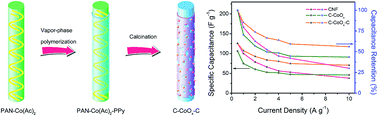
A novel composite nanostructure of C–CoOx–C with CoOx nanoparticles embedded in N-containing porous graphite carbon nanofibers (CNF) is successfully prepared via sintering the electrospun polyacrylonitrile–cobalt acetate tetrahydrate nanofibers covered by a polypyrrole (PPy) sheath that were attained from the chemical vapor-phase polymerization of pyrrole monomers using concentrated nitric acid as both the dopant and oxidant for the first time. The unique configuration with a well-defined morphology possesses a large specific surface area and prominent conductivity contributed to by the catalysis of metallic Co and the external PPy-derived carbon envelope, which could facilitate effective electron transfer and rapid ion penetration in C–CoOx–C, thus improving its electrochemical performance. As expected, when employed as an electrode active material for supercapacitors, the resultant C–CoOx–C showed a more acceptable specific capacitance, better rate capability and higher cycling stability than individual CNF and CoOx nanoparticle-decorated CNF without the coating of a N-doped carbon layer from PPy (C–CoOx).
To overcome the above deficiencies, numerous attempts have been made to incorporate nanostructured cobalt oxides (denoted CoOx) into conductive substrates, such as carbon, metal and conjugated polymer backbones to improve the properties of CoOx.14–16 For instance, a well-aligned CoO@polypyrrole nanowire array and a Co3O4/flocculent graphene hybrid were respectively immobilized onto three-dimensional (3D) nickel foam, which indeed exhibited extraordinarily high specific capacitances with assistance from the inherent electrochemical activity of each component, the excellent electronic conductivity of the scaffold, the short ion penetration pathway in the ordered structures and the unique synergy effects.17,18 Nevertheless, when taking the overall weight of the electrode into consideration, the equated gravimetric capacitance would be very low.19 Normally, for a whole electrode, the larger the content of active material, the more energy the electrode can reserve, but a limit occurs, and increasing the mass loading to a certain extent usually engenders serious loss of the specific capacitance and rate capability, mainly because of enhanced resistance.20 Therefore, it is still challenging and yet imperative to strike a better balance between the electroactive materials and conductive matrix to promote their electrochemical performance.
On the other hand, compared with commercial graphene, carbon nanotubes (CNT) and carbon cloth (CC) with the fiber diameters usually at the micron level, the electrospun carbon nanofibers (CNF) as the electrode materials and supporting frameworks have some advantages in terms of ion transmission, the fabrication process and cost, which are of great concern for practical applications in SCs.21,22 However, the specific surface area (SSA) of CNF prepared using electrospinning is generally 10–30 m2 g−1, one order of magnitude lower than that of graphene (909 m2 g−1) or CNT (272 m2 g−1),22–24 hence limiting their charge–discharge ability at the interface of electrode and electrolyte. More recently, researchers have demonstrated that the in situ combination of CoOx with CNF can create tunable pores for increasing the SSA of the host material, but often with sacrifice of the conductivity, owning to the exudation and decomposition of metal salts during thermal treatments.12,25 Another effective approach to improve the capacitive behavior of carbon electrodes is to embellish the surface functionalities of porous carbonaceous materials with heteroatoms like N, B, S and P,26 among which N is a more plausible dopant as it could substantially ameliorate the conductivity, surface wettability, adhesion and polarity of the carbon as a consequence of the conjugation between N lone-pair electrons and graphitic π-bonds as well as the more active sites and defects imparted by the partial substitution of N for C atoms.27–29 Direct carbonization of N-enriched polymers has been identified as a simple and valid strategy to obtain N-doped carbon with a comparatively high concentration and homogeneous distribution of N, which is able to persist in harsh working conditions.30,31 Polypyrrole (PPy) is an ideal nitrogen source due to its facile conversion of N atoms in pyrrole rings to graphitic N, high nitrogen content and carbon yield.32,33 To the best of our knowledge, chemical vapor-phase polymerization (CVPP) is a solvent-free versatile technique to obtain conducting polymer layers with a desired thickness, smooth surface and good conductivity that are uniformly clad on different insulating substrates, in which the polymerization commonly proceeds at the oxidant-wrapped matrix in the monomer vapor.34
In light of the above reasons, herein, we report a novel nanocomposite (denoted as C–CoOx–C) with CoOx nanoparticles embedded in N-containing porous graphite CNF via sintering the electrospun polyacrylonitrile (PAN)–cobalt acetate tetrahydrate (Co(Ac)2·4H2O) nanofibers covered by a PPy shell that originate from the CVPP of pyrrole monomers using concentrated nitric acid (HNO3) as both the dopant and oxidant for the first time (Scheme 1). As anticipated, when serving as an electrode material for SCs, the obtained C–CoOx–C showed a more acceptable specific capacitance, higher rate capability and better cycling stability than pure CNF and CoOx nanoparticle-decorated CNF without the coating of a N-doped carbon layer from PPy (designated as C–CoOx), which should be attributed to the porous nanostructure with a large SSA, an appropriate equilibrium of exposed graphene edges and defects, outstanding conductivity induced by the external PPy-derived carbon envelope and optimal synergy effects of the constituents.
As illustrated in the representative transmission electron microscopy (TEM) image of C–CoOx–C (Fig. 2A), the majority of the CoOx granules were encapsulated by graphene lamellae, which could not merely firmly hold the nanoparticles, but also notably improve the conductivity of the local amorphous carbon.25 It was measured and calculated from the high-resolution TEM (HRTEM) images of a naked CoOx particle at the transversal section (Fig. 2B) and of the graphene layers (Fig. 2C) that the spacings between two adjacent lattice fringes were 0.24 and 0.34 nm, belonging to the characteristic d311 of cubic Co3O4 phase and d002 of graphite carbon, respectively. The ordered lattice shown in the annotated selected area electron diffraction (SAED) pattern (Fig. 2D) of C–CoOx–C indicated that the bare Co3O4 was of monocrystalline nature. Fig. 2E shows the relevant energy dispersive X-ray (EDX) spectrum, where the signals of Cu and Si should be attributed to the carbon-coated copper grid and the instrument substrate, revealing the presence of C, N, O and Co elements in the specimen C–CoOx–C. The elemental mapping in Fig. 2F shows potent evidence that N atoms were evenly scattered throughout the whole composite nanofiber, while the O element was principally concentrated at the same point as Co.
To validate the crystallographic features of the sintered samples, X-ray diffraction (XRD) patterns are depicted in Fig. 3A. With regard to the obtained CNF, two broad peaks centered at 24.5 and 43.5° were distinctly detected, which are in complete agreement with the (002) and (100) planes Bragg reflection of graphite. In the case of C–CoOx and C–CoOx–C, the just-mentioned peak of (002) facet had narrowed and shifted to a larger diffraction angle (2 theta) of 25.8°, proving that a higher graphitization degree and more exposed graphene edges existed in the carbon matrices of the two products on account of the catalytic and reduction effect of metallic Co.30,36 Besides, all the other peaks could be lightly assigned to the cubic phase of Co3O4 (JCPDS card no. 43-1003) and CoO (JCPDS card no. 48-1719) without any impurities. The Raman spectra of the pure CNF (Fig. 3B) clearly exhibited a pair of peaks at 1345 and 1580 cm−1, corresponding to a defect- or disorder-evoked D band and a crystalline graphitic G band, respectively. As for C–CoOx and C–CoOx–C, analogous patterns except for an increased intensity ratio between the D and G bands (ID/IG) of the latter were observed with the typical vibration modes of Co3O4 and CoO at 190, 466, 508, 606 and 673 cm−1,16,37 which might be chiefly ascribed to the involvement of N heteroatoms having almost no influence upon the species of CoOx. It should be noted that much narrower and sharper D and G bands with relatively low ID/IG values and a new strong 2D or G′ peak (2680 cm−1) appeared for both C–CoOx and C–CoOx–C compared with the synthesized CNF, implying the generation of undisturbed graphitic domains in a larger size due to the metal catalysis in the course of calcination that are absolutely consistent with the XRD data.38,39
The Fourier-transform infrared (FTIR) spectra of the specimens before and after the annealing process were supplemented in Fig. 4 to study their molecular structures. With respect to PAN–Co(Ac)2 (curve b in Fig. 4A), two peculiar absorption peaks of Co(Ac)2·4H2O emerged at 1562 and 1417 cm−1 in contrast to the FTIR spectrum of PAN (curve a in Fig. 4A). As predicted, concentrated HNO3 could oxidize and polymerize pyrrole monomers in a vapor-phase reaction. In detail, the overlapping band in the region of 3000–3700 cm−1 (curve c in Fig. 4A) is caused by the N–H and unsaturated C–H stretching modes of PPy or the hydroxyl groups (O–H) of adventitious water. Moreover, the FTIR spectrum of PAN–Co(Ac)2–PPy also shows the existence of fundamental vibrations of pyrrole rings at 1575 (νCC) and 1452 cm−1 (νC–C), C–N stretching mode at 1315 cm−1, C–H deformation and wagging vibrations at 1047 and 796 cm−1 as well as a doped state of PPy at 942 cm−1.40,41 For the prepared CNF-based products (Fig. 4B), the apparent peak located at 1565 cm−1 was verified as a C
C vibration of graphitic domains, and the weak bulge at around 1220 cm−1 could result from the stretching mode of epoxy C–O–C groups. Meanwhile, double bands at 665 and 576 cm−1 related to Co–O bonds in spinel Co3O4 and CoO were detected for C–CoOx and C–CoOx–C, suggesting the successful implantation of CoOx in or on the carbon frameworks.42,43
![]() | ||
Fig. 4 FTIR spectra of (a) PAN, (b) PAN–Co(Ac)2 and (c) PAN–Co(Ac)2–PPy in (A), and (a) CNF, (b) C–CoOx and (c) C–CoOx–C in (B). |
Further information on the chemical composition and surface electronic state of C–CoOx–C was attained from X-ray photoelectron spectroscopy (XPS) measurements. As displayed in Fig. 5A, the three fitted peaks in the C 1s region could be indexed to C–C/CC (284.7 eV), C–O/C–N (286.2 eV) and C
O (288.6 eV).44,45 The fine-scan N 1s spectrum in Fig. 5B was deconvoluted into three binding peaks pertaining to pyridinic N (398.6 eV), pyrrolic N (400.2 eV) and graphitic N (401.1 eV), which were responsible for the active sites, unambiguously testifying the retention of N atoms in the graphite carbon structure.14,46 The high-resolution XPS spectrum of the O 1s region (Fig. 5C) clearly presents the signals of lattice oxygen in CoOx (530.0 eV), oxygen species in C–OH (532.0 eV) and surface-adsorbed water (533.6 eV).47,48 In addition, we could effortlessly see from Fig. 5D that the dual predominant peaks of Co 2p3/2 and Co 2p1/2 accompanied by two small shake-up satellite lines at 786.2 and 805.3 eV that mostly result from the CoO phase, were immaculately resolved into two spin–orbit doublets positioned at 780.0, 795.1 eV and 781.6, 796.6 eV, separately associated with Co3+ and Co2+ in spinel Co3O4,49,50 attesting to the fact that the CoOx nanoparticles exposed on the surface of C–CoOx–C were prevailingly in the form of Co3O4.
The SSA and pore parameters of C–CoOx–C were estimated using the multipoint nitrogen adsorption–desorption isotherms (Fig. 6) collected after degassing treatment at 150 °C, which resembled the type IV isotherms with characteristic H3 hysteresis loops (IUPAC classification), signifying the formation of mesoporous structures and slit-shaped pores. It is worth emphasizing that the adsorption capacity displayed a sudden rise at high pressures (0.9 < P/P0 < 1) without a plateau, thus hinting at the existence of macropores in the sample.51 The Brunauer–Emmett–Teller (BET) surface area of C–CoOx–C was estimated to be 207.7 m2 g−1, much larger than that of the reported electrospun CNF,22 allowing a sufficient contact of active materials with electrolyte. As could be concluded from the homologous pore size distribution (PSD) curve (inset in Fig. 6), the most probable Barrett–Joyner–Halenda (BJH) desorption pore diameter is 3.82 nm. The mesopores together with macropores were supposed to facilitate mass transfer, and then elevate the electrochemical performance by offering passageways for ions and accommodation for charges.3,35,52
![]() | ||
Fig. 6 Nitrogen adsorption–desorption isotherms of C–CoOx–C. Inset: the PSD curve originating from the desorption branch. |
In order to elucidate the crucial factor playing an important role in the improvement of the electrode properties, electrochemical impedance spectra (EIS) (exhibited in Fig. 7A) were fitted using an equivalent circuit (inset in Fig. 7A) composed of Rs, the intrinsic resistance of the active materials and bulk solution, Rct, the charge-transfer resistance, W, the Warburg impedance, and C1 and C2, the double-layer capacitance and pseudocapacitance. In the low frequency area, the tail sections of the plots for the C–CoOx and C–CoOx–C electrodes inclined nearly with the same slope to the imaginary axis, which were more vertical than that of the CNF electrode, demonstrating that the specimens including CoOx particles possessed an equally smaller diffusion resistance. The Rct of C–CoOx–C calculated from the semicircle diameter at high frequency was lower than that of C–CoOx, indicating that the N-doped carbon modification stemming from the PPy coating could further promote the conductivity and electroactivity of the array. Galvanostatic discharge profiles (Fig. 7B) at a current density of 1.0 A g−1 were used to roughly evaluate the capacitive performance of the obtained electrodes. The specific capacitance was determined in accordance with the following formula: C (F g−1) = (I × Δt) ÷ (m × ΔV), where I (A) is the applied current, Δt (s) is the discharge time, m (g) is the total weight of electrode materials and ΔV (V) is the potential window. In comparison with C–CoOx (74.8 F g−1), C–CoOx–C exerted a larger specific capacitance of 107.3 F g−1, commensurate with that of the pristine CNF (100.1 F g−1) thanks to the more effective electron transfer and rapid ion penetration in C–CoOx–C, mainly due to the CoOx-induced porous graphite nanostructure, the PPy-derived carbon envelope, the higher carbon content (87.75%) than that of C–CoOx (77.93%) and the optimal synergy effect.
Taking the C–CoOx–C electrode as an example, it was conspicuous that a quasi-rectangular enclosure at negative potential coupled with a pair of redox responses at positive potential that were attributed to the possible reversible conversion between Co2+ and Co3+ (CoO + OH− ↔ CoOOH + e−, Co3O4 + OH− + H2O ↔ 3CoOOH + e−) was present in the cyclic voltammetry (CV) curves (Fig. 8A), certifying its double-layer and Faradaic capacitance features.53–55 With the increase of scan rate, there was an upward trend in the peak current and a minor shift of the peak position caused by the weak electrode polarization.56 What’s more, the shape of the CV plots still remained constant even at a higher scan rate of 200 mV s−1, corroborating the good rate capability of the C–CoOx–C electrode. Fig. 8B shows the current density-dependent galvanostatic charge–discharge (GCD) profiles, the plateaus of which match well with the redox peaks in the CV curves. It was realised that the specific capacitance of C–CoOx–C gradually declined from 125.7 to 70.6 F g−1 as the current density changed from 0.5 to 10.0 A g−1, revealing a better rate capability of 56.2% than that of the CNF (30.1%) and C–CoOx (43.6%) electrodes as illustrated in Fig. 8C. According to the literature, the attenuation of the capacitance at extremely high current density might be for the reason that only the outer active sites or pores of the electrode were accessible to electrolyte ions in this circumstance.57,58 Long-term cycling life is another significant aspect in assessing the electrochemical performance of active materials. The specific capacitance values attained from the discharge branches of the GCD curves at 1.0 A g−1 displayed a sluggish decay during the cyclic test partly owing to the mechanical stress introduced by the insertion and deinsertion of ions.59 About 86.0% of the initial capacitance was retained for C–CoOx–C after 1500 cycles, a little higher than that for C–CoOx (75.2%). It is believed that the integration of a smaller charge-transfer resistance and faster ion diffusion was conducive to enhancing the capacitive properties of the C–CoOx–C electrode.
This journal is © The Royal Society of Chemistry 2016 |