DOI:
10.1039/C6RA06775A
(Paper)
RSC Adv., 2016,
6, 57266-57274
Sorafenib-fortified zein–chondroitin sulphate biopolymer nanoparticles as a novel therapeutic system in gastric cancer treatment
Received
14th March 2016
, Accepted 30th May 2016
First published on 31st May 2016
Abstract
Gastric cancer is the second most common cause of cancer related death worldwide and lacks a highly effective treatment for the advanced disease. In this study, we have successfully demonstrated the preparation of core–shell type biopolymer based zein/chondroitin sulphate nanoparticles for the delivery of sorafenib in gastric cancers. The SRF-loaded zein/chondroitin sulphate nanoparticles (SZCS) were nanosized with a spherical morphology and exhibited a higher encapsulation of more than >90%. The biopolymer nanoparticles showed the ability to release the drug in a controlled manner for 120 h, indicating their potential application in systemic delivery. The nanoparticles showed a remarkable uptake in gastric cancer cells in a time-dependent manner. The SZCS displayed an improved cytotoxic effect compared to that of free SRF in the equivalent concentrations in SGC7901 cancer cells. Also, we demonstrated a higher apoptosis and caspase 3/7 activity for the SZCS nanoparticle system. Based on our results, we can conclude that SZCS might hold great potential in the treatment of gastric cancers.
Introduction
Gastric cancer (GC) is the fourth most prevalent cancer in the world with the second most common cause of cancer-related death rates.1 It has been reported that more than 900
000 patients are diagnosed every year with a 5 year survival rate of only 20–25% and the numbers of patients with gastric malignancies are increasing constantly.2 Due to the metastatic nature of GC, the rate of relapse, resistance, and treatment failure are much higher in this cancer and patients are usually diagnosed in the advanced or metastatic stage.3,4 Based on the tumor characteristics, surgery, chemotherapy, radiation therapy, or combination are the common treatment options. Among these, chemotherapy is the mainstay of palliative therapy for advanced or metastatic disease if the anticancer drug reaches the tumor tissues.5 However, systemic anticancer therapy is suffering from severe side effects and has been disappointing with low response rates and high toxicity.6 Therefore, it is of utmost importance to identify potential drugs and explore more efficient therapeutic strategies for the treatment of gastric cancer.
Sorafenib (SRF) is an effective anticancer agent effective against multiple cancers including gastric cancers.7 SRF is a tyrosine kinase inhibitor which targets the e Ras/Raf/Mek/Erk cascade pathway and blocks the tumor cell proliferations. SRF inhibits proliferation, angiogenesis, and invasion of tumor cells.8 However, poor aqueous solubility and undesirable side effects limit the clinical application and local treatment of sorafenib. Besides, systemic administration also results in various drug-related side effects.9 Therefore, in the present study, efforts have been made to increase the therapeutic efficacy of ETP while reducing its associated side effects.
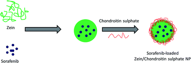
One of the effective and promising ways to overcome the side effects of small-molecule based therapeutics and to increase its therapeutic efficacy is to use polymeric drug delivery system.10 Development of drug delivery carriers have become key for the storage and delivery of anticancer drugs owing to its small particle size and its ability to permeate the tumor cells via the enhanced permeation and retention (EPR) effect.11–13 Recently, colloidal system based on environment friendly biomaterials has attracted the interest of broad range of researchers. Zein is a biodegradable and biocompatible material that contains three parts of lipophilic and one part of hydrophilic residues making it suitable for the encapsulation and delivery of many hydrophobic drugs such as curcumin, vitamin D3, lutein, gitoxin and quercetin.14–16 However, zein particles have poor aggregation stability under certain environmental conditions, such as pH, salt, and temperature. Therefore, it is important to increase the physicochemical stability of zein particles while keeping its property intact. In this regard, formation of polysaccharide–zein combination is expected to offer desired stability to the gastric conditions. For example, chitosan/zein complex has been reported to encapsulate alpha tocopherol and offered protection against harsh gastric conditions. Similarly, carboxymethyl chitosan/zein complex has been developed for the delivery of vitamin. However, chitosan in soluble in the gastric fluids exposing the zein particles, and release the drug in the premature manner.17–19 Therefore, in the present study, we have stabilized zein particles with a naturally derived chondroitin sulphate (CS) (Fig. 1). CS are glycosaminoglycans consisting of repeating units of β-1,4-linked D-glucoronic acid and β-1,3-linked N-acetyl galactosamine and is widely used as a site-specific drug delivery platforms.20,21
 |
| Fig. 1 Schematic illustration of preparation of sorafenib-loaded zein/chondroitin sulphate nanoparticles. | |
Thus far, in this study, SRF-loaded zein nanoparticle was prepared which was then stabilized by chondroitin sulphate as a stealth and protective layer to increase the therapeutic efficacy in gastric cancers. The utilization of CS to coat and stabilize zein nanoparticles was thoroughly investigated. Physicochemical studies were performed to ascertain the particle related parameters. In vitro efficacy was evaluated in gastric cancer SGC7901 cancer cells. Various biological assays including MTT assay, apoptosis assay, Hoechst staining, cellular uptake, and caspase 3/7 activity were carried out.
Results
Physicochemical characterization of zein/chondroitin sulphate nanoparticles
The physicochemical properties of nanoparticles are important for the systemic applications. In the present study, we have prepared the zein nanoparticle with a mean size of ∼120 nm while the size increased to ∼180 nm after chondroitin sulphate assembly on the surface of the zein nanoparticles (SZCS; Fig. 2a). The increase in the particle size indicates the deposition of CS on the particle surface. Additionally, zeta potential decreased from ∼+20 mV of zein nanoparticle to ∼−15 mV after CS coating.
 |
| Fig. 2 (a) Size and size distribution of SZCS nanoparticles (b) transmission electron microscope image of SZCS (c) stability of nanoparticles in different ionic conditions (d) stability of nanoparticles in different pH conditions (e) long term storage stability of nanoparticles. | |
The morphology of nanoparticle was evaluated using TEM (Fig. 2b). The particles were clearly spherical in nature and uniformly distributed in the grid. A polysaccharide layer on the outer particle surface could be easily seen indicating the presence of CS. However, particle size of SZCS observed by TEM was about 150 nm, which was smaller than that determined by DLS. We expected that the particle size determined by DLS represents their hydrodynamic diameter, while TEM is related to the dried state after water evaporation.
Drug encapsulation efficiency (EE)
The drug encapsulation efficiency is an important factor for the drug delivery system designed for cancer targeting. The encapsulation efficiency of SZCS was more than 95% with an active drug loading capacity of ∼10% w/w. The high entrapment efficiency of nanoparticle was attributed to the high hydrophobic nature of zein particles.
In vitro release study
The release profile of SRF from SZCS was performed in PBS and ABS solution at 37 °C (Fig. 3a). As seen, no burst release phenomenon was observed in both the release medium and the nanoparticles exhibited a controlled release profile throughout the release period. Lack of burst release indicates that the entire drug is encapsulated in the core of the nanoparticles. A more uniform steady state sustained release of SRF was observed up to 120 h. Approximately, ∼60% and ∼70% of drug released in PBS and ABS media, respectively after 120 h. Additionally, we have performed release study in pH 2.0 in order to simulate the gastric conditions and to check the stability of zein NP (Fig. 3b). As seen, zein NP was highly unstable in pH 2.0 and exhibited a faster drug release compared to that of CS-stabilized SZCS. It can be seen that 65% of drug released by the end of 24 h whereas only 30% of drug released from SZCS during the same time period. It has been reported that the zein NP is prone degrade in gastric conditions where CS stabilization in the present study effectively controlled the release of encapsulated compounds. Furthermore, slightly higher release was observed for SZCS at pH 2.0 compared to that at pH 7.4 which might be due to slight degradation.
 |
| Fig. 3 In vitro SRF release profile from the SZCS. (a) Release study in phosphate buffered saline (PBS, 0.1 M, pH = 7.4) and acetate buffered saline (0.1 M, pH = 5.5) with 0.1% w/v Tween-80 was employed as the release medium. (b) Release study of SZN and SZCS at pH 2.0 conditions. Data represent mean ± SD. (c) Change in particle size of SZCS in two different pH conditions over 24 h. (d) TEM image of SZCS in pH 7.4 and pH 2.0 conditions. | |
In addition, stability of optimized NP (SZCS) was observed in pH 7.4 and pH 2.0 conditions in terms of particle size over a period of time (Fig. 3c). As seen, particle size remained constant in case of pH 7.4 conditions while particle size of SZCS slightly (relatively) increased when incubated in pH 2.0 conditions. Nevertheless, bio-composite NP remained stable until 24 h study period without any remarkable change. Morphology of NP in two different pH condition was observed using TEM (Fig. 3d). As seen, particles structure did not change and remained spherical in both the pH conditions indicating the excellent stability of SZCS. It is worth noting that surface of particle was observed to be smooth in pH 7.4 conditions while surface was slight rough and corrosive in case of pH 2.0 conditions.
In vitro cellular uptake studies
In this study, we have used rhodamine-B as a fluorescent probe to investigate the cellular uptake of SZCS in cancer cells and used flow cytometer to confirm. The data clearly reveal a typical time-dependent cellular uptake in SGC7901 cancer cells (Fig. 4). A significant amount of nanoparticles were internalized in the first hour of incubation while a 2-fold higher internalization was observed at the end of 4 h incubation.
 |
| Fig. 4 Flow cytometry analysis of human SGC7901 gastric cancer cells incubated with SZCS nanoparticles for different time point. The equivalent of rhodamine-B concentration was 1 μg ml−1. | |
In vitro cytotoxicity assay
The percentage viability of cancer cells upon exposure with blank NP, free SRF, and SZCS was quantified using MTT method. As shown, blank NP did not suppress the proliferation of cancer cells even when treated with the maximum concentration (100 μg ml−1) (Fig. 5a). The cell viabilities remained more than 90% when treated with the maximum concentration indicating its safety profile and non-toxic nature.
 |
| Fig. 5 Cell viability of SGC-7901 treated with (a) blank nanoparticles (b) drug-loaded formulations. The cells were incubated for 24 h at 37 °C and cytotoxic effect was evaluated by means of MTT assay. (c) Morphological imaging of SGC7901 cancer cells. | |
The percentage cell viability of formulations treated group presented in Fig. 5b. As shown, free SRF and SZCS exhibited a typical dose-dependent cytotoxic effect in gastric cancer cells. Detailed analysis showed that SZCS elicited significantly higher anticancer effect in SGC7901 cancer cells compared to that of free SRF in the equivalent concentrations. Consistent with cytotoxicity assay, SZCS treated cell group exhibited a maximum cell death with round and distorted cancer cells, whereas cells in untreated and blank NP treated group remained intact (Fig. 5c).
Hoechst staining
The apoptotic effect of blank NP, SRF, and SZCS on SGC7901 gastric cancer cells were evaluated by Hoechst 33382 staining (Fig. 6). As seen, cells in untreated and blank NP treated group were intact and maintained its typical morphology with its rounded and intact nuclei. On the other hand, SRF did induce a transformation of cells and apoptosis of cancer cells could be seen. Specifically, SZCS treated cells were smaller and brightly stained with condensed chromatin and formed irregular shaped structures. Also, SZCS induced higher number of apoptotic cells and apoptotic bodies.
 |
| Fig. 6 Hoechst 3382 staining of cancer cells. The cells were treated with respective formulations and then treated with Hoechst dye for 10 min. | |
Apoptosis assay
As shown (Fig. 7), blank NP did not have any role in the cell apoptosis. Free SRF did induce the apoptosis of SGC7901 cancer cells, while SZCS exhibited a significantly higher cellular apoptosis of cancer cells.
 |
| Fig. 7 Annexin-V/PI based apoptosis assay. Cells were treated with respective formulations and then washed, collected, and incubated with annexin-V and PI for 15 min and then evaluated using flow cytometer. | |
Caspase activity
The caspase activity was measured in order to further ascertain the anticancer effect of different formulations. As shown in Fig. 8, SZCS exhibited a remarkably higher caspase 3/7 activity than compared to that of free SRF. Consistent with the other biological assays, higher caspase activity of SZCS was mainly attributed to its higher internalization in the cancer cells that might increase its accumulation and resulted in therapeutic effect.
 |
| Fig. 8 Caspase 3/7 activity of SGC7901 cancer cells after treatment with different formulations. | |
Discussion
Advanced gastric cancer is life-threatening and represents a formidable treatment challenge. According to National Comprehensive Cancer Network (NCCN) guidelines for GC in 2012, chemotherapy is the mainstay of palliative therapy for advanced or metastatic disease. Especially, sorafenib (SRF) is an effective anticancer agent effective against multiple cancers including gastric cancers. SRF inhibits proliferation, angiogenesis, and invasion of tumor cells. However, systemic anticancer therapy is suffering from severe side effects and has been disappointing with low response rate and high toxicity. Therefore, it is utmost important to identify potential drugs and explore more efficient therapeutic strategies for the treatment of gastric cancer. In this study, we have developed an environment friendly biomaterials-based zein/chondroitin complex nanoparticle to deliver SRF in gastric cancers. In order to protect the stability and aggregation of zein nanoparticles, chondroitin sulphate was coated on the surface. This polysaccharide–zein combination is expected to offer desired stability to the gastric conditions and anticancer efficacy.
In the present study, we have prepared the zein nanoparticle with a mean size of ∼120 nm while the size increased to ∼180 nm after chondroitin sulphate assembly on the surface of the zein nanoparticles. A nanosized particle of <200 nm diameter suggests that SZCS could be able to selectively accumulate in solid tumors by means of passive targeting, known as enhanced permeability and retention effects (EPR).22
The stability of nanoparticle system is highly dependent on the ionic strength of the medium especially in the gastrointestinal tract. Therefore it is highly important to establish the stability of nanoparticle system in different salt conditions. As seen (Fig. 2c), zein NP (without CS; SZC) immediately aggregated when salt levels were increased, whereas composite biopolymer nanoparticles were relatively stable to aggregation from 0 to 1000 mM NaCl and the particle diameter almost remained unchanged. Low levels of salt may therefore promote particle aggregation through electrostatic screening effects in zein NP. The composite biopolymer nanoparticles are stabilized by both electrostatic and steric repulsion, which means that much higher levels of salt are required to promote particle aggregation. Additionally, it can be seen that absence of CS resulted in the aggregation of SZC at pH 5.0 and pH 6.0 resulting in tremendous increase in the overall particle size (Fig. 2d). This aggregation of zein NP was attributed to the fact that the isoelectric point of zein is around pH 6 that caused in low net charge around this pH. Subsequently electrostatic attraction between particles leads to particle aggregation. Thus, coating zein nanoparticles with CS was able to inhibit the particle aggregation that normally occurs around pH 5 to 6. Consequently, long term stability of both the NP system was assessed at 25 °C (Fig. 2e). It can be clearly seen that particle size of zein NP constantly increased with every time point whereas particle size composite biopolymer nanoparticles remained same throughout the study period until 35 days indicating its high stability.
A more uniform steady state sustained release of SRF was observed up to 120 h. Approximately, ∼60% and ∼70% of drug released in PBS and ABS media, respectively after 120 h. A slightly higher release in acidic medium might due to the disintegration and degradation of outer CS layer that might increase the diffusion of drug in the release medium. This kind of controlled release profile of nanoparticle could be useful for the delivery of anticancer drugs. Furthermore, it can be safely expected that when the nanoparticles reach the tumor site, it will release the payload in the cancer cell and thereby will increase the therapeutic efficacy.
Knowledge of cellular uptake efficiency of nanoparticle system is important as the anticancer drugs or small molecule drugs entrapped inside the nanoparticles could pass through endocytosis process rather than the simple diffusion. The endocytosis of drugs will have a greater impact on the cancer cells than that of simple diffusion. In this study, a significant amount of nanoparticles were internalized in the first hour of incubation while a 2-fold higher internalization was observed at the end of 4 h incubation. Such kind of time-dependent cellular uptake clearly reflects the carrier-mediated endocytosis due to its extra small size. It is well known fact that such time-dependent uptake could not be observed in free diffusion of drugs. Therefore, cellular uptake efficiency of drug-loaded nanoparticles affects the therapeutic effects.
The free SRF and SZCS exhibited a typical dose-dependent cytotoxic effect in gastric cancer cells. The IC50 value which is the concentration required to kill 50% of cells were calculated to quantify the anticancer effect of each formulation. The IC50 value of free SRF and SZCS was 2.89 μg ml−1 and 6.56 μg ml−1, respectively. The superior anticancer effect of SZCS was mainly attributed to the higher uptake of nanoparticles and sustained release of encapsulated small molecules. Results from cellular uptake experiment also confirmed the high cell affinity of nanoparticles. The cellular morphology was further observed after incubation with respective formulations for 24 h.
Annexin-V/PI double staining protocol is one of the most sensitive methods for the detection of cell apoptosis. In this, plasma membrane reorganization indicates the early apoptosis which was detected by positive staining by annexin V-FITC, while later stage apoptosis indicating DNA damage showed positive staining for both annexin V and PI. A significant proportion of cells were present in the early and late apoptosis chamber. The superior anticancer effect of SZCS was mainly attributed to the higher uptake of nanoparticles and sustained release of encapsulated small molecules.23,24
Generally, NPs has the ability to radically change the cancer therapies owing to its superior physicochemical properties. The encapsulation of drug in the biodegradable nanoparticles is expected to accumulate in cancer cells after being internalized by EPR effect. This property of nanoparticles will increase the anticancer effect in gastric cancers while minimizing the associated side effects. In the present study, we mainly focused our attention to use zein as a suitable drug delivery system. However, it is well known fact that the tumor is heterogeneous and therefore additional strategies including targeting moiety would be more effective in the treatment of solid tumors – a subject of our future study.
Conclusion
In conclusion, we have successfully demonstrated the preparation of core–shell type biopolymers based zein/chondroitin sulphate nanoparticles for the delivery of sorafenib in gastric cancers. The SRF-loaded zein/chondroitin sulphate nanoparticles (SZCS) were nanosized with a spherical morphology and exhibited a higher encapsulation of more than >90%. The biopolymer nanoparticle showed the ability to release the drug in a controlled manner for 120 h, indicating its potential application in systemic delivery. The nanoparticles showed a remarkable uptake in gastric cancer cells in a time-dependent manner. The SZCS displayed an improved cytotoxic effect compared to that of free SRF in the equivalent concentrations in SGC7901 cancer cells. Also, we demonstrated a higher apoptosis and caspase 3/7 activity for SZCS nanoparticle system. Based on our results, we can conclude that SZCS might hold a great potential in the treatment of gastric cancers.
Materials and methods
Materials
Zein (Z0001) was purchased from Tokyo Chemical Industry, Co., Ltd. (Tokyo, Japan). Sorafenib, chondroitin sulphate, phosphate buffer solution (PBS) and MTT [3-(4,5-dimethylthiazol-2-yl)-2,5-diphenyl tetrazolium bromide] were purchased from Sigma-Aldrich (St. Louis, MO, USA). Dubelcco's modified Eagle's medium (DMEM), fetal bovine serum (FBS), trypsin–EDTA and penicillin–streptomycin mixtures were from Gibco®BRL (Carlsbad, CA, USA).
Preparation of sorafenib-loaded zein/chondroitin sulphate nanoparticles
To prepare sorafenib-loaded zein/chondroitin sulphate (SZCS) nanoparticles, 100 mg of zein was dissolved in 85% of ethanol and stirred for 60 min. Sorafenib (10% w/w) was dissolved in the ethanolic solution and further stirred for 60 min. Separately, chondroitin sulphate (CS) was dissolved in ultra-pure water and stirred for 2 h, filtered through 0.45 μm filter and kept aside. The aliquot of SRF–zein solution was added to distilled water in a drop-wise manner and mechanically stirred for 5 h until all the ethanol evaporated. Finally, distilled water was added to compensate the loss of ethanol and the drug-zein dispersion was added to the CS solution and further stirred for 3 h at 500 rpm min−1. The resulting drug-loaded nanoparticle was separated from the free drug by high speed centrifugation. The amount of drug encapsulated was determined by HPLC method. Shimadzu HPLC equipped with Phenomenex Luna C18, 5 μm (4.6 × 250 mm) column, two LC-20AD pumps, SCL-10AVP system controller, Rheodyne injector with 50 μl loop, and SPD-20A UV-visible detector was used. A mixture of acetonitrile and water (83.5
:
16.5 v/v) was used as a mobile phase and the effluent was detected at 260 nm.
EE (%) = (Wtotal − Wfree)/Wtotal × 100 |
Particle size analysis
The particle size blank and drug-loaded nanoparticles were determined by dynamic light scattering technique using Zetasizer Nano-ZS (Malvern Instruments, Malvern, UK). The nanoparticle dispersions were appropriately diluted with distilled water and were allowed to equilibrate for 120 seconds before the measurement.
Transmission electron microscopy (TEM)
The morphology of nanoparticle was observed using JEM-100S (Japan) transmission electron microscope (TEM). Briefly, a drop of dispersion was placed on the copper grid and negatively stained with 1% w/v of phosphotungstic acid and dried.
In vitro drug release profile
The in vitro release of the drug from the nanoparticles was assessed in phosphate buffered saline (pH 7.4) and acetate buffered saline (pH 5.0) containing 0.5% Tween 80. The release study was performed by dialysis method. Briefly, 1 ml of drug-loaded nanoparticle dispersion was enclosed in a dialysis bag and placed in the respective release media (25 ml) at 37 °C under gentle agitation. At specific time points, 1 ml of release media was withdrawn and replaced with equivalent amount of fresh media. The amount of drug released was determined using HPLC method as mentioned earlier.
Nanoparticle uptake by tumor cells
A total of 2 × 10⁵ cells were seeded in each well of 12-well plate in DMEM media supplemented with 10% FBS and 1% antibiotic mixture. The cells were allowed to adhere at 37 °C for 24 h. Then, media was removed and replaced with fresh media containing zein/chondroitin sulphate nanoparticles. Rhodamine-B was loaded instead of sorafenib as a fluorescent probe. The nanoparticles were incubated for different time points. The cells were washed three times with PBS and collected and washed again with PBS. The cells suspension was centrifuged and the pellet was resuspended again in PBS evaluated using flow cytometer (FACSCanto II, BD Biosciences). For each sample, 10
000 cells were analyzed using FACSDiva software (version 6).
Cytotoxicity assay
The cytotoxicity assay on SGC7901 cancer cell was performed by 3-[4,5-dimethylthiazol-2-yl]-2,5-diphenyltetrazolium bromide (MTT) assay. MTT assay protocol. 1 × 10⁴ cells per well was seeded in a 96-well plate and incubated for 24 h. Next day, old media was removed and replaced with fresh media containing blank NP, free SRF, and SRZC nanoparticles and incubated for 24 h at 37 °C. Next day, media was removed and washed two times with PBS. Then, MTT solution (5 mg ml−1) was added to each well and cells were incubated for another 4 hours. After removing the unreduced MTT and medium, each well was replaced with 100 μl of dimethylsulfoxide (DMSO) to dissolve the MTT formazan crystals. The absorbance of formazan crystal was measured using a microplate reader (Bio-Rad, CA, USA) at 570 nm. Cell viability is defined as the percentage of the absorbance of the wells containing the cells incubated with the micelles suspension over that of the cells only.
Hoechst staining
The SGC cells were seeded in a 6-well plate and treated with blank NP, SRF, and SRZC nanoparticles and incubated for 24 h. Then, washed twice in phosphate buffer saline (PBS) followed by fixation in cold methonal
:
acetone (1
:
1) for 5 min. After washing thrice in PBS for 5 min, these cells were treated with Hoechst 33382 for 10 min at room temperature. Cells were then washed twice with PBS and examined by fluorescence microscopy.
Apoptosis assay by annexin V/propidium iodide (PI) staining
The apoptosis assay in SGC7901 cancer cell was performed using annexin V/PI double staining protocol. Briefly, cells were seeded in a 6-well plate at a seeding density of 4 × 10⁵ cells per well. After 24 h, cells were exposed with respective formulations and incubated for 24 h. The cells were trypsinized and centrifuged. The pellets were re-dispersed in 100 μl of PBS and incubated with 5 μl annexin V-FITC and 10 μl PI solution for 15 min. The final volume of PBS was made to 1000 μl. The cell apoptosis was detected using flow cytometer.
Caspase 3/7 activity
The caspase 3/7 activity was measured using the Caspase-Glo® 3/7 Assay (Promega, Germany) kit. The seeded cells were treated with respective formulations and incubated for 24 h. Next days, cells were processed as per the manufacturer's protocol. In this assay, cleavage of the caspase substrate was measured by luminescent signal which is proportional to the caspase activity. The luminescence was measured using MicroLumat Plus Microplate Luminometer LB 96V, Germany.
Statistical analysis
Data were presented as mean ± standard deviation (SD). Statistical significance of difference among groups was determined by one-way analysis of variance (ANOVA). The difference between groups was considered significant when p < 0.05.
Acknowledgements
This study was supported from the grant of College of Medicine, National Taiwan University, Taiwan.
Notes and references
- S. C. Oh, Journal of Gastric Cancer, 2013, 12, 3–6 CrossRef PubMed.
- R. Siegel, D. Naishadham and A. Jemal, Ca-Cancer J. Clin., 2012, 62, 10–29 CrossRef PubMed.
- R. E. Schwarz and D. D. Smith, Ann. Surg. Oncol., 2007, 14, 317–328 CrossRef PubMed.
- A. Jemal, F. Bray, M. M. Center, J. Ferlay and E. Ward, et al., Ca-Cancer J. Clin., 2011, 61, 69–90 CrossRef PubMed.
- R. E. Schwarz and K. Zagala-Nevarez, Ann. Surg. Oncol., 2002, 9, 394–400 CrossRef PubMed.
- A. Cappetta, S. Lonardi, D. Pastorelli, F. Bergamo, G. Lombardi and V. Zagonel, Crit. Rev. Oncol. Hematol., 2012, 81, 38–48 Search PubMed.
- D. H. Kim, M. D. Kim and C. W. Choi, et al., Nanoscale Res. Lett., 2012, 7, 91 CrossRef PubMed.
- Y. C. Yang, J. Cai and J. Yin, et al., Carbohydr. Polym., 2016, 136, 782–790 CrossRef CAS PubMed.
- L. Zhang, F. Gong, F. Zhang, J. Ma, P. Zhang and J. Shen, et al., Int. J. Nanomed., 2013, 8, 1517–1524 CrossRef PubMed.
- M. E. Davis, Z. Chen and D. Shin, Nat. Rev. Drug Discovery, 2008, 7, 771–782 CrossRef CAS PubMed.
- K. Kataoka, A. Harada and Y. Nagasaki, Adv. Drug Delivery Rev., 2001, 47, 113–131 CrossRef CAS PubMed.
- T. Ramasamy, J. Y. Choi, H. J. Cho, S. K. Umadevi, B. S. Shin, H. G. Choi and C. S. Yong, Pharm. Res., 2014, 32, 1947–1956 CrossRef PubMed.
- D. W. Kim, T. Ramasamy, J. Y. Choi, J. H. Kim, C. S. Yong, J. O. Kim and H. G. Choi, Int. J. Nanomed., 2014, 9, 3815 CrossRef CAS PubMed.
- F. Dong, G. W. Padua and Y. Wang, Soft Matter, 2013, 9, 5933 RSC.
- Y. Zhang, Y. Niu, Y. Luo, M. Ge, T. Yang and L. L. Yu, et al., Food Chem., 2014, 142, 269–275 CrossRef CAS PubMed.
- H. Chen and Q. Zhong, Food Hydrocolloids, 2014, 35, 358–366 CrossRef CAS.
- A. R. Patel, Y. Hu, J. K. Tiwari and K. P. Velikov, Soft Matter, 2010, 6, 6192 RSC.
- Y. Luo, Z. Teng and Q. Wang, J. Agric. Food Chem., 2012, 60, 836–843 CrossRef CAS PubMed.
- D. Hu, C. Lin, L. Liu, S. Li and Y. Zhao, J. Food Eng., 2012, 109, 545–552 CrossRef CAS.
- S. K. Umadevi and T. Ramasamy, Iran. J. Pharm. Res., 2012, 11, 465–479 Search PubMed.
- V. E. Santo, M. E. Gomes, J. F. Mano and R. L. Reis, J. Tissue Eng. Regener. Med., 2012, 3, s47–59 CrossRef PubMed.
- M. E. Werner, N. D. Cummings, M. Sethi, E. C. Wang, R. Sukumar, D. T. Moore and A. Z. Wang, Int. J. Radiat. Oncol., Biol., Phys., 2013, 86, 463–468 CrossRef CAS PubMed.
- T. Ramasamy, et al., J. Mater. Chem. B, 2015, 2, 6324–6333 RSC.
- T. H. Tran, et al., Int. J. Nanomed., 2015, 10, 5249 CAS.
|
This journal is © The Royal Society of Chemistry 2016 |
Click here to see how this site uses Cookies. View our privacy policy here.