Yibin Yu‡
a,
Hao Pan‡b,
Yingying Wanga,
Wei Xionga,
Qiantao Zhangc,
Kai Chena,
Xiumei Gaia,
Pingfei Lia and
Xinggang Yang*ad
aShenyang Pharmaceutical University, 103 Wenhua Road, Shenyang 110016, China. E-mail: yangxg123@163.com; Fax: +86-24-23953241; Tel: +86-24-23986313
bSchool of Pharmacy, Queen's University Belfast, Belfast BT7 1NN, UK
cShanxi Institute of International Trade & Commerce, Shanxi 712046, China
dState Key Laboratory of New-tech for Chinese Medicine Pharmaceutical Progress, Lianyungang 222001, China
First published on 16th June 2016
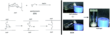
A novel hydrogel based on Auricularia auricular polysaccharide (AAP), was prepared and characterized. AAP hydrogel exhibited a pH-sensitive release of model protein drug (bovine serum albumin, BSA) under simulated gastrointestinal conditions. The results indicate AAP hydrogel has promising potential as an effective vehicle for oral delivery of protein.
Auricularia auricular, one of non-toxic edible mushrooms, has been used as a healthy food for more than 1000 years in East Asian, especially in China and Japan.7 It is also known for its tonic and pharmaceutical effects in traditional Chinese medicine.8,9 The fruit of Auricularia auricular is rich in polysaccharides. Since the pioneering work of Sone et al. in the late 1970s on the isolation and characterization of polysaccharides of Auricularia auricular (AAP), the literature on AAP has exploded.8–14 However, to the best of our knowledge, studies on AAP mainly focus on the characterization and wide-spread biological activities of it, such as hypoglycemic, hypolipidemic, antiviral, anti-aging, antitumor and enhancing immunity, whereas the investigation on its application in drug delivery is rather limited.8,9,11–14 In previous studies, we has reported the novel application of AAP in pharmaceutical area.15 Herein, an AAP-based hydrogel with desirable features was developed and evaluated to deliver drug for the first time.
Along with the rapid development of biotechnology, production of therapeutic protein drugs in large quantities has become feasible currently.16 Unquestionably, the gastrointestinal (GI) tract is the most popular route of drug delivery because of the facility of administration of drugs for compliant therapy, and its large surface area for systemic absorption.17 For designing oral dosage forms, the natural pH environment of GI tract must be considered carefully. The pH of GI tract varies from acidic in the stomach to slightly alkaline in the intestine. It is common knowledge that the stomach pH is about 1.2, while the pH of the small intestine is about 7.0. There are many obstacles of oral protein delivery, including the harsh acidic environment of stomach, protein denaturation or degradation by digestive enzymes in the GI tract and poor epithelial permeability of proteins.16 Therefore, oral delivery of proteins to the GI tract is one of the most challenging issues in the development of therapeutic proteins.
Recently, “smart” hydrogels, as promising biomaterials in the design of oral delivery of protein, have been extensively studied.17–19 Smart hydrogels exhibiting pH-dependent swelling behaviour can protect protein drugs from hostile environments, for instance, enzymatic hydrolysis and low pH in the stomach before protein drugs were absorbed in the intestine, for the reason that the natural pH environment of the GI tract variation of the acid condition (pH 1.2) in the stomach and slightly alkaline condition in the intestine (pH 7.4).17,18,20 Furthermore, hydrogels are able to swell in water and hold a large amount of water while maintaining the structure, which make hydrogels resemble natural living tissue.18 So far, a variety of natural or synthetic polymers with acidic or basic pendant groups have been employed as pH-sensitive hydrogel.17–19 Polysaccharides, with excellent biodegradability, inherent biocompatibility and poor toxicity have been used as hydrogel materials. Comparing with conventional hydrogels, polysaccharides-based hydrogels exhibit minimal toxicity and have excellent potential as drug delivery vehicles.1,21
In this study, a novel pH-sensitive polysaccharide-based hydrogel was constructed for safe and effective protein delivery for the first time. The polysaccharide extracted and purified from the Auricularia auricular was cross-linked with epichlorohydrin (ECH) to form AAP hydrogel. The hydrogel was characterized by infrared spectroscopy, morphology and swelling ratio. Furthermore, bovine serum albumin (BSA) was chosen as a model drug to study the drug release profiles in simulated gastric and intestinal media. The experimental results reveal promising potential of AAP hydrogel as a pH-sensitive and controlled release oral delivery system of protein drugs.
Purified AAP was obtained after hot-water extraction, ethanol precipitation, adsorption–desorption on AB-8 macroporous resins,23 Sevag method, dialysis and lyophilisation. The homogeneity and molecular weight of the purified AAP were determined by gel-permeation chromatography (GPC), with a Sephadex-G100 column (1.1 cm × 70 cm). The peak of AAP was symmetrically sharp, indicating that its molecular weight distribution was relatively narrow (Fig. S1, ESI†). The average molecular weight of AAP was estimated to be 80785.1 Da, when referenced to a calibration curve of dextran T-series standard of known molecular weight (dextran T-5, T-10, T-40, T-70, T-110, T-500, T-2000).
Chemical modification of natural polysaccharides provides the opportunity to combine the typical biocompatibility of natural polymers with the significant advantages of synthetic polymers such as better mechanical properties, chemical versatility, etc. AAP hydrogel was cross-linked by epichlorohydrin (ECH) in basic environment. As a convenient base catalysis cross-linker, ECH has been widely used in polysaccharide chemistry.20,22,24–28 The proposed mechanism for crosslinking reaction of ECH with polysaccharides in basic solution is that each ECH molecule could react with two hydroxyl groups from two chains of different polysaccharides (Scheme 1A). This agent is easy to react in alkaline solution with the hydroxyl group of the polysaccharide molecules. In the first stage of the reaction, through nucleophilic attack of the alcoholate anion, the free chlorohydrin fragments were formed in the side chain of linear macromolecules and the epoxy rings opened simultaneously. Then a new epoxide formed by dehydrochlorinating. Subsequently, a reaction between the new epoxide and another alcoholate anion occurred. Consequently, two neighbouring polysaccharides chains were attached together to form a crosslinking network.27 Proper hydrogel was selected on the basis of the method in Scheme 1(B). The effect of different ratios of the initial solutions of polysaccharide and crosslinker was studied. The concentration of the initial solution of AAP was fixed at 50 mg ml−1; ECH, analytical grade, was changed by volume to control the ratio of ECH to AAP (Table 1). Several volumes of ECH from 0.1 ml to 0.5 ml were attempted to add to 1 ml AAP solution (50 mg ml−1). As illustrated in Table 1, AAP hydrogel could be obtained when the volume of ECH was 0.2, 0.25 or 0.3 ml. The hydrogel formed between 1 ml AAP solution (50 mg ml−1) and 0.25 ml ECH was the most appropriate for further study. In addition, we have investigated the effect of the concentration of sodium hydroxide, crosslinking time and temperature on the crosslinking reaction between AAP and ECH. The repeated experiments indicated that 2.5 mol L−1 NaOH, 3 h and 50 °C were enough for proper hydrogel for further study. Increasing the concentration of sodium hydroxide, reaction time and temperature barely improved the profiles of AAP hydrogel.
![]() | ||
Scheme 1 Crosslinking reaction between AAP and ECH: (A) synthetic procedure of AAP hydrogel. (B) Photographs of AAP hydrogel formation. |
Sample number | Volume of NaOH (ml) | AAP concentration (mg ml−1) | Volume of ECH (ml) | Time (h) | Temperature (°C) | Formation of hydrogel |
---|---|---|---|---|---|---|
1 | 1 | 50 | 0.1 | 3 | 50 | No |
2 | 1 | 50 | 0.15 | 3 | 50 | No |
3 | 1 | 50 | 0.2 | 3 | 50 | Yes |
4 | 1 | 50 | 0.25 | 3 | 50 | Yes, for further study |
5 | 1 | 50 | 0.3 | 3 | 50 | Yes |
6 | 1 | 50 | 0.4 | 3 | 50 | No |
7 | 1 | 50 | 0.5 | 3 | 50 | No |
The FT-IR spectra of AAP and its hydrogel product with ECH were both shown in Fig. 1. The most representative bands around 3800–2700 cm−1 were those assigned to the hydroxyl (O–H) intramolecular and intermolecular stretching modes, to the asymmetric and symmetric methyl and methylene stretching vibration.29 The peak at 3421 cm−1 was recognized as stretching vibration of the large number O–H groups and the peak at 2933 cm−1 and 1418 cm−1 was assigned to the C–H stretching and bending vibrations including C–H, CH2 and CH3.30 Strong band occurring at 1613 cm−1 was the particular absorption of uronic acid, corresponding to the stretching vibration of carboxylate groups COO–.22 Generally, the wave number between 950 and 1200 cm−1 is often called the “fingerprint” region of molecules, which is mostly dependent on the molecular vibrations and molecular structures.13,31 Bands between 1200 cm−1 to 1000 cm−1 might be attributed to the existence of the pyranose ring.30,32 The peak at 1059 cm−1 was characteristics of the glycosidic structures, corresponding to C–O–C of pyranose of sugar units. The peak at 797 cm−1 indicated that α-glycosidic linkages were present between the sugar units and mannose was confirmed by the presence of the band at 617 cm−1 of the spectrum.9 Bands at 3420 cm−1, 2933 cm−1 and 1059 cm−1 could be due to the polysaccharide structure. These bands scarcely changed position when AAP was cross-linked by ECH and they seemed similar in whole. However, the bands were indeed different in detail. In the spectrum of AAP hydrogel, stronger peak at 2926 cm−1 appeared, due to the higher content of C–H and CH2 after crosslinking reaction. Furthermore, the spectrum of AAP hydrogel presented a remarkable increase between 1200 cm−1 to 1000 cm−1, corresponding to the increase in –C–O–C– bands and –CHOH– groups because of crosslinking between AAP and ECH. Therefore, it could be inferred from IR that the network of AAP hydrogel formed.
AAP hydrogel is fully transparent on the macroscopic level. There is gloss of gel in the cross-section torn from lyophilized AAP hydrogel. The interior morphologies of AAP and AAP hydrogel were observed using a scanning electron microscope (SEM, Hitachi S-3400N, Japan), microscopically. The magnification of SEM images (Fig. 2A and B) was 500 times the original size. As shown in the image of AAP hydrogel, irregular pores that interconnect with each other, could be observed clearly. Because AAP solution is the solution of macromolecule, it will be left some pores meanwhile the loss of water after lyophilized. But AAP hydrogel, with some roughness, has a three-dimensional network structure formed via chemical crosslinking, and thus there are much more pores left than AAP. The porous nature of the hydrogel provides more surface area, where the capillary forces help the diffusion of water into the hydrogel, and hence AAP hydrogel has stronger water uptake capacity than AAP.
There are some factors, such as pH, temperature, possibly affecting the swelling properties of hydrogels.17 Thus, swelling ratio (SR), which assesses the water uptake capacity of hydrogels, was measured gravimetrically to evaluate how pH and temperature influenced the swelling behaviour of AAP hydrogel. Time-dependent swelling behaviours have been plotted in Fig. S2 (ESI†). There was a typical biphasic swelling pattern, namely a rapid swelling followed by a slower sustained swelling. When ambient temperatures kept at 25 °C, 37 °C and 45 °C, the equilibrium swelling ratios were 10.77, 9.51 and 10.05, respectively. No significant difference was observed in their swelling profiles. It was indicated that temperature had little effect on the swelling behaviour of AAP hydrogel.
Besides, to study the effects of pH on AAP hydrogel, gels were immersed in solutions of 0.1 mol L−1 HCl (pH 1.2), phosphate buffer (pH 7.4) and 0.1 mol L−1 NaOH (pH 12.5) at 37 °C, respectively. As shown in Fig. 3, the pattern was biphasic, resembling to that of aforementioned temperature assay, with swelling equilibrium in approximately 10 h. Moreover, the swelling behaviour of AAP hydrogel possessed significant pH sensitivity. It was notable that the SR values of AAP hydrogel in media with different pH varied a lot: 5.25 at pH 1.2, 10.66 at pH 7.4 and 8.89 at pH 12.5, separately. AAP hydrogel exhibited apparent discrepancies in swelling that were dominated by external pH changes. According to the curves illustrated in Fig. 3, the swelling ratio of the hydrogel was relatively low at pH 1.2, probably because of the formation of intermolecular hydrogen bonds. At pH 1.2, all the functional groups (carboxyl and hydroxyl) from AAP were in their neutral form (–COOH and –OH), which made the hydrogel network more hydrophobic and hence reduced the water uptake capacity of AAP hydrogel. Nevertheless, at pH 7.4, the SR value was highest, probably due to progressively ionized of carboxyl and hydroxyl (–COONa and –ONa) on the hydrogel structure. The increase of negative charge density inside the hydrogel network positively affected the water uptake capacity between the polymeric chains (by anion–anion electrostatic repulsion), which allowed the hydrogel network to be more hydrophilic. Thus, the swelling of the hydrogel increased highly due to the large swelling force. Interestingly, the swelling ratio of the hydrogel reduced obviously at a higher pH (12.5), which could be hypothesized by Donnan equilibrium theory.33 AAP hydrogel, whose functional groups (–COOH and –OH) can be dissociated in base solution, acts as an ion exchanger. As pH increases, H+ ions will be supplied by dissociated carboxyl and hydroxyl. Simultaneously, the concentration of low-molecular weight cations in the outer solution will also increase. Those cations will be attracted into the hydrogel by electrostatic force, replace the mobile H+ ions, and screen opposite charges on AAP hydrogel chains. Accordingly, the concentration of mobile ions in AAP hydrogel could increase more rapidly than in the outer solution, so that the ion swelling pressure increases. The supply of H+ ions will continue until all the functional groups completely dissociate. Consequently, the ion swelling pressure reaches a maximum in the solution, which is probably in the pH range 7–8.33 It provide a proper explanation for the highest swelling ratio at pH 7.4. When the pH of solution continues increasing, the concentration of mobile ions increases, whereas the completely dissociated AAP hydrogel will not continue dissociating. Any further increase of pH will reduce the ion swelling pressure. Accordingly, the swelling ratio reduces from the maximum as pH increases. Thus, the swelling ratio of AAP hydrogel at pH 12.5 was lower than that at pH 7.4. It is generally accepted that the swelling properties of a hydrogel in gastric and intestinal fluids determine its drug release profile. As shown in Fig. 3, the swelling ratio of the hydrogel was relatively low at pH 1.2 (gastric environment) whereas the swelling ratio was moderate at pH 7.4 (intestinal environment). Therefore, AAP hydrogel could protect the loaded protein drug, such as BSA, insulin from degradation by the low pH environment of the stomach.
BSA was employed as a model protein to observe the smart release behaviour of AAP hydrogel. The drug was loaded into the hydrogel via a swelling method, and the amount of BSA in the medium was quantified by the Bradford method for released BSA at 595 nm using a UV/Vis spectrophotometer.34 From the in vitro drug release profiles presented in Fig. 4, it could be hypothesized that the protein release from AAP hydrogel was sensitive to external pH changes under the simulated GI conditions. Slower release of BSA from AAP hydrogel was observed under low pH condition (pH 1.2, gastric environment); conversely, under high pH condition (pH 7.4, intestinal environment), the hydrogel exhibited a rapid protein release. In particular, at pH 1.2, only approximately 15% of the protein was released from the gel in 10 h, probably related to low degree of swelling of AAP hydrogel. Nevertheless, at pH 7.4, the encapsulated BSA release was obviously higher (about 80.9%) in 10 h, which might be ascribed to significantly increasing of the swelling degree of the hydrogel network. This qualitative correlation between swelling ratio and drug released from hydrogels was first revealed by Kim and Chu35 in the study of doxorubicin release from dextran/methacrylate hydrogels. As Fig. 4A indicated, at the pH of intestine, BSA was released in a sustained and prolonged manner over a period of 10 h. For further imitating gastrointestinal tract environment, two-step releasing study was performed by immersing the BSA-loaded hydrogel in a solution at pH 1.2 for 2 h and afterwards in another solution at pH 7.4 for additional 8 h (Fig. 4B). At pH 1.2, BSA release profiles of the hydrogel were similar to those in the one-step releasing study (Fig. 4A and B). However, at pH 7.4, the maximum amount of BSA release observed in the two-step releasing study was about 76%, slightly lower than that in the one-step releasing study. And the rates of drug release in the two-step releasing study were apparently slower than those in the one-step releasing study. This phenomenon might be ascribed to the progressive change of pH value while AAP hydrogel was transferred from pH 1.2 into pH 7.4 in the two-step releasing study. This change of pH value could slow down the swelling of the hydrogel, and hence delay the release of BSA. The favourable result clearly suggested that AAP hydrogel could be a suitable polymeric carrier for protecting protein from the acid stomach environment and releasing it in a sustained fashion in the intestine.
Footnotes |
† Electronic supplementary information (ESI) available: Experimental details, gel-permeation chromatogram and swelling profiles of the hydrogel at different temperatures. See DOI: 10.1039/c6ra06463a |
‡ These authors equally contributed this work. |
This journal is © The Royal Society of Chemistry 2016 |