Lucrezia Martino*a,
Volga Niknama,
Nathanaël Guigo*a,
Jesper Gabriël van Berkelb and
Nicolas Sbirrazzuolia
aUniversité Nice Sophia Antipolis, CNRS, Laboratoire de Physique de la Matière Condensée (LPMC), UMR 7336, 06100, Nice, France. E-mail: lucrezia.martino@unice.fr; nathanael.guigo@unice.fr
bAvantium Chemicals B.V., Zekeringstraat 29, 1014 BV, Amsterdam, The Netherlands
First published on 16th June 2016
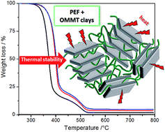
Novel nanocomposites of bio-based poly(ethylene 2,5-furandicarboxylate) (PEF) and organo-modified montmorillonite (OMMT) clays were prepared. Sample morphology was investigated by means of transmission electron microscopy (TEM) and X-ray diffraction (XRD) that revealed a good dispersion of the clay layers in the PEF matrix. Moreover, the effect of clays on the nanocomposites' thermal behavior was determined by differential scanning calorimetry (DSC) and thermogravimetric analysis (TGA). The results show that the presence of clays induces a slight nucleating effect on the PEF crystallization. More remarkably, the clay layers increase significantly the thermal stability of PEF by postponing the initial degradation (+20 °C).
In the recent years there has been an increasing interest in both academic and industrial sectors in polymer nanocomposites where at least the filler has one component carrying one dimension in the nanometer size scale. The dispersion of the nanofiller within the polymer matrix can lead to unique properties, as a result of an immense interfacial surface area between the two components. This new nanocomposite science has been initially sparked by the Toyota research group6,7 by using layered-silicate clays as nanofillers in order to improve the properties of polyamide 6. Since then, different types of both nanofillers (e.g. layered silicates, carbon nanotubes, graphite, nanofibers) and polymer matrixes such as polyamides,8,9 polypropylene,10,11 polyethylene,12,13 polyethylene terephthalate (PET),14–16 poly(lactic acid),17,18 were employed to make polymeric nanocomposites. Many application sectors such as the automotive, packaging and coating are already using polymer nanocomposites in different polymer market areas.19,20 Among the extensive list of available nanofillers, the clays are currently the most popular layered silicates used in polymer nanocomposites production due to their availability, low cost, easy processability and significant enhancements. The clays belong to the family of phyllosilicates and these layered silicates are basically constituted of stacks of platelets. Depending on the relative dispersion of those platelets in the polymer matrix, three extreme morphologies can be described: (i) an intercalated structure if a regular stacking of polymer and silicate layers is generated; (ii) an exfoliated or delaminated morphology if the silicate platelets are homogeneously and individually dispersed in the polymer matrix; (iii) a phase-separated composite if the polymer is unable to intercalate between the silicate layers. Nanocomposite morphology reflects the clay-polymer affinity: strong interactions between polymer and clay surface lead to intercalated/exfoliated structures which generally result in an enhancement of the polymer properties, contrary to what is observed for not well dispersed systems.21 Based on many publications about clay-based nanocomposites, the addition of a small amount of clays (up to about 5 wt%) can significantly improve the material performance in terms of thermal, mechanical, barrier, electrical/electronic properties, flammability resistance.22 Montmorillonite (MMT) which belongs to the 2:
1 layered type of phyllosilicates is one of the most promising and widely used nanoscale filler in the preparation of nanocomposites.23 The structure of MMT consists of a 1 nm-thick layer made of an inner octahedral sheet of either aluminium or magnesium oxide sandwiched between two tetrahedral sheets of silica. Within the interlayer spaces, charge compensating counter-ions such as K+, Na+, Ca2+ and Mg2+ are located so that render clays hydrophilic. The above mentioned alkali or alkali-earth cations can be exchanged with metallic cations (Al3+, Ti4+, Fe3+, Cu2+)24 or organic cations (generally alkyl-ammonium).25 In the latter case, the organophilic montmorillonite clays (OMMT) are more compatible with hydrophobic polymers or monomers which are able to intercalate within the galleries. The MMT clays minerals are intrinsically geo-based natural products which represents a valuable feature when preparing sustainable composite materials. Accordingly, using bio-based polymeric matrix to develop fully bio-sourced composite represents an added value to the production of new materials. In this work, poly(ethylene 2,5-furandicarboxylate), PEF, was used as polymeric matrix to develop nanocomposites for which no study has been reported yet. PEF is a bio-based polyester chemically analogous to the well-known PET. For most of polyesters and especially PET, the interesting effects of montmorillonite dispersion on the polymer thermal behavior (e.g. thermal degradation) are generally observed for mass concentration between 1 and 5%.8,9,13,15,19,22,26,27 Therefore, PEF nanocomposites with organo-modified MMT clay contents of 2 wt% and 4 wt% were prepared in this work. The morphology and thermal properties of the nanocomposite samples were studied and compared to the neat polymer.
Montmorillonite modified with dimethyl benzyl hydrogenated tallow alkyl (2MeBHT), OMMT, under the trade name Cloisite 11 (typical dry particle size: <40 μm (d50), as reported by the manufacturer) was kindly provided by BYK additives and instruments (USA). 1,1,1,3,3,3-Hexafluoro-2-propanol (HFIP) was purchased from Chlorochem.
Thermogravimetric analysis (TGA) was conducted using a Mettler-Toledo TGA 851e. The microbalance has a precision of ±0.1 μg. The measurements were performed by heating the sample from 25 °C to 800 °C at 5 °C min−1 under either air or nitrogen flow. Complementary TGA measurements in nitrogen atmosphere were also performed at 10 and 20 °C min−1 for kinetic analysis. ICTAC recommendations were followed for DSC and TGA measurements and kinetic evaluations.28,29 WAXD analysis of crystallized PEF samples was performed on a Phillips X'Pert X-ray diffraction system with a wavelength of 1.5406 Å (source: Cu Kα). The samples were scanned from 2θ = 1.5° to 2θ = 10°. Space gallery of OMMT clays was determined by using Bragg's law: nλ = 2dsin
θ.
![]() | (1) |
The dependence of the extent of conversion on the temperature (T) is usually expressed as:
![]() | (2) |
![]() | (3) |
![]() | (4) |
In this study, an advanced isoconversional method has been used.32,33 According to this method, for a set of n experiments carried out at different temperature programs, Ti(t), the activation energy is determined at any particular value of α by finding the value of Eα that minimizes the function:
![]() | (5) |
![]() | (6) |
Eα is computed for each value of α generally in the range of 0.02 and 0.98 with a step of 0.02 or smaller. A numerical integration is performed by using the trapezoid rule. For each i-th temperature program, the time tα,i and temperature Tα,i related to selected values of α are determined by an accurate interpolation.34
For complex (multi-step) processes, the same experimental curve can be described by several reaction models; the activation energy and pre-exponential factors may change with extent of conversion, temperature program or reaction model used. In this case, these values are apparent kinetic parameters. An apparent compensation effect exists when the model changes and can be explained by transformation of eqn (1) into (7):
![]() | (7) |
![]() | ||
Fig. 1 WAXS patterns of OMMT (black curve), PEF/OMMT-2 (red curve) and PEF/OMMT-4 (blue curve). Vertical lines indicate 2θ positions of the main reflections. |
The diffractogram of OMMT clays (Fig. 1, black curve) is characterized by a diffraction peak at 2θ = 4.7 (marked on the curve) which corresponds to an interlayer distance d001 of 18.6 Å.
The position of this diffraction peak is shifted toward lower angles in the PEF nanocomposites spectra. Indeed, reflections are located at 2θ = 3.0° and 2θ = 2.8° in the spectrum of PEF/OMMT-2 and PEF/OMMT-4, respectively. Such values correspond to interlayer distances of 29 Å and 31 Å in PEF/OMMT-2 and PEF/OMMT-4 nanocomposites, respectively. The observed increase in the interlayer spacing of OMMT in the nanocomposite samples indicates that PEF chains have been intercalated within the OMMT galleries.35–37 The favorable polar interactions between the modifier (benzyl) and the PEF chains can facilitate the diffusion of the polymer into the galleries. Such interactions result in energetic gains which surpass the entropy losses associated with the penetration of polymer chain in confined areas. Thus, the intercalation between the OMMT silicate layers can result from polar interactions between PEF and the moderate surface polarity of OMMT clays. It has been also shown that the presence of dimethyl benzyl hydrogenated tallow (2MeBHT) between the MMT layers could induce the delamination of OMMT silicate layers in PET/OMMT composites.37
Ultrasonication was used to promote the separation of clay aggregates into fine clay particles with the aim to facilitate the interaction with the polymeric chains during clays and PEF mixing. Fig. 2a shows the swollen layers of MMT clays after dispersion in HFIP by ultrasonication. For the sake of comparison, TEM images in which MMT clay aggregates can clearly be observed after dispersion of the organoclays in HFIP by simple magnetic stirring are also shown (Fig. S2, ESI section†). This result confirms the effectiveness of ultrasonication procedure used in dispersing MMT clays. The TEM images of the PEF nanocomposite films are reported in Fig. 2b and c and show a homogenous distribution of small stacks of silicate layers in the polymer matrix. While nanocomposite films appear dense with platelets stacked parallel to each other (namely tactoid structures), only a sporadic amount of isolated MMT sheets were detected. Layers silicate of length high as 200 nm and low as less than 100 nm where observed by TEM measurements. The same observations hold for PEF/OMMT-2 nanocomposite samples. This result suggests that OMMT allows macromolecular chains of polymers to intercalate into their galleries rather than complete exfoliation, leading to an expansion of the interlayer spacing in concordance with WAXS analysis. Therefore, the analyzed samples show predominantly an intercalated morphology except for the presence of few isolated OMMT platelets that indicate a certain level of exfoliation. The well-dispersed nanocomposite morphology however reflects the favorable interaction between polymer and clay showing in this case a good compatibility between PEF and OMMT clays. As already observed for PET nanocomposites,15 this may result from the polar interactions between PEF and polar groups of both clay surface and MeBHT organo-modifier.
DSC curve of neat amorphous PEF shows glass transition temperature at 87 °C with associated heat capacity change (ΔCp) equal to 0.45 J g−1 °C−1 during heating at 20 °C min−1 (not shown here). DSC curves of PEF nanocomposites with either 2 wt% or 4 wt% of OMMT show identical values of both Tg and ΔCp to those of the neat PEF sample. Therefore, the presence of nanoclays does not change glass transition temperature and ΔCp associated to the polymeric matrix. In all the DSC curves at 20 °C min−1 neither exothermic nor endothermic phenomena were observed, indicating that all the samples are not able to crystallize both during cooling from the melt and heating at the rate of 20 °C min−1. The absence of cold-crystallization and subsequent melting peak for neat PEF at this heating rate is due to the slow kinetic of PEF crystallization,39–42 which also holds for nanocomposite samples.
When performing DSC heating measurements at lower rates such as 2 °C min−1 (Fig. 3a), an exothermic phenomenon followed by an endothermic peak is observed for all the samples analyzed. DSC curve of the neat PEF shows a crystallization peak at Tc = 171 °C with associated crystallization enthalpy (ΔHc) of 25.4 J g−1, followed by a melting peak at Tm = 209 °C. The curves of the PEF/OMMT nanocomposites present a slightly lower cold crystallization temperature (Tc = 168 °C) than neat PEF and no significant variations were observed due to the different clay contents. As concerning the enthalpy of crystallization, PEF nanocomposites show slightly higher ΔHc and thus ΔHm values than the neat polymer. In particular, PEF with 2 wt% of OMMT shows enthalpy of crystallization value (ΔHc = 31.5 J g−1), slightly higher than that of PEF with 4 wt% of OMMT (ΔHc = 27.5 J g−1). A higher crystallization rate is also observed for the nanocomposites as evidenced by the slope of the DSC heat flow which increases in presence of the clays. Such changes of the polymer crystallization behavior have been often observed in nanocomposite samples and attributed to a heterogeneous nucleation effect which leads to an increase of the polymer crystallization rate.15,43–46
In order to further examine the samples thermal behavior, DSC measurements at the cooling rate of 2 °C min−1 were also performed. The obtained cooling curves are shown in Fig. 3b. The DSC cooling curve of the neat PEF shows an exothermic peak due to sample crystallization on cooling with Tc equal to 149 °C and crystallization enthalpy of 9.3 J g−1 (Fig. 3b). The PEF/OMMT-2 and PEF/OMMT-4 present similar thermal behavior exhibiting both samples crystallization temperature at 147 °C and slightly higher ΔHc values than neat PEF. In particular, PEF/OMMT-2 shows, as for cold crystallization (Fig. 3a), slight higher enthalpy of crystallization (ΔHc = 18.0 J g−1) than PEF/OMMT-4 (ΔHc = 14.5 J g−1). As consequence of the higher number of crystals formed in the nanocomposite samples compared to PEF, higher values of enthalpy of melting (ΔHm = 20.1 J g−1 for PEF/OMMT-2; ΔHm = 17.8 J g−1 for PEF/4OMMT) during the subsequent DSC heating scan (Fig. S2 in ESI section†) were also obtained. These results confirm the previous statement that OMMT clays promote the PEF crystallization, and show that this effect is more significant upon cooling than heating. Therefore, both exfoliated and intercalated structures of clay platelets act as heterogeneous sites for PEF nucleation which is a benefit since the PEF crystallization rate is relatively slow compared to PET.40 The slight decrease of the nucleation effect with the clay content increase may be attributed to a saturation of the clay nucleation efficiency at the high loading which does not seem to be related to the clay dispersion quality.
![]() | ||
Fig. 4 TGA (continuous lines) and DTG curves (dashed lines) under (a) nitrogen and (b) air flow of the neat PEF (black curves), PEF/OMMT-2 (red curves) and PEF/OMMT-4 (blue lines). |
Samples | TGA under nitrogen flow | TGA under air flow | |||
---|---|---|---|---|---|
T10% (°C) | Tmax1 (°C) | T10% (°C) | Tmax (°C) | Tmax2 (°C) | |
PEF | 354 | 377 | 347 | 370 | 499 |
PEF/OMMT-2 | 372 | 395 | 370 | 397 | 501 |
PEF/OMMT-4 | 374 | 399 | 370 | 399 | 499 |
TGA results show that while thermal degradation of the analyzed samples in nitrogen can be mainly considered as one step degradation (Fig. 4a) with a broad second step, the samples degrade in two well defined stages in air atmosphere (Fig. 4b). This second weight loss step can be thus attributed to carbonization processes occurring in the sample. It is also shown that PEF degradation starts at lower temperature in air atmosphere (T10% = 347 °C) where both thermal and thermo-oxidative degradation occur, than in nitrogen atmosphere (T10% = 354 °C) where only thermal degradation takes place. Less marked differences are observed when comparing nanocomposites behavior under air environment to those in nitrogen atmosphere, where samples present similar both onset of thermal degradation and temperature of maximum degradation rate (Tmax) (Table 1). In either thermal (Fig. 4a) or thermo-oxidative (Fig. 4b) conditions, TGA analysis shows that the thermal degradation of the neat PEF starts earlier than that of the nanocomposite samples. The PEF decomposition temperature at 10% weight loss (T10%) as well as the temperature of maximum degradation rate (Tmax) shifts to higher temperatures when OMMT clays are added. This shift becomes slightly larger when the amount of OMMT is higher (Table 1). Under nitrogen flow, the TGA curve of PEF shows the temperature at 10% weight loss (T10%) at 354 °C while T10% increases to 372 °C and to 374 °C when 2 wt% and 4 wt% of OMMT are dispersed in the PEF matrix, respectively. Concerning the maximum degradation rate, PEF nanocomposites with 2 wt% and 4 wt% of OMMT exhibit Tmax higher by 18 °C and 22 °C respectively than that of neat PEF (Tmax = 377 °C) (Table 1). This trend is similar under thermo-oxidative conditions where Tmax reaches a maximum value of 399 °C after clays addition, i.e., about 30 °C higher than the neat PEF. Therefore, the analysis of sample thermal degradation behavior shows that the addition of a mere content of OMMT nanoparticles causes a stabilizing effect postponing the degradation of PEF. These positive results for PEF/OMMT can be compared to those obtained in other polyester-based nanocomposites. As reviewed by Bikiaris,47 the nanocomposites containing montmorillonite present conflicting results regarding the effect of the clays on the polymer thermal stability. When a detrimental effect is observed, acceleration of the polyester degradation is often reported to be caused by the catalytic effect of water or the hydroxyl groups on clay platelets. In particular for PET, the chemical analogue of PEF, the dispersion of OMMT generally does not enhance the thermal stability48–50 or even accelerates the polymer decomposition.51,52 When thermally stable surfactants are employed for organophilic modification, improved thermal stability can be observed for PET/OMMT samples.26,27,46 Therein, the significantly improved thermal stabilities of the PEF/clay nanocomposites (∼+20 °C if T10% is considered) were attributed to a shielding effect of the clays which can (i) reduce the mobility of the polymer chains adsorbed on the clay surface lowering degradation kinetic, (ii) hinder the diffusion of the volatile compounds produced that are then barely liberated from the polymeric matrix, (iii) slow down the rate of heat transfer in the material. In regard to the thermo-oxidative degradation behavior, clays can also play a barrier role for the oxygen diffusion within the sample retarding the polymer decomposition. However, it has also been shown that a trapping effect of the radicals generated by polymer chain-scission may also play a role.53
In Fig. 4, the DTG curves of the PEF/OMMT nanocomposite are shifted in temperature but they present similar variations in comparison with the DTG curve of neat PEF. It indicates that the rates of degradation are comparable between all the samples which would mean that the dispersion of clay layers is only affecting the initiation of the decomposition but the polymer degradation pathway itself is not influenced. The inorganic platelets can relocate at the air–polymer interface and act as a heat shield thus slowing down the heat transfer from the external environment to the core material. In thermo-oxidative conditions, the final carbonization process marked by the second peak in the DTG curve is not affected by the presence of clays (Fig. 4b and Table 1).
In order to have some information on thermal degradation mechanisms and activation energy (Eα) values during the sample thermal degradation, TGA analysis was carried out at three different rates (5, 10 and 20 °C min−1) and the advanced isoconversional kinetic analysis32,33 as described in the Experimental section was applied to TGA data obtained under oxidative conditions. The variation of the extent of decomposition (α) as function of the temperature is reported in Fig. S3 in ESI.† The resulting effective activation energy and pre-exponential factor dependencies on the extent of decomposition are presented in Fig. 5.
Fig. 5 shows quasi-constant values of both the effective activation energy and pre-exponential factor values in the range 0 < α < 0.70, followed by values increasing at higher extent of degradation. This behavior is observed for both the pure PEF sample and the PEF nanocomposite samples suggesting that the samples may degrade following a similar decomposition pathway. The neat PEF and the PEF/OMMT-4 show similar Eα values for 0 < α < 0.70 that are around 226 ± 12 kJ mol−1 and 216 ± 7 kJ mol−1, respectively. The logarithm of the pre-exponential factor varies from 35 ± 2 (1.6 × 10¹⁵ s−1) for the neat PEF to 32 ± 2 (7.9 × 10¹³ s−1) for nanocomposite showing no significant difference between samples either. This would indicate that the increase in the thermal stability observed due to the presence of the clays cannot be attributed to a higher energetic barrier (E) nor to a steric effect or to an entropic stabilization as changes in Aα should have otherwise indicate. It should be noted that, despite being slightly higher, the Eα values obtained for PEF decomposition are in good agreement with those obtained in a previous report by the Friedman kinetic method.54 In Fig. 5, the only difference which can be observed is a slight shift of the Eα and lnAα-dependencies to higher temperature for PEF/OMMT sample with respect to the pure PEF (inset of Fig. 5), in agreement with the delayed onset of degradation observed for nanocomposite samples (Fig. 4). As previously discussed, such results confirm that the presence of clays delays the PEF thermal decomposition by a purely physical effect, such as a shielding effect, without modifying the intrinsic degradation mechanism of PEF.
Footnote |
† Electronic supplementary information (ESI) available. See DOI: 10.1039/c6ra09114h |
This journal is © The Royal Society of Chemistry 2016 |