Dipankar Panda,
Anupam Nandi,
Swapan K. Datta,
Hiranmay Saha and
Sanhita Majumdar*
Centre of Excellence for Green Energy and Sensor Systems (CEGESS), Indian Institute of Engineering Science and Technology (IIEST), P.O. Botanic Garden, Shibpur, 711103, Howrah, West Bengal, India. E-mail: email2sm@gmail.com; Fax: +91-33-2668-4564; Tel: +91-33-6455-1644
First published on 5th May 2016
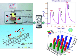
Graphene materials have been widely explored for fabrication of gas sensors because of their atom-thick two-dimensional conjugated structures, high conductivity and large specific surface area. Thin graphene layers with attached functional groups desirable for gas sensing applications were synthesized by a wet chemical route (modified Hummers' method). Sensing performances of reduced graphene oxide (rGO) against carbon monoxide (CO) were studied in terms of percent sensitivity (sensor response), response and recovery times and I/V characteristics at room temperature as well as at elevated temperatures. Sensitivity data indicate the highest activity (∼71% sensitivity against 30 ppm CO) occurs at room temperature (RT), indicating that the sensor is best operated at RT. Sensor response is quick (within 30 s) even to a trace amount (0.001% or, 10 ppm) of CO gas. Selectivity of the sensor was demonstrated by using different n-type reducing gases (like carbon monoxide, ammonia, methane and hydrogen), at different concentrations, showing negligible sensitivity towards gases other than CO. The synthesized material proves to be good as a selective room temperature sensor for harmful and poisonous carbon-monoxide gas.
The first experimental study of graphene for gas sensing was performed by Novoselov et al.1 They demonstrated the gas-sensing potential of graphene to detect water or ethanol vapors or ammonia gas. However, it was Schedin et al.5 who demonstrated that graphene is an ideal material for high sensitive gas detection. Of late, experimental and theoretical assessments of graphene's performance in gas and/or vapor sensing have been made in several reports.6–8
Sensing is a complex function that requires the integration of a number of properties from interface accessibility to transduction efficiency, molecular sensitivity and mechanical or electrical robustness. It appears that graphene fulfills many of these requirements and the realization of highly sensitive graphene sensors could illustrate how the previously described properties can be brought in synergy to this specific objective. Indeed, graphene is a pure interface with almost all atoms exposed to the analyte of interest, since it is only few layers thick.9 Moreover, local destruction of the sp2 lattice preserves its mechanical robustness and does not jeopardize its 2D delocalized transport properties unlike carbon nanotubes. Electronic and mechanical properties can be exploited to perform the transduction of the sensing signal.10
Since the last few decades, metal oxide based solid state gas sensors are being extensively used, but they suffer from various major drawbacks like poor selectivity, high operating temperatures, lack of stability etc. High operating temperature restricts their use in remote places. Moreover, high power consumption necessary to achieve high temperature for their operation may cause danger for gases which are flammable/explosive. Poor selectivity makes these sensors unsuitable in places where more than a gas species is available. The aim of the present study was to overcome these problems by using graphene (rGO) as the working material for gas sensors.
Amongst various gases, the detection of CO gas is especially important because it is toxic to humans and other animals. Moreover, being a colorless, odorless and tasteless gas which is slightly less dense than air, it is difficult to recognize in normal way. Carbon monoxide is highly toxic to human beings; the maximum time weighted average (TWA) exposure value or permissible exposure limit ascribed by the United States National Institute of Occupational Safety and Health, is 35 ppm over an 8 h period.11 Inhaling CO over TWA can cause headache, nausea, vomiting, dizziness and fatigue. CO combines irreversibly with hemoglobin (iron-center) of blood and produces carboxy-hemoglobin, which prevents delivery of oxygen to tissues. Thus carbon monoxide is constantly in the public eye, largely because the home is such a susceptible place for carbon monoxide poisoning.
In this work the synthesis of graphene layers by inexpensive wet chemical route (through modified Hummer's method) has been reported with layer thickness of ∼2 nm (detected by AFM height profile measurement). The sensing properties of rGO are employed for detecting low-concentration of CO gas, under the most practical environment, viz., room temperature and atmospheric pressure, in presence of ambient humidity. Suitable sensing sites have been established through prolonged refluxing. Sensors fabricated using synthesized rGO show as high as 71% sensitivity at room temperature against 30 ppm carbon monoxide gas. Significantly, sensitivity decreases with increasing operating temperature. Low cost fabrication of the device prototype was, thus, feasible.
:
10 wt ratio and sonicated in an ultrasonic bath (Wensar WUC Series-12L) for 30 min, followed by drop-wise addition of hydrazine hydrate (20%) with constant stirring for 2 h. The liquid turned into black graphene (or, reduced graphene oxide, rGO).
In the Hummers method, a combination of potassium permanganate (KMnO4) and sulfuric acid (H2SO4) is used. Though permanganate is commonly used as oxidant (e.g., de-hydroxylation), the active species is, in fact, di-manganese heptoxide (Mn2O7), generated in situ. The bimetallic heptoxide is far more reactive than its monometallic tetraoxide counterpart.
etc.) on the surface. During prolonged reflux, condensation reactions are responsible for introduction of the desired functional groups which ultimately facilitate the gas sensing behavior by changing the resistivity/conductivity upon exposure to the analyte gas.
The sample was preserved as dispersed in DI water for future experiments and characterization.
![]() | ||
| Fig. 1 (a) Real image of graphene sensor sample, drop coated on a glass substrate with attached Cu-wires. (b) Schematic diagram of gas measurement system. | ||
| Samples | Contact angle (degree) | Crystallite size from RAMAN (DRAMAN) (nm) | Lattice strain calculated from XRD |
|---|---|---|---|
| Graphene or, reduced graphene oxide (rGO) | 138 | 16 | 0.26 |
| Graphene oxide (GO) | 46.5 | 18 | 0.03 |
| Graphite | 115 | — | 0.01 |
| Functionalized (refluxed) rGO | 98.2 | 19 | — |
Fig. 3 shows the Raman spectra of GO, rGO and functionalized (refluxed) rGO taken at 515 nm LASER excitation (green LASER) along with the reference of bare glass (substrate) in the range of 500–2000 cm−1. It is a powerful non-destructive tool to distinguish ordered and disordered crystal structures of carbon. Each of the de-convoluted Raman spectra shows two peaks; disorder-induced (D) band and tangential (G) band. G band is usually assigned to the E2g phonon of C sp2 atoms, while D band is a breathing mode of κ-point phonons of A1g symmetry.13 Two distinct peaks at ca. 1360 and 1608 cm−1 correspond with D and G band of GO whereas, the same at ca. 1345 and 1600 cm−1 correspond with those of rGO. On the other hand, the D and G band of functionalized rGO appear at ca. 1375 and 1615 cm−1, respectively. There are some minor peak shifts of GO and functionalized rGO with respect to rGO probably caused by stress.14 However, the peak shifting for GO and functionalized rGO with respect to rGO are in the same direction indicating the presence of oxygen containing groups for both the species with respect to rGO. Intensity of both the bands of GO increases substantially, indicating the decrease in size of the in-plane sp2 domains, possibly due to the extensive oxidation and ultrasonic exfoliation as, when the exfoliated GO is chemically reduced, the intensity of the D and G band increases further.15 This can be due to defects introduced during preparation procedure.14 The intense ‘D’ peak indicates that the graphene sheets have structural disorder. Increased intensity ratio of D and G band (ID/IG) in rGO (1.04) compared with GO (0.91) suggests a decrease in the average size of sp2 domain due to the reduction process.
![]() | ||
| Fig. 3 Raman spectra of (a) bare glass (used as the substrate of the samples) (b) reduced graphene oxide (rGO), (c) functionalized (refluxed) rGO and (d) graphene oxide (GO). | ||
The mean crystallite size (DRAMAN) of GO, functionalized rGO and rGO layers were calculated using the following relation16
| DRAMAN = (2.4 × 10⁻¹⁰)λl4(ID/IG)−1 | (1) |
rGO, GO, functionalized (refluxed) rGO and graphite thin films were also used to test their wettability by measuring their static contact angle with water (shown in Fig. 4). It was found that the static contact angle between graphite film and water is 115° (Table 1). Because graphite consists of carbon atoms without any polarity, it demonstrates hydrophobic properties. For GO film and water, measured static contact angle is 46.5°, much less than 90°. Hence, GO shows a good hydrophilic property, as GO surface was presumably grafted with hydroxyl and epoxy groups. These polar groups change the graphite properties from hydrophobic to hydrophilic. After chemical reduction of GO by hydrazine hydrate, hydroxyl and epoxy groups are mostly eliminated, which is exhibited through the measured static contact angle of 138° which is larger than 90°.
![]() | ||
| Fig. 4 Static contact angle measurement with water and (a) graphene oxide, (b) functionalized (refluxed) reduced graphene oxide (c) graphite and (d) reduced graphene oxide. | ||
It is interesting to note that this contact angle is larger than that of graphite film and water. Even though isolated graphene also shows hydrophobic property, its degree of aqueous wettability is less than that of graphite. This can be understood easily as non-polarity of rGO is comparatively greater than graphite due to preferential reduction involving de-oxygenation and/or, de-hydroxylation during reduction by hydrazine hydrate. For this reason, graphite preserves more oxygen containing functional groups than rGO. This can be further experimentally evidenced by FTIR analyses (provided later on). On the other hand, contact angle between functionalized rGO and water is 98.2°, which is also hydrophobic in nature but the hydrophobic tendency is much lower than rGO and graphite. Hence, from the experimental data we may conclude that wettability trend of these C-forms follow the order: GO > functionalized (refluxed) rGO > graphite > rGO.
Graphite, GO, rGO and functionalized rGO were characterized by FTIR in the mid-infrared range, and the results are shown in Fig. 5. The sharp peaks appearing at ca. 1050 and ca. 1389 cm−1 were characteristic peaks of νC–O and the δC–OH stretching vibrations, respectively.17 The peak at ca. 984 and 3441 cm−1 can be attributed to O–H stretching vibrations of adsorbed water molecules or interlayer water molecules and structural –OH groups. This particular peak is almost absent in rGO as, it is preferentially de-oxygenated or, de-hydroxylated during the reduction process by hydrazine hydrate. On the contrary, graphite shows this peak (at 3441 cm−1) though, the magnitude is much less than that of GO or functionalized rGO. This de-oxygenation or, de-hydroxylation process of rGO is also confirmed by the wettability trend observed by the contact angle studies discussed earlier. In GO sample, the peak at ca. 1685 cm−1 can be attributed to O–H bending vibrations18,19 while, peak at ca. 1582 cm−1 reflects the skeletal vibration of GO.20 The presence of oxygen or –OH indicates that during the oxidation process of the graphite powder with KMnO4 in concentrated sulfuric acid medium, the original extended conjugated π-orbital system of graphite was destroyed and oxygen-containing functional groups were inserted into carbon skeleton i.e., the presence of these vibrations is indicative of the attachment of epoxide and hydroxyl groups. After reduction to rGO by hydrazine hydrate, these functional groups derived from the intensive oxidation have been eliminated (evidenced from Fig. 5). Disappearance of these two peaks (1582 and 1685 cm−1) in rGO sample indicate that the graphene oxide (GO) have been successfully exfoliated to graphene sheets (rGO). However, some peaks especially those originating from –OH functional group (like, peaks at ca. 984 and/or, 3441 cm−1) re-appear in the skeleton of functionalized rGO (by prolonged refluxing) indicating the presence of structural deformities, which ultimately facilitate the sensing performances.
![]() | ||
| Fig. 5 FTIR spectra of graphite, graphene oxide (GO), reduced graphene oxide (rGO) and functionalized rGO (refluxed rGO). | ||
A few representative TEM, HRTEM and related images of rGO thin films prepared by modified Hummers' method are presented in Fig. 6(a–d). Fig. 6(a) and (b) depict the bright field images showing uniform thin layers in nano-metric dimension. Electron diffraction pattern is depicted in a selected area (SAED) suggesting the crystalline nature of the nanostructure [Fig. 6(c)]. The fig. is indexed with 〈002〉 diffraction plane which corroborate well with Braggs' plane obtained from the XRD patterns and satisfy the reciprocal relationship between Miller indices (h, k, l planes) and SAED rings diameter.21 However, among the two techniques, the SAED technique is more authentic than X-ray diffraction because the dimensions of areas concerned are as small as several hundred nanometers whereas, X-ray diffraction deals with areas typically the samples' area, the dimensions of which is to be measured in several centimeters. The high-resolution transmission electron microscopic (HR-TEM) image of rGO is presented in Fig. 6(d) in which, those lattice planes (fringes) are visible that possess inter-planar spacing greater than the lateral spatial resolution limits of the instrument. Here, the HR image displays a d-spacing value of 3.6 Å corresponding to the 〈002〉 crystal plane.
![]() | ||
| Fig. 6 (a) and (b) TEM micrograph of rGO (c) SAED patterns with indexed plane and (d) indexed fringes of HR-TEM image. | ||
DC current–voltage (I/V) characteristics of as-fabricated graphene sensors at bias voltage ranging between −5 and +5 V at thirteen different operating temperatures between 30 °C (room temperature) and 150 °C (at 10 °C intervals) were recorded in air with a high resistance meter and the I/V characteristic plots obtained, presented in Fig. 7. The I/V characteristic curves demonstrate a good ohmic behaviour, indicating that the metal–carbon junction between gold (Au electrode) and graphene and the carbon–carbon junction between graphene layers are both ohmic in nature. This is of prime significance as sensitivity of gas sensors can be maximized when these junctions are ohmic or have negligible junction resistance. Fig. 7 also shows that at a particular voltage, the current between the electrodes increases continuously with increase in temperature, which is the characteristic property of semiconductors and is very common in metal–semiconductor contact systems. The semiconducting behaviour of graphene is thought to originate from the polarization of adsorbed molecules (e.g., water and oxygen sp.) and/or to defects introduced into the graphene sheets during the preparation22 or even to the influence of the supporting substrate.23
![]() | ||
| Fig. 7 Current vs. voltage (I–V) characteristic plots of functionalized graphene sensor films measured at temperatures between room temperature (30 °C) and 150 °C (at 10 °C intervals) in air. | ||
In presence of oxidising or reducing gases, adsorbed species on the surface of graphene sheets acting as electron acceptor or donor, respectively. Thus, like many semiconducting gas sensors, sensitivity of gas sensors made of sheets of graphene, is also a surface phenomenon and charge transportation through adsorbed species is likely to be a function of sheet thickness and orientation with respect to ‘dirac point’ which is comparable with crystallite size24 in poly-crystallites. However further explorations to elucidate the transport mechanism in graphene film, have not been made in this study.
| S = [(Rgas − Rair)/Rair] × 100 | (2) |
| Temperature | Sensor response (%S) | |||||
|---|---|---|---|---|---|---|
| 30 ppm CO | 20 ppm CO | 10 ppm CO | 100 ppm NH3 | 1000 ppm CH4 | 1000 ppm H2 | |
| a *ND: Not Detectable. | ||||||
| Room temperature (RT) | 71 | 52 | 44 | 7 | *ND | *ND |
| 60 °C | 68 | 65 | 57 | 15 | *ND | *ND |
| 90 °C | 64 | 48 | 40 | 28 | *ND | *ND |
| 100 °C | 60 | 45 | 35 | 33 | 3.1 | 5.6 |
| 120 °C | 51 | 43 | 23 | 42 | 3.9 | 6.2 |
Fig. 8(b) and (c) also shows that the sensor response is quick (within 30 s) even to a trace amount (0.001% or, 10 ppm) of CO gas, however, the recovery is not so swift (>15 min) probably because of slow oxidation of gas molecules on the surface of the sensor films.
It has been observed that, the sensors are not able to recover 100% in normal way. However, the sensors may recover almost abundantly and come to their initial baseline value if exposed to UV-light, after removal of the target gas [Fig. 8(c)]. For UV exposure, the LED UV flashlight (385 nm) is exposed (LED-120 Inspection Light; Groz Engineering) during the recovery period for 1 min, when the CO gas is turned off; although the UV light does not speed up the recovery process. In this study, a number of experiments have been carried out to measure the sensitivity as a function of operating temperature and time. In all the cases, sensitivity of the sensor element showed approximately constant values, indicating the repeatability of the sensor.
![]() | ||
| Fig. 9 Relative humidity (RH) effects on (a) the prolonged refluxed functionalized rGO [at room temperature and at 120 °C temperature] and (b) without refluxing rGO films. | ||
| Sl. No. | Material(s) sensing CO gas | Sensing parameter(s) | Selectivity to CO | Ref. |
|---|---|---|---|---|
| 1. | Nano-crystalline LaFeO3 and LaFe0.8Co0.2O3 particles | Sensitivity measured against 100 ppm CO | Selective to CO at 100 °C operating temperature | 27 |
| 2. | Pd doped SnO2 | Sensitivity measured in presence of humidity in the temperature range between 200 and 400 °C | Selectivity tests not performed | 28 |
| 3. | Nanogravimetric sensors made by [Pd(η2-ol)(8-(di-tert-butylphosphinooxy)quinoline)] and [Pd(η2-ol)(8-(di-tert-butylphosphinooxy)-2-methylquinoline)] | Sensitivity measured against 125 and 250 ppm CO | [Pd(η2-ol)(8-(di-tert-butylphosphinooxy)quinoline)] is selective to CO, even in presence of humidity | 29 |
| 4. | Rb2CO3 promoted In2O3 | Responds well to 200–4000 ppm CO in wet air at 300 °C and exhibits low cross sensitivities to H2, CH4, C3H6, NO and CO2 | Selective to CO at elevated temperature (200–300 °C) | 30 |
| 5. | Al doped graphene | CO sensitivity enhancement shown via introducing large amount of shallow acceptor states by forming Al–CO bond | Selectivity tests not performed | 31 |
| 6. | Al-doped ZnO films deposited onto SiO2/Si substrates | Maximum sensitivity of 61.6% reported for 65 nm ZnO:Al film thickness at the operating temperature of 400 °C. Sensitivity and response time improved by increasing the operating temperature | Selectivity tests not performed | 32 |
| 7. | Au- and Pt-doped SnO2–In2O3 nanocomposites | Showed superb sensitivity for both reducing and oxidizing gases like CO, NO and NO2 | Not selective to CO | 33 |
| 8. | CuO- and ZnO-doped pellet-type SnO2 | Sensitivity measured for 200 ppm CO and H2 in the temperature range of 80 to 450 °C | Showed high selectivity to CO at elevated (∼160 °C) operating temperature | 34 |
| 9. | 25% NiFe2O4–75% La0.8Pb0.2Fe0.8Co0.2O3 composite nano-powders | Exhibits excellent sensitivity toward CO at operating temperatures between 125 and 175 °C | Selective to 250 ppm CO at ∼175 °C operating temperature in presence of other gases like, 500 ppm C4H10, 500 ppm H2 and 5 ppm NO2 | 35 |
| 10 | Functionalized (18 h refluxed at 100 °C) reduced graphene oxide (rGO) | Showed the highest sensitivity (∼71%) against 30 ppm CO at room temperature, in presence of ambient humidity. Sensitivity decreases with increasing operating temperature | Selective to CO. Other n-type reducing gases (like, 100 ppm ammonia, 1000 ppm methane and 1000 ppm hydrogen) showed negligible sensitivity compared to CO | Present study |
| O2 (gas) ↔ O2 (ad) ↔ O2− (ad) ↔ O− (ad) ↔ O2− (ad) ↔ O2− (lattice) | (3) |
![]() | ||
| Fig. 10 Schematic representation of gas sensing mechanism of CO adsorption on graphene surface (carbon atom in black, oxygen in red and hydrogen atom in white). | ||
In this study, the prolonged reflux method helped to attach hydroxyl groups (–OH) into the reduced graphene oxide skeleton which ultimately increases the number of electrons in the conductance band leading to an augmented depletion layer. The attached OH− is responsible for generation of oxygen ion (O−) which remains chemisorbed on the surface of the material and gradually transforms into some other forms of anionic oxygen (like, O
) by extracting free electrons from the conduction band, resulting in an electron depleted surface layer and a consequent rise in resistance. The relevant chemical species exist in equilibrium39–41 as follows:
| 2OH− → H2O + O− (chemisorbed) + e− | (4) |
O− + e− → O (chemisorbed)
| (5) |
When the sensor film surface is exposed to the analyte gas (like CO), it gets physisorbed:
| CO (g) → CO (physisorbed) | (6) |
| CO (physisorbed) + O− → CO2 + e− | (7) |
CO (physisorbed) + O → CO2 + 2e−
| (8) |
| CO (physisorbed) + OH− → CO2 + H˙ | (9) |
| OH− + H˙ → H2O | (10) |
The processes indicate the in situ generation of atomic/ionic oxygen, which makes the process irreversible in nature. These chemisorbed charged oxygen species at the grain boundaries are responsible for the space charge appearance and band modulation. Therefore, the variation of the chemisorbed molecule density is supposed to be the main factor responsible for the electrical response whereas the rGO phase remains chemically stable. For this reason, such sensors can be used repeatedly and has prolonged shelf-life (unless humidity or other external interfering agents affect the sensor surface).
In the decomposition reactions of analyte gases in presence of surface adsorbates, the resultant products are carbon dioxide (CO2) and water (H2O) though the reaction proceeds through several intermediates. These reactions decrease device conductivity as a function of time44 and hence increase the resistance (p-type behavior) of the device prototype. As conductivity is proportional to the product of charge-carrier density and mobility, it is evident that changes in the number density or mobility of carriers, or both, must be responsible for the change in conductivity.45 Graphene's linear band structure around the Dirac points (the six κ points in the Brillouin zone) is responsible for the gas adsorption, which possibly increase the number of electrons as the gas is a donor one. Closed-shell adsorbates such as CO do not directly change the band structure of graphene, but rather alter the distribution of charges within graphene.46 Adsorbed water is a prevalent surface “impurity”, especially when located between the graphene and the substrate, which can shift the impurity bands of the substrate in the vicinity of graphene's Fermi level and so cause indirect doping of the graphene. Indeed, even if not initially present between the graphene and substrate, water molecules might diffuse into this region upon exposure to humid air.47 Recent theoretical studies also support this conclusion by demonstrating that gas molecules adsorb only weakly on pristine graphene, but adsorb more strongly on defective graphene (i.e., graphene that contains a vacancy due to point defect or incomplete removal of epoxy/oxygenated functional groups, that may generate during the reduction process or attachment of desired functional groups by prolonged condensation reaction through reflux method) surface.9,48 These defects and foreign functional groups remarkably affect the electronic conductivity of graphene based devices like gas sensors. The relative position of HOMO and LUMO of the adsorbate CO molecule with respect to Dirac point in graphene is presented in Fig. 11, which determines the direction of charge transfer.
![]() | ||
| Fig. 11 The relative position of HOMO and LUMO of the adsorbate CO molecule with respect to Dirac point in graphene (C atom in black and O atom in red). | ||
Usually CO molecule acts as a donor but the sites of the charge transfer only depends on the orientation of the molecules with respect to the surface. The differences in charge transfer are due to differences in orbital overlap between the HOMO (5σ; weakly antibonding) of the CO molecule and graphene. The LUMO (2π; strongly antibonding symmetry) seems to play no important role in the electron doping process although it is closer to the Dirac point than the HOMO. In this case, HOMO is thus the more important orbital and confers transferability of charge to graphene. Because the HOMO is mainly located on the C atom, the charge transfer is largest when the C atom is closest to the surface, smallest when the O atom is closer to the surface, and intermediate when both atoms are at (almost) equal distance from the surface. Graphene also exhibits dangling edge-bonds during CO adsorption.49 However, such edge effects, though providing energetically favourable sites for gas absorption, contribute only to a small extent (∼2%) of the total electrical response of a typical microscale graphene device.50 Stronger adsorption arises from either defects or doping with suitable components.48
Footnote |
| † Electronic supplementary information (ESI) available: AFM image of a multilayer graphene flake with height profile; FESEM micrograph of RGO; UV-Visible spectra. See DOI: 10.1039/c6ra06058g |
| This journal is © The Royal Society of Chemistry 2016 |