Matrix-assisted diffusion-ordered spectroscopy

Robert Evans *a and Iain J. Day b
aChemical Engineering and Applied Chemistry, Aston University, Birmingham, B4 7ET, UK. E-mail: r.evans2@aston.ac.uk
bSchool of Life Sciences, University of Sussex, Falmer, Brighton BN1 9QJ, UK

Received 29th February 2016 , Accepted 25th April 2016

First published on 26th April 2016


Abstract

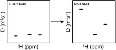
Diffusion NMR is a potentially routine tool in the analysis of mixtures, from industrial and synthetic outputs to natural products. However, the technique struggles to resolve species of similar size. Matrix-assisted DOSY offers a flexible approach to resolving such ambiguities on the basis of the chemical structures involved and on their interactions with a larger co-solute or matrix. The use of chromatographic supports, surfactants and polymers, in particular, is illustrated. The resolution of a wide range of different analyte mixtures, on the basis of differences in chemical structure and in stereochemistry, is demonstrated.


image file: c6ra05380g-p1.tif

Robert Evans

Robert Evans is Lecturer of Physical Chemistry at Aston University. He holds a MChem and PhD from the University of Oxford. He moved to Aston following postdoctoral work at Ecole Polytechnique, with Stefano Caldarelli, and the University of Manchester, first with Gareth Morris and Mathias Nilsson and then in the group of Nicola Tirelli. His research interests involve the use of Nuclear Magnetic Resonance in the analysis of polymers, soft materials and complex mixtures.

image file: c6ra05380g-p2.tif

Iain J. Day

Iain Day is Lecturer in Chemistry and NMR Spectroscopy at the University of Sussex. He studied for his MChem and DPhil at the University of Oxford. He was a postdoctoral fellow at the University of Massachusetts Medical School with Robert Matthews, before joining Medway Sciences, a joint initiative of the University of Greenwich and Pfizer Global R&D as a postdoctoral fellow with Adrian Davis, John Mitchell and Martin Snowden. In 2007 he was appointed Lecturer at Sussex. His research interests include the application of NMR spectroscopy to small molecule aggregation, chromatographic NMR and sensitivity enhancement for structure elucidation.


Introduction

The analysis of mixtures using Nuclear Magnetic Resonance (NMR) is a common yet difficult problem. Peaks produced by different species in the mixture can overlap with each other and it is difficult to assign any one peak in the spectrum to a particular species. Diffusion-ordered spectroscopy (DOSY) is a NMR method specifically designed for this purpose.1 In these experiments, a series of pulsed field gradient (PFG) experiments is used to estimate the diffusion coefficients of individual signals in a spectrum. A pseudo two-dimensional spectrum is produced in which individual NMR signals are correlated with the calculated diffusion coefficients. DOSY is therefore not a physical separation of the species present in the sample, as in chromatography, but a pseudo-separation which can be interpreted in a similar manner. As all the spins in a given species will be moving at the same speed, their NMR signals will all be found along a common horizontal line in the DOSY spectrum, corresponding to the diffusion coefficient of that species. Challenges occur when there is insufficient separation in the diffusion dimension or overlap in the spectral dimension.

Diffusion NMR

While there have been many advances in the design of diffusion NMR pulse sequences, they all share a number of common features.2,3 A series of r.f. and field gradient pulses are applied to the sample to wind the magnetisation into a helix, spatially encoding the position of the spins within the sample; a delay to allow the species to move according to Brownian motion; and a second series of r.f. pulses and field gradients which serves to refocuses the magnetisation helix prior to spectrum acquisition. Any species that has moved will therefore experience a difference between the encoding and decoding gradients and the magnetisation will not be completely refocused. Hence, the signals in the sample are attenuated in proportion to the speed at which they are moving during the delay period. The attenuation of the signals is given by the Stejskal–Tanner equation4 (eqn (1)).
 
S = S0eD[δγg]2Δ (1)

The Stejskal–Tanner equation relates the intensity of a signal in the presence of pulsed field gradients to the gyromagnetic ratio of the spins being observed, γ, key experimental parameters of the pulse sequence used, δ and Δ′, the strength of the magnetic field gradients, g, and the diffusion coefficient of the species of interest, D. The precise form of the Stejskal–Tanner equation (i.e. Δ′) depends on the pulse sequence used, but general cases have been reviewed.5 This review is based on the application of PFG-NMR techniques, and a full review of PFG-NMR, the Stejskal–Tanner equation and their use in measuring diffusion coefficients is beyond its scope. However, a number of key reviews and book chapters are highlighted, which may be of general interest.6–9

The processing of the echo attenuation data can be performed using a number of different algorithms, depending on the information required, and the resulting data presented in various ways.11–14 Of these, a DOSY spectrum, where the chemical shifts and diffusion coefficients are arranged in a two dimensional presentation, is the most visual and immediate way of showing this information. Software for the processing of this data in this manner is supplied by all major NMR manufacturers and stand-alone software is also available. A DOSY spectrum is used in a semi-quantitative manner, separating out the components of a mixture according to their respective diffusion coefficients. Most of the NMR spectra presented in this review take this form, with Fig. 1 showing a typical DOSY spectrum of a mixture of three alcohols. While the sample is not physically separated, the pseudo-separation of the signals of different components in a mixture may be regarded as analogous to the physical separation carried out in chromatography.


image file: c6ra05380g-f1.tif
Fig. 1 (a) DOSY presentation of a 1[thin space (1/6-em)]:[thin space (1/6-em)]2[thin space (1/6-em)]:[thin space (1/6-em)]2 mixture of methanol, ethanol and n-pentanol in D2O. (b) Shows an enlargement of the region between 3.2 and 3.6 ppm. Reprinted with permission from Morris et al., Anal. Chem., 66 (1994) 211–215. Copyright 1994 American Chemical Society.

However, the assignment of NMR signals to specific components in a mixture is hindered by overlap of chemical shifts, a topic that is the focus of much research interest, for example pure-shift DOSY,15 and when the components of the mixture have similar sizes and hence diffusion coefficients. The modulation of the diffusion coefficients through interaction with some additive to the solvent system, i.e. matrix-assisted DOSY or MAD (also referred to as chromatographic NMR and chrom-NMR16), is the basis of this review. Elements of this work have been reviewed elsewhere, as indicated in the preceding text. This review aims to make the use of diffusion NMR and its enhancement by addition of well-chosen additives relevant and accessible to a wider audience. Spectrometers are now routinely equipped with reliable pulsed field gradient systems, due to their ability to improve spectral quality and speed-up the acquisition of two-dimensional experiments by obviating the need for extensive phase cycling.10 This should make the use of all of the techniques described in this review routine.

From diffusion coefficient to size and association constants

It is possible to obtain quantitative information from the diffusion coefficients, if some care is taken both with the experimental execution and the subsequent treatment of the data.17 The relationship between measured diffusion coefficient and molecular size is deceptively straightforward at first glance. The Stokes–Einstein equation (eqn (2)) balances the thermal energy of random molecular motion against the friction acting upon a hard sphere, with a hydrodynamic radius rH, moving through a continuous fluid of viscosity, η, at temperature, T.
 
image file: c6ra05380g-t1.tif(2)

However, molecules are not typically hard spheres and solvents are not continuous. Eqn (2) is valid for molecules significantly larger than the solvent, e.g. proteins, but for the small molecules used in the experiments highlighted in this review, it typically under-estimates the diffusion coefficient.

A number of modifications have been suggested, taking into account the assumptions implicit in eqn (2). For example, the change in the friction factor for elliptical species can be estimated using the Perrin shape factors,18 which add an additional term to the denominator of eqn (2), accounting for the fact that the species will no longer undergo isotropic motion due to the presence of major and minor axes. A number of alternatives and modifications to the Stokes–Einstein equation, such as empirical fitting functions19 and analytical corrections for non-continuous solvents,20 have been suggested to obtain both hydrodynamic radii and molecular weights for species in solution.

Eqn (3)21 is a recently developed relationship that parameterises various deviations from a diffusion coefficient predicted by the Stokes–Einstein equation. In eqn (3), α is the cube root of the ratio of sample molecular weight to solvent molecular weight, η is the viscosity of the solvent and ρeff is an effective density of the solvated species, allowing for packing effects, geometry, solvation and flexibility, obtained by a single parameter fit for a set of compounds. A value of ρeff ≈ 620 kg m−3 was found to fit a large test set of molecules. Eqn (3) only acts as an approximate model as it does not handle the effects of factors such as shape, flexibility, and solvation explicitly, however; it does perform well enough to address chemical problems and has been used to predict the diffusion coefficients of a wide range of species.22,23

 
image file: c6ra05380g-t2.tif(3)

Both eqn (3) and the Stokes–Einstein equation show that similarly sized molecules, such as isomers, will have very similar diffusion coefficients. The typical resolution of a diffusion NMR experiment is such that it becomes practically impossible to resolve the small differences between their diffusion coefficients. This is especially true in the case where signals in the original NMR spectrum overlap. The addition of a matrix results in a differential interaction between the various components of the mixture and the matrix, and therefore the apparent diffusion coefficient, as measured in the NMR experiment, is reduced according to eqn (4).

 
Dobs = ffreeDfree + fboundDbound (4)
Dfree and Dbound correspond to the diffusion coefficients in the free and bounds states, with fractional populations given by ffree and fbound respectively. This equation is at the heart of all the experiments that follow. The analyte species interact with the larger, matrix, species and the diffusion coefficients observed are apparent diffusion coefficients, the weighted average of the motion of the species during the experiment. A stronger interaction leads to more time spent in the bound state and therefore a lower apparent diffusion coefficient.

Early results

The idea of adding a co-solute in order to perturb the diffusion behaviour of the sample is well established.24 The DOSY spectrum of a mixture of methanol, ethanol and pentan-1-ol shows all species resolved (Fig. 1). However, a mixture of methanol, iso-pentanol, neo-pentanol and tert-butanol could not be fully resolved without the addition of the cationic surfactant DTAB (dodecyltrimethylammonium bromide). While toluene, benzylalcohol and tetraethylene glycol could be resolved, the observed diffusion coefficients could be altered by the addition of the anionic surfactant, sodium dodecyl sulfate (SDS), as demonstrated in Fig. 2. The ordering of the species was reversed from the un-modified solution. The ordering of apparent diffusion coefficient in the micellar solution matched the trend in partition coefficient between oil and water (log[thin space (1/6-em)]P). Partition coefficients are a measurement of a molecules hydrophobicity, or otherwise,25 based on the ratio of equilibrium concentrations of the species in a immiscible mixture of oil, typically octanol, and water. The observed ordering in the presence of SDS can now be related to the structure and properties of the molecule.
image file: c6ra05380g-f2.tif
Fig. 2 (a) DOSY presentation of an equimolar mixture of toluene, benzyl alcohol and tetraethylene glycol in D2O. (b) Shows the same mixture in the presence of 150 mM SDS. Reprinted with permission from Morris et al., Anal. Chem., 66 (1994) 211–215. Copyright 1994 American Chemical Society.

A similar early use of PFG-NMR was in the study of potential interactions between small molecules and larger ones.26,27 This technique, known as affinity NMR, is a method for screening large numbers of ligands for interactions with their receptor molecules. A mixture of possible ligand molecules is added to a likely receptor and the diffusion-edited NMR spectrum acquired. As a ligand forms a complex with the bulky receptor, its Brownian motion is slowed. A diffusion filter is applied in the affinity NMR experiment to remove the fast diffusing components, i.e. the non-binding small molecules, and only the slow moving receptor–binding ligands (and the receptor itself) are left in the spectrum. The binding ligands can therefore be easily identified by their characteristic chemical shifts. While affinity NMR spectra are rarely presented in a two-dimensional format, the technique relies on the same principles as outlined in the previous section.

Likewise, the addition of chiral molecular micelles to electrokinetic chromatography has been shown to be capable of resolving enantiomers. The interaction between enantiomer and micelle can be studied with a range of NMR techniques including diffusion NMR. One such study28 probed the interaction of enantiomers such as (R) and (S) 1,1′-bi-2-napthol with poly(sodium N-undecanoyl-L-leucylvalinate) molecular micelles using a mixture of diffusion NMR and nuclear Overhauser effects to quantify the strength of interaction between micelle and enantiomer, and to identify enantiomers binding site on the surface of the micelles. The experimental methods and approach used by Morris et al. are similar to those used in later studies and could be important in probing the nature of the interactions between analytes and added matrices.

Matrix-assisted DOSY

Use of chromatographic supports

With the analogy to chromatography already made, chromatographic material such as silica, and functionalised silica, is an obvious choice for a suitable matrix. A wide range of different materials are regularly used in column chromatography. Hyphenated techniques such as HPLC-NMR couple the output of the chromatographic column directly to the NMR probe, acquiring NMR spectra of the eluents as they come off the column29 have been demonstrated but are technically challenged and have not found routine use.

Also known as chromatographic NMR, the addition of a chromatographic support such as fused silica to the sample in an NMR tube aims to replicate traditional chromatographic techniques in which the sample for analysis, containing a mixture of different compounds, is injected into a mobile phase which is flowed over a stationary phase, such as silica, typically packed in a column. Differential interactions with the silica selectively slow down the components of the mixture as they travel along the column resulting in different retention times for each component. In chromatographic NMR experiments, the support is added directly to an NMR tube containing the sample. The components of the mixture will establish an equilibrium between those molecules in free solution and those interacting with the silica stationary phase, giving rise to a reduced apparent diffusion coefficient and ideally, enhanced separation based on chemistry and structure.

An unfortunate side effect of using an insoluble stationary phase, such as a silica, to modify the diffusion properties of the mixture is that the sample inhomogeneity results in significant broadening of the spectral resonances, typically due to susceptibility broadening, potentially leading to peak overlap and a loss of information even for small particle sizes. Two methods have been proposed which can reduce this line broadening.

The first is high resolution magic angle spinning (HR-MAS). The measurement of diffusion coefficients in a MAS NMR experiment has some associated problems.30 Spinning a sample at high speeds induces an additional source of motion in the sample and increases in observed diffusion coefficient have been observed in water samples at high spinning rate. For low-viscosity solvents, such as acetonitrile, adverse effects of spinning are observed at even low spinning rates. The effect of spinning can be reduced by the use of a small sample volume and higher viscosity solvents. Care is also typically required to synchronise the diffusion labelling period and gradient pulses with the rotor spin rate.31

The use of MAS in matrix-assisted DOSY experiments with silica stationary phases has been successfully demonstrated by Calderelli and co-workers with the clear separation of mixtures of naphthalene, ethanol and dec-1-ene and dichlorophenol, ethanol and heptane using both bare and functionalised ‘reverse-phase’ C18-silica respectively as demonstrated in Fig. 3.32


image file: c6ra05380g-f3.tif
Fig. 3 DOSY spectra for two mixtures showing the effect of adding a chromatographic stationary phase. Naphthalene, ethanol and dec-1-ene (A) are separated using C18-functionalised silica (B), while dichlorophenol, ethanol and heptane (C) are separated used fused silica (D). Reprinted from Viel et al., Proc. Natl. Acad. Sci. U. S. A., 100 (2003) 9696–9698. Copyright (2003) National Academy of Sciences, USA.

The use of bare silicas on samples made with deuterated solvents reproduced a separation of a homologous series of aromatic compounds and also separated a methanol/iso-propanol/phenol/ethylene glycol mixture.33 As with standard chromatography, the selectivity of the chromatographic NMR experiment can be modified by changing the nature of the silica, with a wide range of commercial silicas available, with numerous different functionalities. The selectivity can also be modified by changing the composition of the solvent used, in an analogous manner to modifying the mobile phase in traditional HPLC experiments. Using a mixed solvent “mobile” phase, resolved spectra were obtained in diffusion NMR experiments for compounds that were not resolved using standard HPLC techniques. In an experiment intended to resemble hydrophobic interaction chromatography (HILIC), a test mixture of aromatic homologues in a mixed solvent of acetonitrile and water was separated in the diffusion domain by bare silica, whereas the HILIC experiment that it sought to reproduce showed only a single broad peak.34 Reproduction of the expected chromatographic result was also achieved using a polar reversed-phase silica, with a sulphonamide group, and a set of linear polyaromatic compounds.35

The role of the solvent in chromatographic NMR experiments is also important. Traditional flow chromatography experiments are usually performed using standard (i.e. proteo-) solvents, which would introduce large peaks into the NMR spectra if used directly. The high cost of deuterated solvents makes hyphenated techniques uneconomic for all but the simplest common solvent systems.29 In contrast, matrix-assisted DOSY in the form of chromatographic NMR is easy to perform and requires no more solvent than a standard NMR experiment.

The retention of different species by a chromatography column depends, in part, on the composition of the mobile phase. Varying the constituents of the solvent system can modulate the effect of the chromatographic support on its ability to separate the components of the mixture. This sensitivity to solvent composition can be reproduced in the NMR experiments. Altering the composition of an acetonitrile/water mixture changed the measured apparent diffusion coefficients of a number of mixtures in contact with octadecylsilyl bonded (C18) silica but the ordering of the compounds remained the same.36

It is also possible to modulate the observed diffusion coefficients in the presence of chromatographic supports by altering the ratio of solution to solid. A number of experiments have already shown the ability to separate mixtures in diffusion NMR experiments while similar separations were either not observed or less well resolved in traditional liquid chromatography experiments. It has also been reported that benzene has been observed to diffuse faster in the presence of a silica stationary phase than in the bulk solution.34 It would be expected that, even in the absence of any additional interactions, diffusion in a confined space, i.e. pores in the silica, would reduce the distance a species can diffuse during the diffusion encoding period of an experiment and hence result in a smaller apparent diffusion coefficient. The observed faster diffusion can explained by including the vapour phase of the benzene solvent and invoking an additional evaporation–condensation mechanism contributing to the motion of the benzene in and around the silica support.37 This work highlighted that, while the main interaction thought to be responsible for the reduction in apparent diffusion coefficient is absorption of the solute onto the stationary silica phase, additional solute-solvent and solvent-stationary phase interactions need to be considered.

The importance of the ratio of solid-to-liquid on mass transport has been known for the motion of single species through a chromatographic support.38 The effect on separation in matrix-assisted DOSY experiments was demonstrated in a number of experiments using both napthalene/aniline/phenol and polycyclic aromatic hydrocarbon mixtures in the presence of LiChrospher Si100 silica with the ratio of solution-to-solid increased from just under 1 to almost 7. For the homologous series of linear aromatics, separation was only observed at low values of the phase ratio, while the opposite was true for the napthalene/aniline/phenol sample, with the high phase ratio results reproducing observed chromatographic separations.39 It is far easier to alter the phase ratio in matrix-assisted DOSY experiments than in “normal” chromatography experiments, making it an important experimental parameter to consider. Seemingly unsuccessful chromatographic NMR experiments might be a result of a poor choice of solvent, co-solvent or solvent-solid ratio.

The second approach to reducing broadening caused by the addition of a stationary phase aims to match the magnetic susceptibility of the solvent to that of the stationary phase employed. Highly chlorinated and brominated solvents have large volume magnetic susceptibilities and using combinations of solvents, the susceptibilities of the solid support and the solvent can be matched, reducing the line widths observed in the NMR spectra. A limitation of this technique is that the stationary phase must remain stable in the solvent system employed. This is not always the case in the presence of highly brominated or iodinated solvents. This approach has been shown to work well with both bare silica gels and modified reverse phase silica.40 The two types of silica gel exhibited different selectivity for different functional groups on the analytes, with the bare silica interacting more strongly with more polar compounds and the opposite behaviour exhibited by the reverse phase material. This expected selectivity matches that observed in traditional chromatographic experiments. The mechanism of interaction was investigated using a large number of compounds with a wide range of chemical structures and functionalities. In the NMR experiments using bare silica, the number of bonds between sample and bare silica surface was an important factor, as was the extent of hydrogen bonding present in the molecules, in determining the strength of the interaction. Where species formed a similar number of bonds with the silica, the relative positions of the groups proves important. For example, ethylene glycol was slowed down considerably more than propan-1,3-diol and compounds that formed intramolecular hydrogen bonds also interacted more strongly with the silica surface.41

An alternative to susceptibility matching through use of solvent mixtures can be achieved by changing the structure of the silica particles themselves. The effect of adding silica to the NMR sample is to disturb the magnetic field homogeneity of the sample, with the magnetic field experienced by the spins being a sum of the external field and the superposition of the dipolar fields generated by randomly distributed silica particles. Using hollow silica spheres reduces the magnetic dipole of the silica, and large reductions in the magnetic dipoles of silica particles are achieved when the shell thickness is 5% of the sphere radius. Successful separation of a benzoic acid/benzyl alcohol/benzaldehyde sample was demonstrated with hollow silica spheres with shell thickness of 13% of the sphere radius.42

Chromatographic supports are not limited to silica-based materials. Thin layer chromatography can be achieved using paper supports. Zirconia-packed columns can also be used, exhibiting a different selectivity to silica.43 Size-exclusion chromatography (SEC, also known as gel permeation chromatography, GPC) is widely used in polymer chemistry. Using a cross-linked dextran support, macromolecules such as polymers or proteins can be separated on the basis of size. As the mixture passes through the porous polymer gel, the smaller species are able to fit into the pores and become trapped for longer periods of time while the larger species are less impeded. The larger species are therefore eluted first, with a separation based mainly on the hydrodynamic radius of the species involved. Typical applications of SEC include obtaining details on polymer molecular weight profiles and the analysis of the multimeric states of proteins.

The use of these materials has been demonstrated in MAD experiments for a range of polymers as solutes. Proof of principle was achieved by using poly(styrene 4-sulfophonate) and poly(N-isopropyl acrylamide) of differing molecular weights. A range of commercially available molecular weight reference standards covering the fractionation range of the stationary phase were then used to further characterise the observed changes in diffusion coefficient.44 While the overall effect of adding an SEC support was to reduce the diffusion resolution of a sample, as the effect on smaller species (with initially larger diffusion coefficients) is greater than on larger species, a number of applications have been demonstrated. It aids in the analysis of polymer sizes, as both diffusion coefficients before and after addition can be related to log(Mw) of the polymer. This effect is observed for simple samples of a single polymer44 and also for mixtures of polymers, both with similar and different molecular weights.45

The diffusion coefficients obtained in both the absence and presence of the support can be interpreted using an empirical equation, indicating the effect of the size-exclusion on the diffusion behaviour of the polymer44,45 as shown in Fig. 4. The technique has also been applied to the azo-dye sunset yellow, which self-assembles in solution. Once the assemblies grow beyond a certain size, they can no longer enter the pores, therefore a partitioning between smaller aggregates within the pores and larger assemblies in free solution is established.46


image file: c6ra05380g-f4.tif
Fig. 4 Observed diffusion coefficients for a range of poly(styrene sulfonate) molecular weight reference standards in the absence and presence of Sephadex G-50 stationary phase. The straight lines are fits to: log[thin space (1/6-em)]M = a0a1D. Reprinted from J. Magn. Reson., 220, R. E. Joyce and I. J. Day, Chromatographic NMR with size exclusion chromatography stationary phases, 1–7, Copyright (2012) with permission from Elsevier.

The accepted mechanism for size exclusion chromatography involves the solute molecules in the species diffusing in and around the porous stationary phase. Smaller molecules spend more time in the pores than larger species. A dynamic equilibrium forms between the species in the pores and those in free solution, and this allows the use of a slight modification of eqn (4).

 
Dobs = ffreeDfree + fporeDpore (5)
 
Kav = ffree/fpore (6)

The observed diffusion coefficient in a constrained pore of approximate radius a can be understood in simple cases and parametrised according to the relationship:47

 
image file: c6ra05380g-t3.tif(7)
D is the diffusion coefficient and Δ is the diffusion labelling period. At one extreme, where ξ ≪ 1, the effect of pores is minimal, the gel has little effect on the species in the sample and the measured diffusion coefficient is that of the species in free solution. At the other, where ξ ≫ 1, the size and shape of the pores governs the diffusion, reducing it by an amount related to the porosity of the stationary phase. In this case, the path of the species within the pore ensures it collides with the walls and reducing the path taken. The effect of this is to reduce Dpore by an amount related to the porosity of the material. In this limit, D is independent of the diffusion labelling time, Δ. This is not the case for intermediate values of ξ where the measured diffusion coefficient is a function of Δ. This allows some prediction of the effect of size-exclusion chromatographic supports on a sample. The behaviour of the sample can be estimated from the properties of analyte, chromatographic support and experimental parameters. If ξ is large enough, then the effect of the support on a given polymer or protein can be estimated from its porosity.

Use of surfactants

An alternative method by which the diffusion properties of species in an NMR sample can be modified is by the use of surfactants. A typical surfactant, such as sodium dodecyl sulfate, consists of a hydrophobic chain and a polar head group. The wide variety of different chain structures and head groups gives great potential in matrix-assisted DOSY experiments as there is a change in hydrophobicity across the length of the molecule. The variation of the concentration of co-surfactants and oils to modify the final emulsion structure allows a wide degree of control of the matrix and modulation of the interaction between the matrix and the analyte.

A number of reports have demonstrated the use of diffusion NMR in studying how surfactants, such as SDS, can solubilise peptides, hydrophobic drugs and as controlled release scaffolds.48–50 This concept has been thoroughly reviewed by Silber et al.51 and provides a good theoretical underpinning of the thermodynamics of the process and of the selection mechanism.

Multicomponent micro-emulsions typically contain three phases: an oil phase, a water phase and a surfactant phase. A micro-emulsion is a structured mixture of two immiscible liquids that spontaneously form nanometre-sized droplet structures that are thermodynamically stable. The droplets are almost mono-dispersed and usually smaller than 50 nm. The final micro-emulsion and its structure depend on the composition of the material.

The advantages of surfactants over solid supports were summarised by Hoffmann et al.52 as:

• Use of a regular solution-state NMR spectrometer. In the absence of a solid support, magic angle spinning capability is not required.

• As the sample can be studied without MAS the measured diffusion rate is not affected by sample spinning.

• There is no need for brominated or iodinated photosensitive solvents that have a sufficiently high magnetic susceptibility to match with the silica phase. In fact, the magnetic susceptibility of the micro-emulsion and the very small size of the droplets leads to good field homogeneity and the resulting line widths are comparable to normal NMR spectra.

• Silica suspensions tend to precipitate out of solution over a period of minutes to hours while micro-emulsions can be stable for years in sealed ampules/NMR tubes.

In all of these surfactant MAD experiments, the same general rules apply. Any component that is trapped in the smaller phase is expected to have a slow diffusion coefficient, being confined and restricted. A component that is located in the continuous phase is free to move long distances and can therefore diffuse relatively quickly. The components located at the interface of bi-continuous micro-emulsions are expected to diffuse the slowest. As the micro-emulsion structure is changed, for example, by increasing the water content, the different phases will change in size and nature, leading to changes in the observed diffusion coefficient.

In order to reduce the overlap of the emulsion with the mixture signals of interest, differing amounts of deuteration and fluorination of the surfactant species were used. Initially, to demonstrate the modulation of diffusion coefficients by a micro-emulsion, an solution consisting of an oil phase of R(+)-limonene and ethanol (1[thin space (1/6-em)]:[thin space (1/6-em)]1 w/w), an aqueous phase of water and polyethylene glycol (1[thin space (1/6-em)]:[thin space (1/6-em)]1 w/w), and Tween 60 (ethoxylated sorbitan monostearate) as the surfactant was used.53 A test mixture of model fluorinated compounds was chosen: inorganic polar NaF, non-polar perfluorohexane which is very hydrophobic, and 2,4-dinitrofluorobenzene which has intermediate polarity. At low water concentrations, where small water droplets are dispersed in the oil/surfactant mixture, the fluoride ions would be expected to diffuse slowly. However, rapid relaxation of the signals made accurate determination of diffusion coefficients difficult. Increasing water content converted the sample to a bicontinuous mesophase and then into an oil in water micro-emulsion, with a resulting increase in measured fluoride diffusion coefficient. The reverse behaviour was observed for the lipophilic component(s).

The use of these microemulsions was initially demonstrated on commercial drug formulations such as Dexamol and Advil. Such formulations are supplied as capsules or tablets containing a range of other compounds including organic and inorganic excipients used stabilise the active pharmaceutical ingredient and confer the desired release profile. In each case, a previously unresolved DOSY spectrum was resolved into a number of constituent spectra, corresponding to the components of the formulation. Typically resolved spectra were for the active ingredient and the stearic acid and sucrose excipients.53 Fig. 5 shows the DOSY spectrum of Advil in the presence of a typical microemulsion system.


image file: c6ra05380g-f5.tif
Fig. 5 DOSY spectrum of Advil in the presence of a microemulsion of SDS-d25/n-butanol-d10/D2O/cyclohexane-d12 (12.4[thin space (1/6-em)]:[thin space (1/6-em)]24.8[thin space (1/6-em)]:[thin space (1/6-em)]7.5[thin space (1/6-em)]:[thin space (1/6-em)]55.4 w/w). Reprinted with permission from Pemberton et al., Langmuir, 27 (2011) 4497–4504. Copyright 2011 American Chemical Society.

The technique was subsequently demonstrated with a wide selection of flavour and fragrance compounds, all of which were of a similar size and possessed similar structures and functional groups.52 Two different micro-emulsions were used, an oil-in-water system comprising lithium perfluorododecanoate, propan-2-ol-d8, deuterium oxide, and perfluorohexane with a weight ratio of 7.97[thin space (1/6-em)]:[thin space (1/6-em)]28.46[thin space (1/6-em)]:[thin space (1/6-em)]61.65[thin space (1/6-em)]:[thin space (1/6-em)]1.92 and the other, water-in-oil composed of SDS-d25, butan-1-ol-d10 deuterium oxide and cyclohexane-d12 with a weight ratio of 12.38[thin space (1/6-em)]:[thin space (1/6-em)]24.79[thin space (1/6-em)]:[thin space (1/6-em)]7.63[thin space (1/6-em)]:[thin space (1/6-em)]55.20. All of the analyte species had their measured diffusion coefficients reduced by the application of both microemulsion solutions. However, of the sample set, only the diffusion coefficient of sucrose was reduced more by addition of the W/O microemulsion. The remainder of the flavour/fragrance compounds exhibited the reverse behaviour, where the smaller diffusion coefficient was observed in the O/W microemulsion. An explanation for the observed selectivity was proposed in terms of where the analyte molecules bound to the micelles. The role of the sample as co-surfactant was also considered. In these experiments, the lipophilicity of the sample compounds, a property that increases with the size of the non-polar part and decreases with added polar groups, determines where the compound is solubilised. This is modulated by the size and shape of the compound and its ease of packing into the surfactant. The explanation was tested by the analysis of the diffusion behaviour of a number of similarly sized molecules from the previous set. For example, 2-trans-4-trans-decadienal, menthol and β-citronellol all contain ten carbon atoms and one oxygen-containing functional group. Differences in the shape of the molecules (cyclic vs. branched vs. linear) and the number of methyl groups give rise to differences in lipophilicity and therefore in the diffusion coefficient measured in the matrix-assisted DOSY solution.

Similar results can be obtained by using micelles in water. The basic effect is similar to that of micellar solubilisation of species, a well-established and demonstrated phenomenon, with applications from delivery of poorly soluble drugs54 to the day to day washing of laundry. The first use of both anionic and cationic surfactants was demonstrated very early on in the development of diffusion NMR, as noted earlier in this review.24

However, most of the early work resolved samples consisting of functionally different but similarly sized species. Using sodium dodecyl sulphate micelles in aqueous solution, a mixture of three isomers of dihydroxybenzene was resolved, separating the compounds on the basis of their structure, and hence, their hydrophobicity/hydrophilicity (Fig. 6). The ordering of apparent diffusion coefficient in the micellar solution matched the trend in partition coefficient between oil and water (log[thin space (1/6-em)]P).55 The analogous set of methoxyphenols was also separated using this approach.56


image file: c6ra05380g-f6.tif
Fig. 6 Oneshot DOSY spectra of an equimolar mixture of catechol (C), resorcinol (R) and hydroquinone (H). Top is in D2O, below is with 150 mM SDS micelles. Reprinted with permission from Evans et al., Anal. Chem., 81 (2009) 4548–4550. Copyright 2009 American Chemical Society.

The range of mixtures that can be studied using micelles as a matrix is not limited to structural isomers of substituted phenols. The cis/trans isomers maleic and fumaric acid were resolved using SDS and AOT (sodium bis[2-ethylhexyl] sulfosuccinate), with the trans isomer interacting more strongly with SDS. This selectivity is reversed upon addition of AOT.57 Mixtures of structurally similar flavonoids, such as those found in green tea extract, were also resolved using SDS in mixed solvents containing DMSO-d6 and D2O.58 Catechin, fisetin and quercetin are all effectively the same size and have very similar structures. The small structural differences between the three compounds allow their unambiguous resolution using SDS micelles in a 50[thin space (1/6-em)]:[thin space (1/6-em)]50 (v/v) mix of DMSO-d6 and D2O. This suggests a high level of specificity in the interaction between solute and micelle. The diffusion behaviour of the species could be modified by simply changing the composition of the solvent, but the measured diffusion coefficients are a simple matter of size and solvent viscosity. The smaller flavone molecules are observed to move faster than fisetin and catechin. The addition of SDS separates catechin from fisetin, a pair of molecules that differ in size by only a few mass units. The additional hydroxyl group in catechin, effectively replacing the carbonyl group of fisetin may be important in the separation process. At the larger end of the scale, a microemulsion was used to separate out a mixture of oligomers of the detergent Igepal ca-520. As the polyethoxy chain increases in length, the molecule is more likely to be found in the polar region at the centre of the microemulsion reverse micelles and hence observed at increasingly smaller diffusion coefficients.59 A linear relationship between the partition coefficient and the number of ethoxy units was obtained.

While MAD experiments have been performed with both anionic and cationic surfactants, it is also possible to use non-ionic species such as the Brij family of surfactants. These consist of an hydrophobic alkyl chain connected to a hydrophilic polyether. Brij surfactants in a mixed DMSO-d6/D2O solution were able to resolve the mixture of structurally similar natural products quercetin, fisetin and catechin.60

All surfactants described thus far in this review have long alkyl chains, which introduce additional signals to the initial NMR spectrum. While these are typically found in the same region of the spectrum, around 1–2 ppm, and the large size of the micelles ensures that they appear at low diffusion coefficients, the overlap of signals in a diffusion NMR experiment significantly increases the difficulty of processing the experimental data and reduces the accuracy of any measured diffusion coefficients.15 A number of strategies exist that can reduce the influence of these signals. Deuteration of the alkyl chain of SDS is an effective way of removing all of the micelle signals and SDS-d25 has been shown to separate similar sized peptides on the basis of the amino acids present.61 Fluorinated surfactants have also been used to similar effect.52,53

The mechanism of the resolution of species by micelles was studied using a large range of experimental conditions, with useful separation of many compounds observed over a wide range of sample and micelle concentrations. Separation was even observed in unlikely cases – when the surfactant concentrations were at low ratios compared to the solute and even when lower than the critical micelle concentration, CMC, of the surfactant. This suggests that the solute can aid in the formation of an appropriate matrix, reducing the amount of surfactant required. It is possible to model the system by considering the formation of micelles from single surfactant models and then considering the equilibrium, K, between bound and free solute molecules.

 
image file: c6ra05380g-t4.tif(8)
 
image file: c6ra05380g-t5.tif(9)
 
image file: c6ra05380g-t6.tif(10)

These equations work well for surfactants that form well-defined, reasonably monodisperse micelles and do not change size too much upon absorbing a solute molecule. They can also be used to obtain a value of the equilibrium constant between bound and free solute. This analysis was performed for a range of small alcohols, linear and branched, primary, secondary and tertiary, for SDS and AOT and the final values of log[thin space (1/6-em)]K were compared with the values of log[thin space (1/6-em)]P for the species as plotted in Fig. 7.62


image file: c6ra05380g-f7.tif
Fig. 7 Scatter plot of log[thin space (1/6-em)]K vs. log[thin space (1/6-em)]P for a range of alcohols in SDS (open symbols) or AOT (filled symbols) micelles. The dotted line has unity slope, while the solid line is the linear regression for the SDS data. Reprinted with permission from Tormena et al., Magn. Reson. Chem., 50 (2012) 458–465. Copyright (2012) John Wiley and Sons Ltd.

There is an essentially linear correlation between log[thin space (1/6-em)]P and log[thin space (1/6-em)]K for SDS micelles, strongly suggesting that the main driving force for the separation due to this micelle system is the differential association into different environments with different hydrophobicities. The deviations from this correlation are due to steric effects, with secondary and branched chain alcohols exhibiting lower association constants than their primary counterparts, and the amphiphilicity of the solute molecules, as the solute molecules play some role in the formation of the micelles. There is no such clear trend for AOT, a surfactant that does not show a well-defined CMC and forms polydisperse aggregates.

Use of polymers

Polymers offer an attractive alternative to the previous supports described so far in this section. In theory, any functional group can be synthesised into a block co-polymer, with different loadings of the group dependent on the details of the synthesis. The large number of repeating units gives the matrix a large weight and therefore a small diffusion coefficient.

The use of a copolymer of methyl methacrylate and a small proportion of methacrylate functionalised with undecanoic acid was first reported many years before a significant proportion of the results discussed in this review.63 This copolymer was used for screening a compound library on the basis of recognition and host–guest properties, following the same principles as matrix-assisted DOSY. The diffusion behaviour of a large set of compounds was modified by the addition of a polymer, and stronger interactions were revealed by larger decreases in the diffusion coefficient. Across the series of compounds used in this test, most only showed a small change in diffusion coefficient, indicating only a small interaction with the polymer. One compound, a hydroquinine, however, showed a large interaction and analysis of the NMR spectra indicated that the saturated quinuclidine ring was protonated under these conditions, causing association between the cation and the anionic polymer. Weaker bases in the set of compounds, typically not protonated under these conditions, showed no interaction.

Polymers can also be synthesised to take advantage of specific interactions. A block copolymer of polystyrene and a chiral polystyrene derivative were used on a sample containing a range of chiral species. Only one of the selected species, α-methoxyphenylacetic acid, interacted with the chiral polymer, almost saturating the binding sites. Analysis of the NMR spectrum of the bound species shows a splitting of its NMR signals, in particular the methyl and the α-proton, indicating the specific nature of the interaction.63

A range of commercially available polymers have also been used in MAD experiments. Applied to a mixture of similarly sized molecules, polyvinylpyrrolidone (PVP) was shown to interact strongly with phenols, intermediately with linear alcohols and not interact at all with either toluene or benzaldehyde.64 Poly(ethylene glycol) (PEG) exhibited a similar profile of interactions with a larger set of molecules.65 Phenols and carboxylic acids showed a strong interaction, linear alcohols showed an intermediate interaction while amines and non-polar species showed very little interaction with the polymer. Typically, the selectivity follows the polarity of the species. There was a marked difference in interaction between the isomers of nitrophenol, with the ortho-isomer barely interacting with the PVP polymer and the meta- and para-isomers interacting strongly. This suggests intramolecular hydrogen bonding within the ortho-isomer prohibits interaction with the polymer matrix and gives further indication to the nature of the analyte–matrix interactions.

The performance of the PVP matrix was further analysed in order to better understand the chromatographic processes occurring in the samples.66 Diffusion experiments, with an additional T2 filter, were used to obtain the diffusion coefficients, with the polymer signals removed from the spectra. Increasing the concentration of the polymer increases the number of interactions, decreasing the diffusion coefficient by a larger amount, but this is offset by the increased sample viscosity. The amount of separation plateaus at higher added polymer concentrations. A similar effect was observed with increasing molecular weight of the polymer.

In analogy with both chromatography and MAD experiments involving chromatographic supports as described above, the selectivity of the matrix will depend not only on the sample species but also on the solvent. The use of solvents with different polarities has analogy to normal and reversed-phase chromatography and could allow the fine tuning of selectivity for different interactions in a similar manner to that described above for silica stationary phases.

Diffusion NMR sequences with appropriate relaxation filtration of signals can produce final DOSY spectra without the influence of the polymer signals. However, NMR spectra without matrix signals, or with matrix signals far from the sample signals, would be an ideal solution. The successful use of polydimethylsiloxane demonstrates this, as the silicon atoms ensure that the methyl group signal appears at low chemical shift, typically close to 0 ppm, while the oxygen atoms bridging between the dimethylsilyl groups impart enough functionality to enable interactions with the sample under investigation. Proof of principle was demonstrated with a mixture of propane derivatives, with increasing numbers of hydroxyl groups increasing the strength of the interaction with the PDMS polymer. Separation of a range of different samples has been demonstrated in Fig. 8.67


image file: c6ra05380g-f8.tif
Fig. 8 DOSY spectra of mixture of propan-1,2-diol, propan-1-ol and propylene oxide. (a) Is in CDCl3 and (b) is with the addition of 80 mg of polydimethylsiloxane. Reprinted with permission from Huang et al., Angew. Chem., Int. Ed., 53 (2014) 11592–11595. Copyright (2014) John Wiley and Sons Ltd.

In order to demonstrate practical application of the technique, a mixture mimicking the Suzuki reaction68 – phenylboronic acid, iodobenzene and biphenyl – was studied. The three components were separated by the PDMS matrix, with the polar phenylboronic acid interacting most strongly and experiencing the largest decrease in its diffusion coefficient. A number of additional examples of the utility of this matrix across other common reactions have been presented, illustrating the significant potential of this approach.67

Other systems

Matrix-assisted DOSY experiments have been reported using a number of other matrices beyond chromatographic supports, surfactants and polymers. Crown ethers and cyclodextrins are known to solubilise fragrance molecules,69 with a modified cyclodextrin the active ingredient in a number of commercial fabric refreshers.70 The use of these cyclic oligomers in MAD experiments has been demonstrated on a number of species. Both α- and β-cyclodextrins have been shown to resolve a number of structural isomers, such as positional isomers of aminobenzoic acid, benzenedicarboxylic acids and fumaric/maleic acid.71 Cyclodextrins, cyclic oligomers of various sugars, are chiral molecules and are already used for chiral separations in chromatographic columns.72 The application of cyclodextrins in matrix-assisted DOSY can also resolve epimers. The change in chirality of a single stereogenic centre is enough to distinguish (2R) and (2S) naringin using β-cyclodextrin as the matrix, as shown in Fig. 9.73
image file: c6ra05380g-f9.tif
Fig. 9 Oneshot DOSY spectra of naringin in (a) D2O and (b) in the presence of 4.7 mM β-cyclodextrin showing the resolution of the 2R and 2S epimers. Reproduced from ref. 64. with permission from The Royal Society of Chemistry.

In this experiment, the resolving species is about the same size as the analyte, suggesting a large difference in binding strength between the two epimers. This is reflected in the large changes observed in chemical shift, indicating a large interaction between epimer and cyclodextrin cavity. The use of cyclodextrins has also been demonstrated to separate a number of catechin-like compounds typically found in green tea, and also in concentrated samples of green tea extract where individual catechins were resolved and identified.74

Crown ethers are simpler cyclic oligomers, made up of repeating ether units, and also have a long record in enantio-selectivity75,76 and have been used in the mobile phase in some chromatography applications.77–82 Among the compounds resolved in DOSY experiments using crown ethers as a matrix were structural isomers of substituted anilines, a phenol and an aniline and the R and S isomers of 2-methylpiperidine, although only some of the peaks were sufficiently well resolved in the chemical shift dimension to allow resolution of the diffusion coefficients.83

Lanthanide shift reagents (LSRs) have been widely used in the resolution of crowded spectra, as the interaction between spins and the paramagnetic centre of the complex leads to large changes in chemical shifts.84 The method of interaction is typically via the formation of equilibrium between the paramagnetic complex and the compound of interest. For a shift reagent such as Eu(fod)3, the complex is large and bulky, therefore allowing for differences in measured diffusion coefficient to be observed. The use of LSRs as a matrix in DOSY experiments was demonstrated using a mixture of n-hexane, hexan-1-ol and n-heptanol.85 The additional resolution in the chemical shift dimension ensures that the signals of the individual are well-resolved and the three compounds in the mixture were clearly resolved in the diffusion domain.

The solvent itself plays an important factor in all of these experiments. The interactions that drive the separation in the observed diffusion coefficients are based around the partitioning of a molecule between two different environments. A co-solute that, for example, interferes with the hydrogen bonding structure found in water will therefore alter this equilibrium. By modifying the solvent, such as water, with a co-solute, such as an alcohol, separation of the isomers of dihydroxybenzene was achieved without the addition of a large matrix species. The most clearly resolving ethanol/water mixture was achieved with an ethanol mole fraction of 0.8. Similar results were observed for two other primary alcohols, but not for ethylene glycol. These results suggest that a carefully chosen co-solute could support matrix-assisted DOSY experiments, with an additional degree of separation possible.86

A recent related development is the use of nanoparticles in detection of dissolved ions. There are a number of methods for the detection of a single, selected compound from a complex mixture. All of the methods work by having a specific interaction between the molecule of interest and some change in property that can be measured. Nanoparticles can be coated with functional thiols, and the interactions with target molecules can be observed using 1D NMR experiments involving nuclear Overhauser effect. The nanoparticles in this work were large enough to perturb the diffusion of smaller species but are not so large as to produce the magnetic field inhomogeneity that hinders the chromatography-based MAD experiments. A mixture of functionalised aromatic compounds was separated using the nanoparticles, corroborating the results of a series of NOE experiments.87 Given the highly selective interaction between nanoparticle and small molecule demonstrated in the 1D NOE experiments, it is likely this will be replicated in the diffusion NMR experiments.

Finally, returning to the principles of affinity NMR, the protein BSA has been used as a matrix in the separation of catechins. The addition of an extra OH group on an aromatic ring in epigallocatechin reduced the interaction between sample and protein matrix enough for the species to be resolved from the pair of isomers, catechin and epicatechin.74

Conclusions

This review is intended to demonstrate the use of additional co-solutes in resolving previously ambiguous mixture spectra. Diffusion NMR has the potential to be a routine tool in chemical analysis, with most modern NMR spectrometers capable of running the experiments and processing tools available.13 However, interpretation of the data is limited by overlap of signals and similarity in diffusion coefficients. The results presented here will act as a resource for use of matrix-assisted DOSY on real chemical problems.

In all of the experiments, the matrix has an effect on the spectrum of interest, either adding signals of its own or broadening the peaks of the analyte mixture. Advances in the field seem likely to come from matrices that do not overlap with or cause broadening of the analyte signal.

This review has outlined the history and fundamental theory behind matrix-assisted DOSY and demonstrated a number of different possible matrices. This thorough summary of successful uses of the technique should result in increased use of both diffusion NMR techniques and matrix-assisted DOSY in the wider chemistry community.

Acknowledgements

RE thanks Sarah Lee for valuable comments during the preparation of the manuscript and Aston University for funding. IJD thanks the EPSRC (EP/H025367/1) and the University of Sussex for financial support.

Notes and references

  1. C. S. Johnson, Prog. Nucl. Magn. Reson. Spectrosc., 1999, 34, 203–256 CrossRef CAS.
  2. M. D. Pelta, H. Barjat, G. A. Morris, A. L. Davis and S. J. Hammond, Magn. Reson. Chem., 1998, 36, 706–714 CrossRef CAS.
  3. M. D. Pelta, G. A. Morris, M. J. Stchedroff and S. J. Hammond, Magn. Reson. Chem., 2002, 40, S147–S152 CrossRef CAS.
  4. E. O. Stejskal and J. E. Tanner, J. Chem. Phys., 1965, 42, 288–292 CrossRef CAS.
  5. D. Sinnaeve, Concepts Magn. Reson., Part A, 2012, 40, 39–65 CrossRef.
  6. W. S. Price, Concepts Magn. Reson., 1997, 9, 299–336 CrossRef CAS.
  7. W. S. Price, Concepts Magn. Reson., 1998, 10, 197–237 CrossRef CAS.
  8. G. A. Morris, in Encyclopedia of Nuclear Magnetic Resonance, ed.D. M. Grant and Harris R. K., John Wiley & Sons Ltd., Chichester, 2002, pp. 34–44 Search PubMed.
  9. T. D. Claridge, in High-Resolution NMR Techniques in Organic Chemistry, Elsevier, 2009, ch. 9, pp. 303–334 Search PubMed.
  10. T. Parella, Magn. Reson. Chem., 1998, 36, 467–495 CrossRef CAS.
  11. S. W. Provencher, Comput. Phys. Commun., 1982, 27, 229–242 CrossRef.
  12. M. Nilsson and G. A. Morris, Anal. Chem., 2008, 80, 3777–3782 CrossRef CAS PubMed.
  13. M. Nilsson, J. Magn. Reson., 2009, 200, 296–302 CrossRef CAS PubMed.
  14. R. Huo, R. Wehrens, J. v. Duynhoven and L. M. C. Buydens, Anal. Chim. Acta, 2003, 490, 231–251 CrossRef CAS.
  15. M. Nilsson and G. A. Morris, Chem. Commun., 2007, 933–935 RSC.
  16. S. Caldarelli, Annu. Rep. NMR Spectro., 2011, 73, 159–173 CAS.
  17. A. Macchioni, G. Ciancaleoni, C. Zuccaccia and D. Zuccaccia, Chem. Soc. Rev., 2008, 37, 479–489 RSC.
  18. F. Perrin, J. Phys. Radium, 1936, 7, 1–11 CrossRef CAS.
  19. H. C. Chen and S. H. Chen, J. Phys. Chem., 1984, 88, 5118–5121 CrossRef CAS.
  20. A. Gierer and K. Wirtz, Z. Naturforsch., 1953, 8, 532–538 Search PubMed.
  21. R. Evans, Z. Deng, A. K. Rogerson, A. S. McLachlan, J. J. Richards, M. Nilsson and G. A. Morris, Angew. Chem., Int. Ed., 2013, 52, 3199–3202 CrossRef CAS PubMed.
  22. S. Litters, E. Kaifer, M. Enders and H.-J. Himmel, Nat. Chem., 2013, 5, 1029–1034 CrossRef CAS PubMed.
  23. T.-P. Lin and J. C. Peters, J. Am. Chem. Soc., 2014, 136, 13672–13683 CrossRef CAS PubMed.
  24. K. F. Morris, P. Stilbs and C. S. Johnson, Anal. Chem., 1994, 66, 211–215 CrossRef CAS.
  25. A. Leo, C. Hansch and D. Elkins, Chem. Rev., 1971, 71, 525–616 CrossRef CAS.
  26. M. Lin, M. J. Shapiro and J. R. Wareing, J. Org. Chem., 1997, 62, 8930–8931 CrossRef CAS.
  27. J. S. Gounarides, A. D. Chen and M. J. Shapiro, J. Chromatogr. B: Biomed. Sci. Appl., 1999, 725, 79–90 CrossRef CAS.
  28. K. F. Morris, B. A. Becker, B. C. Valle, I. M. Warner and C. K. Larive, J. Phys. Chem. B, 2006, 110, 17359–17369 CrossRef CAS PubMed.
  29. J. C. Lindon, J. K. Nicholson and I. D. Wilson, Prog. Nucl. Magn. Reson. Spectrosc., 1996, 29, 1–49 CrossRef CAS.
  30. S. Viel, F. Ziarelli, G. Pages, C. Carrara and S. Caldarelli, J. Magn. Reson., 2008, 190, 113–123 CrossRef CAS PubMed.
  31. J.-M. Wieruszeski, G. Montagne, G. Chessari, P. Rousselot-Pailley and G. Lippens, J. Magn. Reson., 2001, 152, 95–102 CrossRef CAS PubMed.
  32. S. Viel, F. Ziarelli and S. Caldarelli, Proc. Natl. Acad. Sci. U. S. A., 2003, 100, 9696–9698 CrossRef CAS PubMed.
  33. C. Carrara, S. Viel, C. Delaurent, F. Ziarelli, G. Excoffier and S. Caldarelli, J. Magn. Reson., 2008, 194, 303–306 CrossRef CAS PubMed.
  34. G. Pages, C. Delaurent and S. Caldarelli, Angew. Chem., Int. Ed., 2006, 118, 6096–6099 CrossRef.
  35. C. Carrara, C. Lopez and S. Caldarelli, J. Chromatogr. A, 2012, 1257, 204–207 CrossRef CAS PubMed.
  36. G. D. Pages, C. Delaurent and S. Caldarelli, Anal. Chem., 2006, 78, 561–566 CrossRef CAS PubMed.
  37. C. Carrara, G. Pagés, C. Delaurent, S. P. Viel and S. Caldarelli, J. Phys. Chem. C, 2011, 115, 18776–18781 CAS.
  38. D. Majolino, C. Corsaro, V. Crupi, V. Venuti and U. Wanderlingh, J. Phys. Chem. B, 2008, 112, 3927–3930 CrossRef CAS PubMed.
  39. C. Carrara and S. Caldarelli, J. Phys. Chem. C, 2012, 116, 20030–20034 CAS.
  40. R. E. Hoffman, H. Arzuan, C. Pemberton, A. Aserin and N. Garti, J. Magn. Reson., 2008, 194, 295–299 CrossRef CAS PubMed.
  41. C. Pemberton, R. Hoffman, A. Aserin and N. Garti, J. Magn. Reson., 2011, 208, 262–269 CrossRef CAS PubMed.
  42. T. Gonzalez-Garcia, T. Margola, A. Silvagni, F. Mancin and F. Rastrelli, Angew. Chem., Int. Ed., 2016, 55, 2733–2737 CrossRef CAS PubMed.
  43. J. Nawrocki, M. P. Rigney, A. McCormick and P. W. Carr, J. Chromatogr. A, 1993, 657, 229–282 CrossRef CAS PubMed.
  44. R. E. Joyce and I. J. Day, J. Magn. Reson., 2012, 220, 1–7 CrossRef CAS PubMed.
  45. G. Lucena Alcalde, R. E. Joyce and I. J. Day, Magn. Reson. Chem., 2014, 52, 760–763 CrossRef CAS PubMed.
  46. R. E. Joyce and I. J. Day, J. Phys. Chem. C, 2013, 117, 17503–17508 CAS.
  47. W. S. Price, NMR studies of translational motion: principles and applications, Cambridge University Press, 2009 Search PubMed.
  48. A. R. Bilia, M. C. Bergonzi, F. F. Vincieri, P. Lo Nostro and G. A. Morris, J. Pharm. Sci., 2002, 91, 2265–2270 CrossRef CAS PubMed.
  49. B. A. Begotka, J. L. Hunsader, C. Oparaeche, J. K. Vincent and K. F. Morris, Magn. Reson. Chem., 2006, 44, 586–593 CrossRef CAS PubMed.
  50. K. R. Deaton, E. A. Feyen, H. J. Nkulabi and K. F. Morris, Magn. Reson. Chem., 2001, 39, 276–282 CrossRef CAS.
  51. J. J. Silber, A. Biasutti, E. Abuin and E. Lissi, Adv. Colloid Interface Sci., 1999, 82, 189–252 CrossRef CAS.
  52. R. E. Hoffman, A. Aserin and N. Garti, J. Magn. Reson., 2012, 220, 18–25 CrossRef CAS PubMed.
  53. C. Pemberton, R. E. Hoffman, A. Aserin and N. Garti, Langmuir, 2011, 27, 4497–4504 CrossRef CAS PubMed.
  54. G. S. Kwon and T. Okano, Adv. Drug Delivery Rev., 1996, 21, 107–116 CrossRef CAS.
  55. R. Evans, S. Haiber, M. Nilsson and G. A. Morris, Anal. Chem., 2009, 81, 4548–4550 CrossRef CAS PubMed.
  56. C. F. Tormena, R. Evans, S. Haiber, M. Nilsson and G. A. Morris, Magn. Reson. Chem., 2010, 48, 550–553 CrossRef CAS PubMed.
  57. S. R. Chaudhari and N. Suryaprakash, J. Mol. Struct., 2012, 1017, 106–108 CrossRef CAS.
  58. J. Cassani, M. Nilsson and G. A. Morris, J. Nat. Prod., 2012, 75, 131–134 CrossRef CAS PubMed.
  59. F. Asaro and N. Savko, Magn. Reson. Chem., 2011, 49, 195–198 CrossRef CAS PubMed.
  60. M. G. S. Vieira, N. V. Gramosa, N. M. P. S. Ricardo, G. A. Morris, R. W. Adams and M. Nilsson, RSC Adv., 2014, 4, 42029–42034 RSC.
  61. M. E. Zielinski and K. F. Morris, Magn. Reson. Chem., 2009, 47, 53–56 CrossRef CAS PubMed.
  62. C. F. Tormena, R. Evans, S. Haiber, M. Nilsson and G. A. Morris, Magn. Reson. Chem., 2012, 50, 458–465 CrossRef CAS PubMed.
  63. P. Hodge, P. Monvisade, G. A. Morris and I. Preece, Chem. Commun., 2001, 239–240 RSC.
  64. J. S. Kavakka, I. Kilpelainen and S. Heikkinen, Org. Lett., 2009, 11, 1349–1352 CrossRef CAS PubMed.
  65. J. S. Kavakka, V. Parviainen, K. Wahala, I. Kilpelainen and S. Heikkinen, Magn. Reson. Chem., 2010, 48, 777–781 CrossRef CAS PubMed.
  66. S. Huang, R. Wu, Z. Bai, Y. Yang, S. Li and X. Dou, Magn. Reson. Chem., 2014, 52, 486–490 CrossRef CAS PubMed.
  67. S. Huang, J. Gao, R. Wu, S. Li and Z. Bai, Angew. Chem., Int. Ed., 2014, 53, 11592–11595 CrossRef CAS PubMed.
  68. A. Suzuki, Angew. Chem., Int. Ed., 2011, 50, 6722–6737 CrossRef CAS PubMed.
  69. H. Dodziuk, in Cyclodextrins and Their Complexes, Wiley-VCH Verlag GmbH & Co. KGaA, 2006, pp. 1–30 Search PubMed.
  70. M. Ferreira, F.-X. Legrand, C. Machut, H. Bricout, S. Tilloy and E. Monflier, Dalton Trans., 2012, 41, 8643–8647 RSC.
  71. S. R. Chaudhari, Srinivasa and N. Suryaprakash, J. Mol. Struct., 2013, 1033, 75–78 CrossRef CAS.
  72. A. Berthod, H. L. Jin, T. E. Beesley, J. D. Duncan and D. W. Armstrong, J. Pharm. Biomed. Anal., 1990, 8, 123–130 CrossRef CAS PubMed.
  73. R. W. Adams, J. A. Aguilar, J. Cassani, G. A. Morris and M. Nilsson, Org. Biomol. Chem., 2011, 9, 7062–7064 CAS.
  74. J. Xu, T. Tan, L. Kenne and C. Sandström, New J. Chem., 2009, 33, 1057 RSC.
  75. T. J. Wenzel and J. D. Wilcox, Chirality, 2003, 15, 256–270 CrossRef CAS PubMed.
  76. T. J. Wenzel and C. D. Chisholm, Prog. Nucl. Magn. Reson. Spectrosc., 2011, 59, 1–63 CrossRef CAS PubMed.
  77. T. Nakagawa, H. Mizunuma, A. Shibukawa and T. Uno, J. Chromatogr. A, 1981, 211, 1–13 CrossRef CAS.
  78. T. Nakagawa, A. Shibukawa and T. Uno, J. Chromatogr. A, 1982, 239, 695–706 CrossRef CAS.
  79. T. Nakagawa and A. Shibukawa, J. Chromatogr. A, 1983, 254, 27–34 CrossRef CAS.
  80. T. Nakagawa, A. Shibukawa and H. Murata, J. Chromatogr. A, 1983, 280, 31–42 CrossRef CAS.
  81. T. Nakagawa, H. Murata, A. Shibukawa, K. Murakami and H. Tanaka, J. Chromatogr. A, 1985, 330, 43–53 CrossRef CAS.
  82. T. Nakagawa, A. Shibukawa, A. Kaihara, T. Itamochi and H. Tanaka, J. Chromatogr. A, 1986, 353, 399–408 CrossRef CAS.
  83. S. R. Chaudhari, Srinivasa and N. Suryaprakash, RSC Adv., 2012, 2, 8689 RSC.
  84. J. Peters, J. Huskens and D. Raber, Prog. Nucl. Magn. Reson. Spectrosc., 1996, 28, 283–350 CrossRef CAS.
  85. A. K. Rogerson, J. A. Aguilar, M. Nilsson and G. A. Morris, Chem. Commun., 2011, 47, 7063–7064 RSC.
  86. D. J. Codling, G. Zheng, T. Stait-Gardner, S. Yang, M. Nilsson and W. S. Price, J. Phys. Chem. B, 2013, 117, 2734–2741 CrossRef CAS PubMed.
  87. M. V. Salvia, F. Ramadori, S. Springhetti, M. Diez-Castellnou, B. Perrone, F. Rastrelli and F. Mancin, J. Am. Chem. Soc., 2015, 137, 886–892 CrossRef CAS PubMed.

This journal is © The Royal Society of Chemistry 2016