Morphology engineering: dramatic roles of serine and threonine in supramolecular assembly

M. B. Bijesh, Rituraj Mishra, Narayanan D. Kurur and V. Haridas*
Department of Chemistry, Indian Institute of Technology Delhi, Hauz Khas, New Delhi-110016, India. E-mail: h_haridas@hotmail.com; haridasv@iitd.ac.in

Received 27th February 2016 , Accepted 16th April 2016

First published on 20th April 2016


Abstract

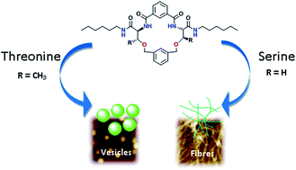
A series of lipidated macrocycles M1–M4 based on serine and threonine was designed and synthesized. Interestingly, threonine containing macrocycles assemble to vesicles, while serine-based macrocycles prefer to form fibrillar assemblies. Serine macrocycles with a 1,3-benzene dicarbonyl spacer forms fibrils while those with a biphenyl spacer resulted in a morphological change to vesicles.


Design and synthesis of molecules that self-assemble to a definite architecture is an intensely investigated area in supramolecular chemistry. Chemists have designed and synthesized a large number of molecules which self-assemble to various supramolecular assemblies such as coils,1 vesicles,2 toroids,3 nanotubes4 and nanofibers.5 The study of self-assembling molecules with predictable architectures will enable chemists to design new functional materials with desirable properties, and can also provide information about the relationship between molecular structure and self-assembling patterns.6 Cyclophanes, a class of cyclic molecules with aromatic units in its backbone have drawn the fascination of chemists due to their constrained geometry and unique self-assembling properties. Design of hybrid cyclopeptides consisting of amino acid residues and aromatic linkers is a promising approach towards the construction of self-assembling molecules.7

Herein we report the design, synthesis and unusual self-assembling properties of a series of lipidated, cyclic and acyclic peptides. Amino acids serine and threonine are tactically utilized for the synthesis of macrocyclic compounds, since both these amino acids have easily functionalisable side chains. The amide linkages, aromatic spacers and lipid units were expected to participate in non-covalent interactions leading to unique self-assembled structures. Cyclopeptides M1, M2 and M4 contain serine, while M3 contains threonine in the macrocyclic backbone. M1 and M2 consisting of two serine residues linked between two aromatic units and lipid chains on the exterior of the cyclic framework were prepared from their acyclic precursors A1 and A3. The spacers such as p-xylylene and m-xylylene units are used to link the two serine residues through the side chain –OH to obtain A1 and A3 respectively. The amino groups are used for the final step cyclization via benzene 1,3-dicarbonyl dichloride. The use of threonine instead of serine yielded the acyclic precursors A2/A4 with p/m-xylylene moieties as spacers. The final step macrocyclization of A4 gave M3, while cyclization was unsuccessful in the case of A2 (Fig. 1).


image file: c6ra05218e-f1.tif
Fig. 1 Structures of acyclic intermediates A1–A4 and macrocycles M1–M4.

To synthesize cyclophanes M1–M4, Boc-serine/threonine was reacted with hexylamine under standard coupling conditions to yield compounds Boc-L-Ser/Thr hexylamides S1/S2 (Scheme 1), which upon further reaction with p-xylylene dibromide in the presence of NaOH and tetrabutylammonium bromide (TBABr) as phase-transfer catalyst yielded A1/A2. A1 was deprotected and the resulting amine was reacted with benzene 1,3-dicarbonyl dichloride to yield M1, while deprotection of Boc group from A2, followed by the reaction with benzene 1,3-dicarbonyl dichloride yielded a complex mixture. The similar reaction of Boc-L-Ser/Thr hexylamides with m-xylylene dibromide yielded A3/A4 respectively. Deprotection of A3/A4 using trifluoroacetic acid and further reaction with benzene 1,3-dicarbonyl dichloride yielded macrocycle M2/M3. Amine obtained by the deprotection of A3 was reacted with diphenic acid dichloride to yield M4 with serine and m-xylylene in the framework.


image file: c6ra05218e-s1.tif
Scheme 1 Synthesis of macrocycles M1–M4.

The self-assembling trends of the macrocycles were studied by scanning electron microscopy (SEM), high-resolution transmission electron microscopy (HR-TEM) and atomic force microscopy (AFM).

Macrocycle M1 with a benzene 1,3-dicarbonyl unit and p-xylylene formed a gel in chloroform (31 mM) and also in a mixture of chloroform/hexane (15 mM) indicating that the self-assembling properties of M1 are dependent upon the solvent system. SEM analysis of the gels however did not show any specific morphology. Various solvents were screened to find a suitable one for self-assembly. SEM analysis of a dried sample of M1 (1 mM) in methanol (Fig. 2a) or ethylacetate (Fig. S1a, ESI) showed fibrillar morphology. The fibres obtained from methanol solution were of <100 nm thickness and 2–6 μm length (Fig. 2a). Like in the case of chloroform and chloroform/hexane mixture, M1 formed gels but of no specific morphology in a mixture of chloroform and methanol (Fig. S1b, ESI), while it showed disk-shaped morphology in isopropanol. These results support the high solvent dependent assembly of M1 (Fig. S1c, ESI).


image file: c6ra05218e-f2.tif
Fig. 2 SEM images of (a) 1 mM solution of M1 in methanol (b) 1 mM solution of M2 in methanol (c) 1 mM solution of M3 in methanol (d) 0.5 mM solution of M4 in methanol. The samples were prepared by drop-casting the respective solutions on glass. Inset shows the HR-TEM images of the respective samples on copper grid.

To know the involvement of the various functional groups in the self-assembly, we carried out concentration-dependent NMR studies. Concentration dependent 1H NMR of M1 in CDCl3 revealed changes in chemical shift of the amide and aromatic protons implying aggregation (Fig. S2, ESI). The aromatic CHs showed an upfield shift (Δδ 0.1 ppm) upon increasing concentration, which is often attributable to π–π stacking of the aromatic units. The peripheral amide NHs (Δδ 0.25 ppm) and backbone amide NHs (Δδ 0.2 ppm) were shifted however downfield indicating their involvement in the self-assembly. AFM analysis of M1 showed long fibers in methanol (Fig. 3a and S3, ESI). The thickness of fibres were of ∼100 nm and were of ∼2–3 μm in length. Analysis of unstained HR-TEM images show thin, long fibres of 20 nm diameter, which combine together to form thicker fibres (Fig. 2a inset and S4, ESI).


image file: c6ra05218e-f3.tif
Fig. 3 (a) AFM image of a 1 mM solution of M1 in methanol. (b) AFM image of a 1 mM solution of M2 in methanol. (c) AFM image of a 1 mM solution of M3 in methanol. (d) AFM image of a 1 mM solution of M4 in methanol. (e) Schematic representation of the formation of spheres (left) and fibres (right) from M3 and M2 respectively.

We then analyzed the self-assembly properties of M2, the m-xylylene counterpart of M1, for understanding the effects of the spacer units. The self-assembling patterns of M1 and M2 were similar and is in agreement with our previous report.8 SEM images of a 1 mM solution of M2 in methanol showed thin and long fibres of approximately 100 nm thickness and several micrometers length (Fig. 2b). M2 showed fibrillar assembly in a concentration range of 0.1–1 mM (Fig. 2b and S5, ESI). Fibrillar assembly was further supported by AFM and HR-TEM analysis (Fig. 3b and S6a and b, ESI). The height of a single fibre calculated using AFM was ∼15 nm with a width of ∼60 nm. HRTEM images revealed that the fibres are solid as indicated by the uniformly darkened fibres (Fig. S6c, ESI). Interestingly, SEM of a 1 mM solution of A1 displayed small plate like morphology, whereas A3 did not form any particular morphology (Fig. S7, ESI). These observations indicate the importance of a cyclic framework of M1 and M2 in the formation of fibrillar assembly.

Similar to M1, 1H NMR spectrum of M2 displayed single set of signals for each type of proton, implying a symmetrical structure. Concentration-dependent 1H NMR studies showed down-field shifted amide NHs of the cyclic as well as the peripheral NHs (Δδ = 0.25 ppm) upon increasing the concentration (Fig. S8, ESI). 1H NMR spectra of M2 and M1 clearly showed that these macrocycles assemble into fibres by virtue of the π–π interaction of aromatic moieties and hydrogen bonding via amide NHs.

To understand the effects of the additional chiral centre, we analyzed M3, in which serine was replaced by threonine. Interestingly, a 1 mM solution of the threonine-based macrocycle M3 in methanol showed spherical vesicles (Fig. 2c). A majority of the vesicles were of 1.2 μm in diameter, which is based on the analysis of 100 vesicles from several samples of M3 (Fig. S9, ESI). Concentration-dependent studies revealed that M3 formed nearly spherical vesicles in concentrations ranging from 0.1 mM to 1 mM (Fig. S9, ESI). These results were further confirmed by AFM analysis (Fig. 3c). The diameter of the vesicle is higher than the height of the vesicle due to the flattening of the vesicle at the poles. HR-TEM images of unstained sample confirmed the vesicular morphology (inset of Fig. 2c). The vesicles were of ∼1 μm in size and the contrast between the periphery and interior of the vesicles shows that the vesicles are hollow. M3 formed a gel in chloroform (∼57 mM). 1H NMR studies in CDCl3 showed a downfield shift of amide NHs with increase in concentration (Fig. S10, ESI), establishing the role of intermolecular hydrogen bonding. We analyzed the acyclic precursors A2 and A4 for getting more information about vesicular assembly. Surprisingly, the control compound A2 with a p-xylylene and two threonine residues do not assemble to form vesicles, but A4 with m-xylylene units and threonine residues formed vesicles with ∼600 nm in diameter (Fig. S11, ESI). These results show that m-xylylene unit coupled with threonine makes the acyclic precursor favor vesicular assembly, whereas m/p-xylylene in combination with serine could not induce the right curvature for vesicular assembly.

Based on the analysis of M2 and M3, we envisaged that the spacer units which could induce non-planarity in the macrocyclic structure may facilitate spherical vesicular assembly. We analyzed M4, a 19-membered macrocycle containing two serine residues, a m-xylylene spacer and a biphenyl unit. The biphenyl unit was chosen as a spacer due to its unique ability to induce non-planarity in the structure. M4 can be considered as an expanded version of the macrocycle M2 – an 18 membered macrocycle. SEM image of M4 (0.5 mM, methanol) showed spherical vesicles with ∼800 nm to 1.5 μm in diameter (Fig. 2d and S12, ESI), which was further supported by AFM analysis (Fig. 3d). Introduction of a biphenyl moiety changed the fibrillar self-assembly pattern of serine containing macrocycle to vesicles. CD spectra of M2 and M3 in CHCl3 showed a positive Cotton effect at around 240 nm (Fig. S13a, ESI). The negative Cotton effect observed for M1 is attributed to the π–π stacking of the aromatic rings.9 CD spectra recorded in methanol showed a characteristic positive band at 223 nm for M2 and M3 (Fig. S13b, ESI). The p-xylylene based M1 showed a positive band at 231 nm, whereas M4 with a biphenyl moiety displayed a band at 234 nm. CD spectra ruled out the presence of any secondary structure in solution.

The UV spectroscopic studies on M1–M4 in methanol showed a red-shift upon increasing the concentration, indicative of self-assembly by π-stacking (Fig. S14, ESI).10 Further proof of supramolecular association in solution was obtained by diffusion ordered spectroscopy (DOSY).11 The diffusion coefficient values of M1–M4 showed a regular decrease upon an increase of concentration (Fig. S15–S18 and Table S1, ESI).

Our findings point that Thr favors vesicular assembly, while serine along with a 1,1′-biphenyl unit also facilitates vesicular assembly. The two chiral centres in threonine might be responsible for the right curvature required for vesicular assembly, while such a curvature to the assembly is achieved when serine along with biphenyl unit is present in the macrocyclic backbone. We envisioned that biphenyl unit could induce a turn in the peptide backbone due to the orthogonal arrangement of the phenyl rings in the biphenyl. We believe that serine embedded in the constrained macrocyclic framework does not provide adequate curvature for the vesicular assembly. An additional conformational lock is required for achieving the curvature required for vesicular self-assembly.

Conclusions

In summary, we designed and synthesized a series of lipidated cyclophanes incorporating serine/threonine amino acid residues. These designer macrocycles contain spacers such as m/p-xylylene, isophthaloyl or 1,1′-biphenyl units. The macrocycles containing Ser with isophthaloyl and m/p-xylylene moieties self-assemble to nano fibres. The replacement of Ser with Thr changed the morphology from fibres to vesicles. Replacement of the phenyl unit by biphenyl changes the self-assembly of Ser-containing macrocycles from fibrils to vesicular assembly.

Acknowledgements

We acknowledge the Department of Science and Technology (DST Grant No. SB/S1/OC-23/2014) India for funding. We thank the Nano Research Facility (NRF), IITD for AFM and AIIMS, New Delhi for HR-TEM measurements. We acknowledge the Department of textile technology, IITD for SEM. BMB thanks University Grants Commission, India for JRF.

Notes and references

  1. (a) H. Engelkamp, S. Middelbeek and R. J. M. Nolte, Science, 1999, 284, 785–788 CrossRef CAS PubMed; (b) H.-Y. Lee, H. Oh, J.-H. Lee and S. R. Raghavan, J. Am. Chem. Soc., 2012, 134, 14375–14381 CrossRef CAS PubMed.
  2. (a) J. H. Jung, Y. Ono, K. Sakurai, M. Sano and S. Shinkai, J. Am. Chem. Soc., 2000, 122, 8648–8653 CrossRef CAS; (b) M. Yang, W. Wang, F. Yuan, X. Zhang, J. Li, F. Liang, B. He, B. Minch and G. Wegner, J. Am. Chem. Soc., 2005, 127, 15107–15111 CrossRef CAS PubMed; (c) A. Ajayaghosh, R. Varghese, S. Mahesh and V. K. Praveen, Angew. Chem., Int. Ed., 2006, 45, 7729–7732 CrossRef CAS PubMed; (d) S.-H. Tung, H.-Y. Lee and S. R. Raghavan, J. Am. Chem. Soc., 2008, 130, 8813–8817 CrossRef CAS PubMed; (e) B. Tian, X. Tao, T. Ren, Y. Weng, X. Lin, Y. Zhang and X. Tang, J. Mater. Chem., 2012, 22, 17404–17414 RSC.
  3. (a) J.-K. Kim, E. Lee, Z. Huang and M. Lee, J. Am. Chem. Soc., 2006, 128, 14022–14023 CrossRef CAS PubMed; (b) S. Yagai, M. Yamauchi, A. Kobayashi, T. Karatsu, A. Kitamura, T. Ohba and Y. Kikkawa, J. Am. Chem. Soc., 2012, 134, 18205–18208 CrossRef CAS PubMed.
  4. (a) A. K. Mehta, K. Lu, W. S. Childers, Y. Liang, S. N. Dublin, J. Dong, J. P. Snyder, S. V. Pingali, P. Thiyagarajan and D. G. Lynn, J. Am. Chem. Soc., 2008, 130, 9829–9835 CrossRef CAS PubMed; (b) R. Hourani, C. Zhang, R. van der Weegen, L. Ruiz, C. Li, S. Keten, B. A. Helms and T. Xu, J. Am. Chem. Soc., 2011, 133, 15296–15299 CrossRef CAS PubMed; (c) L. Ziserman, H.-Y. Lee, S. R. Raghavan, A. Mor and D. Danino, J. Am. Chem. Soc., 2011, 133, 2511–2517 CrossRef CAS PubMed; (d) Z. Liu, G. Liu, Y. Wu, D. Cao, J. Sun, S. T. Schneebeli, M. S. Nassar, C. A. Mirkin and J. F. Stoddart, J. Am. Chem. Soc., 2014, 136, 16651–16660 CrossRef CAS PubMed.
  5. (a) R. C. Claussen, B. M. Rabatic and S. I. Stupp, J. Am. Chem. Soc., 2003, 125, 12680–12681 CrossRef CAS PubMed; (b) K. L. Niece, J. D. Hartgerink, J. J. J. M. Donners and S. I. Stupp, J. Am. Chem. Soc., 2003, 125, 7146–7147 CrossRef CAS PubMed; (c) J. del Barrio, L. Oriol, C. Sánchez, J. L. Serrano, A. Di Cicco, P. Keller and M.-H. Li, J. Am. Chem. Soc., 2010, 132, 3762–3769 CrossRef CAS PubMed.
  6. (a) G. M. Whitesides and B. Grzybowski, Science, 2002, 295, 2418–2421 CrossRef CAS PubMed; (b) A. M. Kushner and Z. Guan, Angew. Chem., Int. Ed., 2011, 50, 9026–9057 CrossRef CAS PubMed; (c) C.-A. Palma, M. Cecchini and P. Samorì, Chem. Soc. Rev., 2012, 41, 3713–3730 RSC.
  7. (a) D. Ranganathan, C. Lakshmi, V. Haridas and M. Gopikumar, Pure Appl. Chem., 2000, 72, 365–372 CrossRef CAS; (b) J. Becerril, M. I. Burguete, B. Escuder, S. V. Luis, J. F. Miravet and M. Querol, Chem. Commun., 2002, 738–739 RSC; (c) W. S. Horne, C. D. Stout and M. R. Ghadiri, J. Am. Chem. Soc., 2003, 125, 9372–9376 CrossRef CAS PubMed; (d) J. Becerril, M. I. Burguete, B. Escuder, F. Galindo, R. Gavara, J. F. Miravet, S. V. Luis and G. Peris, Chem.–Eur. J., 2004, 10, 3879–3890 CrossRef CAS PubMed; (e) S. E. Gibson and C. Lecci, Angew. Chem., Int. Ed., 2006, 45, 1364–1377 CrossRef CAS PubMed; (f) V. Haridas, K. Lal, Y. K. Sharma and S. Upreti, Org. Lett., 2008, 10, 1645–1647 CrossRef CAS PubMed.
  8. M. B. Bijesh, N. U. Dheepthi, A. R. Sapala, A. Shandilya, K. Khare and V. Haridas, Molecular Systems Design & Engineering, 2016 10.1039/c6me00009f.
  9. I. Alfonso, M. Bru, M. I. Burguete, E. García-Verdugo and S. V. Luis, Chem.–Eur. J., 2010, 16, 1246–1255 CrossRef CAS PubMed.
  10. H. Wu, L. Xue, Y. Shi, Y. Chen and X. Li, Langmuir, 2011, 27, 3074–3082 CrossRef CAS PubMed.
  11. (a) K. F. Morris and C. S. Johnson Jr, J. Am. Chem. Soc., 1992, 114, 3139–3141 CrossRef CAS; (b) T. Megyes, H. Jude, T. Grósz, I. Bakó, T. Radnai, G. Tárkányi, G. Pálinkás and P. J. Stang, J. Am. Chem. Soc., 2005, 127, 10731–10738 CrossRef CAS PubMed; (c) Z. Zhang, Y. Luo, J. Chen, S. Dong, Y. Yu, Z. Ma and F. Huang, Angew. Chem., Int. Ed., 2011, 50, 1397–1401 CrossRef CAS PubMed; (d) N.-L. Strutt, H. Zhang, M. A. Giesener, J. Leia and J. F. Stoddart, Chem. Commun., 2012, 48, 1647–1649 RSC.

Footnote

Electronic supplementary information (ESI) available: Experimental details, NMR, UV, mass spectra, and microscopic images. See DOI: 10.1039/c6ra05218e

This journal is © The Royal Society of Chemistry 2016
Click here to see how this site uses Cookies. View our privacy policy here.