Seunghee Lee,
D. Amaranatha Reddy and
Tae Kyu Kim*
Department of Chemistry and Chemical Institute for Functional Materials, Pusan National University, Busan 609-735, Republic of Korea. E-mail: tkkim@pusan.ac.kr
First published on 8th April 2016
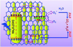
Graphene-based nanocomposites have attracted considerable attention in photocatalytic research owing to their remarkable potential for the photodegradation of environmental pollutants. However, despite the progress made in this field, the development of visible-light-active photocatalysts with high activity and durability remains a challenge. In this work, bunches of Nb3O7(OH) nanorods wrapped in reduced graphene oxide (RGO) nanosheets were prepared by using a hydrothermal method. The photocatalytic activity of the as-synthesized Nb3O7(OH)-RGO nanocomposites was significantly enhanced compared to that of the pure Nb3O7(OH) nanostructures owing to their improved visible-light absorption and separation of photogenerated electron–hole pairs. Photoluminescence studies strongly supported the proposed charge separation and charge transport mechanism. Moreover, the photocatalytic efficiency was strongly dependent on the concentration of RGO in the nanocomposites. The highest photodegradation rate was obtained using the nanocomposite prepared with a graphene loading of 3 mg mL−1, and when the RGO loading exceeded 3 mg mL−1, the photodegradation efficiency decreased. This occurred because excess RGO nanosheets aggregated and hindered the absorption of incident light. We believe that this work provides invaluable information for the design of new efficient visible-light-active reduced graphene oxide-based photocatalysts to be used in water remediation through the oxidative degradation of organic dyes and toxic phenols.
Over the past few years, several novel visible-light-active nanostructures, such as Ag3PO4, AgI, BiOCl, AgCl, and Nb3O7(OH), have been synthesized, and show superior photocatalytic activity under visible-light irradiation.6–10 Among these photocatalysts, Nb3O7(OH) nanostructures are a promising material for catalytic applications because of their non-toxicity, high stability under light irradiation, and high corrosion resistance in both acidic and alkaline media.11,12 However, the visible-light photocatalytic activity of bare Nb3O7(OH) is generally inadequate owing to its relatively low surface area and the high recombination rate of its photogenerated electron–hole pairs. It has been generally recognized that for an efficient photocatalyst, long term stability against light irradiation, high life time chare carriers, appropriate energy level offsets and charge trapping centers play an important role for efficient photocatalytic selective degradation reactions. Thus, ameliorating the usage efficiency of photogenerated electrons and prolonging the life time of carriers are expected to improve the catalytic activity.
To overcome the practical issues in Nb3O7(OH), the design of Nb3O7(OH)-based nanocomposites containing other low-band-gap semiconductor nanostructures or metal-free nanostructures, such as g-C3N4, MoS2, or reduced graphene oxide (RGO), is a novel stagey to reduce the recombination of charge carriers and improve stability and photocatalytic activity.13–15 Among these, the synthesis of RGO-based nanocomposites has gained increasing attention, and the nanocomposites formed are widely used for the degradation of various organic compounds in water, taking advantage of the excellent adsorption efficiency, high electrical conductivity, and large surface area of RGO sheets.16 When RGO nanosheets are used as a support for semiconductor nanostructures, the RGO nanosheets act as a mat between active semiconductor nanostructures that can help to shuttle the photogenerated charge carriers to retrain the electron–hole recombination.16 Additionally, the large two-dimensional layered RGO structures prevent the aggregation of the semiconductor nanostructures through interfacial interactions, thus increasing the number of catalytically active sites.16 However, to the best of our knowledge, there are no reports on the visible-light photocatalytic properties of Nb3O7(OH)-RGO nanocomposites.
Motivated by the lack of research on RGO-based Nb3O7(OH) nanostructures and their photocatalytic dye degradation for water remediation, here we present the first report of the design and preparation of a nanocomposite comprising Nb3O7(OH) nanorods wrapped in RGO nanosheets as a highly efficient visible-light-induced photocatalyst. The Nb3O7(OH)-RGO nanocomposite, prepared using hydrothermal methodology, exhibits enhanced photostability and visible-light photocatalytic activity toward degradation of the colored organic pollutant rhodamine B (RhB) and the colorless organic pollutant phenol compared to those of bare Nb3O7(OH) nanostructures. Moreover, the catalytic efficiency is strongly dependent on the RGO content of the nanocomposites, with the highest photodegradation rate being obtained using a nanocomposite prepared with a graphene loading of 3 mg mL−1. Furthermore, a possible photocatalytic mechanism for the Nb3O7(OH)-RGO nanocomposites is proposed with the aid of photoluminescence (PL) studies and reactive-species-trapping experiments.
The microstructural properties of the materials were investigated using a JEOL JEM-2100F transmission electron microscope (TEM) at an accelerating voltage of 200 kV. Phase determination of the as-prepared powders was performed using a Bruker D8 Avance X-ray diffractometer with CuKα as the X-ray source. X-ray photoelectron spectroscopy (XPS) measurements were recorded using a monochromatic AlKα X-ray source (hν = 1486.6 eV) at an energy of 15 kV/150 W. Ultraviolet-visible diffuse reflectance absorbance spectra (UV-Vis DRS) of the solid powder samples were recorded in the range from 200 to 800 nm using a UV-1800 double-beam spectrophotometer (Shimadzu) with BaSO4 as the reflectance standard. The Fourier transform infrared (FTIR) spectra were recorded in transmission mode from 400 to 4000 cm−1 at a resolution of 4 cm−1 using a Nicolet 380 FTIR spectrometer. In addition, PL measurements were performed at room temperature using a Hitachi F-7000 fluorescence spectrophotometer.
The photocatalytic activities of the Nb3O7(OH) and Nb3O7(OH)-RGO nanocomposites were evaluated via the degradation of RhB under simulated solar irradiation. A solar simulator equipped with an AM 1.5G filter and a 150 W Xe lamp (Abet Technologies) fitted with a <425 nm cut-off filter was used as the visible-light source. The photocatalyst (50 mg) was suspended in a 100 mL aqueous solution of RhB or phenol with an initial concentration (Co) of 10 mg L−1. Before turning on the light source, the reaction system was magnetically stirred in the dark for 30 min to allow the absorption–desorption equilibrium between the photocatalyst and dye to be established. Magnetic stirring was continued after irradiation in order to keep the photocatalyst particles suspended throughout the measurements. At given illumination-time intervals, ca. 2 mL aliquots of the mixture were removed and centrifuged at 5000 rpm for 15 min to separate the photocatalyst from the reaction solution. A UV-visible spectrum of the supernatant was then recorded to monitor the adsorption and degradation behavior. The characteristic absorption peak for RhB at 554 nm was used to assess the extent of degradation.
The microstructures and growth characteristics of the Nb3O7(OH) nanostructures and Nb3O7(OH)-RGO nanocomposites were further investigated by TEM and HRTEM. Fig. 1(d) and (e) show the TEM and HRTEM images of the pure Nb3O7(OH). From Fig. 1(d), it can be clearly seen that the morphology of the nanorods is regular and uniform, but that their lengths and diameters vary. The nanorods are measured to be 30–200 nm in length, 3–5 nm in thickness, and 2 nm in width. The HRTEM image in Fig. 1(e) shows that all the Nb3O7(OH) nanostructures are highly crystalline. The spacing between adjacent fringes is measured to be 0.271 nm, which is close to the (111) inter-planar distance of orthorhombic Nb3O7(OH) nanostructures. The TEM and HRTEM images of Nb3O7(OH)-RGO nanocomposites (Fig. 1(i)–(l)) clearly show that the nanorods are well-wrapped in RGO nanosheets. These results confirm that the nanocomposites are composed of both RGO and Nb3O7(OH) nanorods. The HRTEM image in Fig. 1(i) shows that the spacing between adjacent fringes is 0.375 nm, which is close to the (110) inter-planar distance of orthorhombic Nb3O7(OH) nanostructures.
Fig. 2(a) shows the XRD patterns of Nb3O7(OH) and Nb3O7(OH)-RGO nanocomposites with different RGO loadings synthesized hydrothermally at 210 °C for 24 h. It is evident that all the diffraction peaks in the pattern of the as-synthesized Nb3O7(OH) and Nb3O7(OH)-RGO nanocomposites can be fully indexed as standard orthorhombic Nb3O7(OH) (JCPDS file 31-0928), with the characteristic reflection phases (200), (400), (001), (110), (600), (310), (510), (111), (601), (511), (1000), (002), and (020). Moreover, due to the small amount of RGO and the relatively low diffraction intensity of the RGO in the nanocomposites, no distinctive diffraction peaks for the carbon species are observed. Furthermore, no new peaks related to secondary phases of niobium are observed, indicating that the synthesized composites have high purity. The growth direction can be ascertained using the intensity of the diffraction signals due to different planes. The patterns clearly show that in pure Nb3O7(OH) nanostructures, the diffraction peak at 2θ = 32.41 is much stronger and narrower than the other peaks, whereas in the nanocomposites, the diffraction peak at 2θ = 23.94 is much stronger, indicating that the preferred growth directions for the bare Nb3O7(OH) and the Nb3O7(OH)-RGO nanocomposites are along the (111) and (110) crystal planes of the orthorhombic structure, respectively. Average nanocrystallite sizes were estimated using the Debye–Scherrer formula, D = 0.89λ/β
cos
θ, where λ, β, and θ are the wavelength of the CuKα radiation, the diffraction angle, and the full width at half maximum (FWHM) of the diffraction peak for the major diffraction plane, respectively. The estimated average crystallite sizes are in the range of 50–95 nm, and are slightly smaller for the nanocomposites, suggesting that the presence of a certain amount of RGO improves the distribution of the nanoparticles, due its large surface area and thin, two-dimensional layered structure.20
The reduction of GO during the hydrothermal process and the oxidation states of the synthesized nanostructures was further characterized by XPS. Typical survey scan XPS spectra of the GO and the Nb-RGO-3 nanocomposite are shown in Fig. 2(b). There are two dominant peaks in the GO spectra, which represent the graphitic C 1s and O 1s orbitals. However, for Nb-RGO-3, in addition to the C 1s and O 1s peaks, a new peak related to Nb is observed. On further analysis, the C 1s region contains peaks at 284.56, 286.48, and 288.83 eV, assignable to the non-oxygenated ring C, the C in C–O bonds, and the carboxylate carbon (O–C
O) respectively (Fig. 2(c)).21 The presence of similar species is also indicated in the C 1s region of Nb-RGO-3 (Fig. 2(d)). However, the peak intensities of the various oxygenated carbon species drastically decrease compared to those in GO. Moreover, the peak corresponding to the C
O group vanishes, and new peak at 289.36 eV, ascribed to the O–C
O group, is observed.22 This suggests that the GO has been reduced to RGO during the formation of the nanocomposites under hydrothermal conditions. The high-resolution Nb 3d XPS spectrum shows peaks at 210.43 and 207.63 eV, which correspond to Nb 3d3/2 and Nb 3d5/2, respectively (Fig. 2(e)).23 Fig. 2(f) shows the O 1s deconvolution narrow scan spectra. Two intense peaks are observed at binding energies of 531.23 and 532.88 eV, which are assigned to oxygen vacancies and dissociated oxygen species, respectively.24 These results confirm the existence of Nb3O7(OH) and RGO in the composites.
In order to determine the presence of RGO and investigate the reduction phenomena during the hydrothermal process, the GO and nanocomposite was further characterized by FT-IR spectroscopy. The results of the FT-IR analysis of the as-prepared GO and Nb-RGO-3 samples are shown in Fig. 3(a). The characteristic peaks of GO appear at 3409 (O–H stretching band), 1742 (C
O vibrations of the –COOH groups), 1625 (C–C skeletal vibrations of the aromatic domains), 1348 (bending absorption of the O
C–O carboxyl group), 1248 (O–H bending vibrations), and 1065 cm−1 (C–O stretching vibrations).25 All of these vibrations reveal the existence of a high number of oxygen-containing functional groups at the edges and basal plane of the nanosheets. For the FTIR spectra of Nb-RGO-3 nanocomposites, the GO-related stretching bands of the C–O and carboxyl groups are completely absent, and the peaks for C–O and C–C are still observed after reduction, but are much smaller than those in GO.26 These results indicate that GO is effectively reduced by Nb3O7(OH) to generate RGO nanosheets during the hydrothermal reaction.
To investigate the optical properties and estimate the band gap the as-synthesized Nb3O7(OH), Nb3O7(OH)-RGO nanocomposites and pure RGO nanostructures were analyzed by UV-Vis diffuse reflectance spectra (DRS) and the results are shown in Fig. 3(b). The spectrum of the bare Nb3O7(OH) nanostructures shows the absorption edge at 436.5 nm (2.84 eV).10 This behaviour indicates that the nanostructures can absorb visible light radiation of wavelengths less than 440 nm, while the pure RGO absorbs UV and visible light at wavelengths less than 800 nm. Nb3O7(OH)-RGO nanocomposites exhibit much stronger response in the visible region compared to the Nb3O7(OH) nanostructures, with stronger absorbance from 450–800 nm, which indicates that the synthesized nanocomposites may utilize more visible light during the photocatalytic reaction. The enhanced visible-light absorbance range in the Nb3O7(OH)-RGO nanocomposites may be due to the cooperative effect between the RGO and the Nb3O7(OH) nanostructures.
Room temperature PL spectra of pure Nb3O7(OH) and Nb3O7(OH)-RGO nanocomposites were recorded in order to investigate the possible interactions between the Nb3O7(OH) nanorods and the RGO nanosheets, and are presented in Fig. 3(c). It is clear that the bare Nb3O7(OH) nanostructure exhibits two emission bands: one in the UV region at ca. 400 nm and the other in the blue region at ca. 464 nm. The observed emission peaks may be attributed to electron transitions from the valence band to the conduction band, trap sites, and oxygen vacancies.12 The emission spectra of Nb3O7(OH)-RGO nanocomposites also exhibit similar emission bands to those of Nb3O7(OH). However, the emissions are greatly suppressed compared to those of the bare nanostructures, which is probably due to charge transfer from the Nb3O7(OH) nanostructures to the RGO nanosheets. In general, the PL emission occurs from the recombination of excited electrons and holes under light irradiation, with a higher PL intensity indicating a higher recombination rate of photoexcited electrons and holes, and a commensurate decrease in the photocatalytic activity. Conversely, a lower PL intensity indicates a lower recombination rate of photoexcited electrons and holes, suggesting that more photoexcited holes and electrons can participate in the oxidation and reduction reactions, thus enhancing photocatalytic performance. In our case, the decrease in PL intensity is due mainly to the presence of RGO nanosheets in the nanocomposites. The RGO sheets act as photoluminescence-quenching centers, and effectively inhibit electron–hole pair recombination owing to their high specific surface area and high electrical conductivity, resulting in non-radiative recombination.27
The photocatalytic activities of the as-synthesized bare Nb3O7(OH) and Nb3O7(OH)-RGO nanocomposites were investigated by the degradation of RhB (10 mg L−1) under visible-light (λ > 425 nm) irradiation. Fig. 4(a) shows the UV-Vis absorption spectra of RhB solutions irradiated under visible light for 5 h in the presence of bare Nb3O7(OH) and Nb3O7(OH)-RGO nanocomposites. It is evident that the absorption peak at 554 nm drastically decreased in the presence of all the nanocomposites. The spectra confirm that the synthesized nanostructures are effective for dye degradation under visible-light irradiation. In the absence of the photocatalyst (control experiment) showed that the concentration of RhB was marginally reduced when exposure to the experimental radiation for 5 h. This indicates that the degradation of RhB was negligible in the absence of the photocatalyst. To further systematically investigate the photocatalytic activities of the synthesized samples, their degradation efficiencies towards RhB, given by
| D = (A(RhB)0 − A(RhB))/A(RhB)0 × 100 |
Further, to improve the understanding of the reaction kinetics of RhB organic degradation over all the photocatalysts, the results were fitted to a pseudo-first order correlation (ln(Co/Ct) = kt, where k is the apparent reaction-rate constant, and Co and Ct are the RhB concentrations initially and at time t, respectively). The calculated apparent k values for bare and RGO nanocomposites are shown in Fig. 4(c). The highest photocatalytic mineralization performance was achieved with the Nb-RGO-3 nanocomposite: its k value is about 5.14 times greater than that of the bare Nb3O7(OH) nanostructures. Therefore, we can conclude that the formation of the RGO nanocomposite enhances the visible light photocatalytic activity.
To further study the photocatalytic mechanism and identify the main oxidative species (h+, ˙O2−, and ˙OH) in the photocatalytic process, we studied the effect of active-species scavengers on the decolorization of RhB, and the results are presented in Fig. 4(c). Ethylenediaminetetraacetic acid (EDTA), benzoquinone (BQ), and t-butyl alcohol (TBA) were used as scavengers for h+, ˙O2− radicals, and ˙OH radicals, respectively. It is clear from Fig. 4(d) that the addition of EDTA slightly affected the photocatalytic degradation of RhB over the Nb-RGO-3 nanocomposite, suggesting that the h+ reactive species is only partially involved in the photocatalytic process. Conversely, the degradation efficiency decreased considerably after the addition of BQ and TBA, which indicates that ˙O2− and ˙OH radicals are the main active species in the photocatalytic degradation of RhB under visible-light irradiation.31
Considering the effect of scavengers on the photocatalytic reaction, the observed PL quenching, and the band gap structure of Nb3O7(OH), the enhanced photodegradation ability of the nanocomposites is rationalized as follows: under visible-light irradiation, the synthesized nanostructures utilize light energy to generate electrons and holes. The photogenerated electrons and holes in Nb3O7(OH) can move freely into the RGO nanosheets owing to their close interfacial contact. Hence, coupling with the RGO provides different transfer possibilities for the photogenerated holes and electrons. These promote the effective separation of the photoexcited electron–hole pairs, decreasing the probability of electron–hole recombination at the interface of the nanocomposites, and promoting high photocatalytic performance. The photogenerated electrons participate in reduction reactions with electron acceptors, such as adsorbed oxygen molecules, to generate hydroxyl and superoxide radicals upon protonation. The photoinduced holes either can oxidize organic compounds directly or be trapped by electron donors, such as OH−, to produce OH radicals. The superoxide radical anions and hydroxyl radicals are responsible for the decomposition of the RhB dye into non-toxic products, such as CO2, NO3, NOx, and mineral acids.32 A schematic illustration of the charge transfer and enhanced photocatalytic activity of the Nb3O7(OH)-RGO nanocomposites is shown in Fig. 5.
![]() | ||
| Fig. 5 Illustration of the proposed reaction mechanism for the photocatalytic degradation of RhB in aqueous solution over Nb3O7(OH)-RGO nanocomposites under visible-light irradiation. | ||
In addition to their photocatalytic activity, the stability of nanostructures is an important criterion for photocatalysts. Therefore, we carried out stability tests on the as-synthesized Nb-RGO-3 nanocomposite using four successive recycling experiments. As shown in Fig. 4(e), after four consecutive runs the degradation efficiency of the sample is still 77.99%. This remarkable performance is attributed to the higher number of reaction sites and the chemical interactions between the RGO nanosheets and the Nb3O7(OH) nanostructures.
To rule out the photosensitization effect under visible-light irradiation, we also evaluated the photocatalytic performance of Nb3O7(OH) and Nb3O7(OH)-RGO nanocomposites toward the degradation of phenol, which is colorless, because the photosensitization effect of phenol is negligible under visible light.33,34 Fig. 4(f) shows the absorption spectra of aqueous solutions of phenol (5 mg L−1) after visible-light irradiation for 5 h in the presence of 50 mg of one of the synthesized catalysts. The characteristic absorption peak of phenol at 270 nm was chosen to monitor the progress of the photocatalytic degradation process. From Fig. 4(f), it can be clearly seen that the absorption peaks corresponding to phenol decrease with irradiation time, which indicates rapid phenol decomposition. Fig. 4(g) shows the phenol degradation rate. Pure Nb3O7(OH), Nb-RGO-1, Nb-RGO-2, Nb-RGO-3, and Nb-RGO-4 exhibit degradation efficiencies of 45.6%, 62.8%, 73.4%, 83.1%, and 76.7% respectively. The rate constants were 0.0166, 0.02263, 0.0293, 0.03555 and 0.03293 min−1 for Nb3O7(OH), Nb-RGO-1, Nb-RGO-2, Nb-RGO-3, and Nb-RGO-4, respectively (Fig. 4(h)). It is clear that the Nb-RGO-3 nanocomposite promotes a significantly higher level of phenol photodegradation after 300 min of irradiation compared with the bare Nb3O7(OH) nanostructures. The enhanced photocatalytic activity mechanism for phenol is similar to that for the organic dyes.
| This journal is © The Royal Society of Chemistry 2016 |