Structure analysis of a glycosides hydrolase family 42 cold-adapted β-galactosidase from Rahnella sp. R3

Yuting Fanab, Jiang Yic, Xiao Huaa, Yinghui Fenga, Ruijin Yang*a and Yuzhu Zhang*b
aState Key Laboratory of Food Science and Technology, School of Food Science and Technology, Jiangnan University, 214122 Wuxi, China. E-mail: yilin58299@163.com; yrj@jiangnan.edu.cn; Fax: +86-510-85919150; Tel: +86-510-85919150
bUS Department of Agriculture, Agriculture Research Service, Western Regional Research Center, Pacific West Area, Albany, California 94710, USA. E-mail: yuzhu.zhang@are.usda.gov; Fax: +1-510-5595818; Tel: +1-510-5595981
cCollege of Chemistry and Environmental Engineering, Shenzhen University, 518060 Shenzhen, China

Received 20th February 2016 , Accepted 7th April 2016

First published on 7th April 2016


Abstract

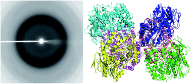
The β-galactosidase isolated from a psychrotrophic bacterium, Rahnella sp. R3 (R-β-Gal), exhibits high activity at low temperature and has potential in the dairy industry. R-β-Gal is a member of the glycoside hydrolases family 42 (GH42), and it forms a 225 kDa trimeric structure in solution. The crystals of R-β-Gal were acquired via hanging-drop vapor-diffusion method, and the X-ray crystal structure of the native R-β-Gal was determined at a 2.5 Å resolution. It is the first structure of a cold-adapted GH42 enzyme. In the crystallographic asymmetric unit, there were two homotrimers of the enzyme. Each monomer consists of three domains, an N-terminal catalytic domain which is a (β/α)8 barrel, a mixed β-sheet and α-helices domain, and a C-terminal β-sandwich domain. Two putative residues might be involved in catalysis, a proton donor E157 and a nucleophile E314, were superimposed well with the catalytic residues of other β-galactosidases. Site-directed mutagenesis targeting these residues abolished the activity of the enzyme. Structure and sequence comparison of R-β-Gal with two mesophilic β-gals and a thermophilic β-gal indicated that intramolecular force and higher structural flexibility might result in the cold-adaptation of R-β-Gal.


1. Introduction

Lactase is a β-galactosidase (β-gal, EC 3.2.1.23) necessary to digest lactose and an important industrial enzyme in the dairy industry. One of the major sources of energy in the milk of numerous species is lactose1–3 which is a disaccharide sugar. It is composed of, and can be hydrolyzed into one galactose and one glucose molecule. In humans the lack of adequate lactase or lactase nonpersistence causes lactose malabsorption which can result in bacterial fermentation of undigested lactose in the colon. This, in turn, produces symptoms of lactose intolerance which is the syndrome of abdominal pain, flatulence, diarrhea, and/or bloating. Although the true prevalence of lactose intolerance is not known4 and milk intolerance are not necessarily all lactose intolerance,5 many individuals with real or perceived lactose intolerance avoid milk and dairy products. This may predispose them to osteoporosis and decreased bone accrual because of inadequate amounts of calcium and vitamin D. Abdominal discomfort caused by fermentation of lactose also impacts the utilization and marketing of dairy products.

β-gals can hydrolyze the β (1–3) and β (1–4) linkage in oligo- and disaccharides (such as lactose) and simultaneously transfer galactosyl to different acceptors. β-gals from many organisms have been widely used in food manufacturing, such as in dairy processing to hydrolyze lactose. Currently, the major commercial β-gals used in lactose hydrolysis include Lactozym Pure® (Novozymes) and Maxilact® (DSM Food Specialties). These enzymes are usually optimally active at moderate temperatures (around 37 °C) but display low activity at 4 °C. The dairy industry is in great need of an enzyme with optimal β-gal activity at the conditions compatible with dairy processing, i.e. at temperatures close 4 °C, pH 6.5, and is not inhibited by sodium or calcium. β-gals are divided into six distinct glycoside hydrolases families, namely GH1, GH2, GH35, GH42, GH50 and GH59.6 Three dimensional (3D) structures are essential to fully understand of a protein. The structures of four other GH42 members have been reported. These include a thermostable β-gal from Thermus thermophiles A4 (A4-β-gal, PDB: 1KWG),7 a thermostable β-gal from Geobacillus stearothermophilus (Gan42B, PDB: 4IOF),8 a mesophilic β-gal from Bacillus circulans sp. alkalophilus (Bca-β-gal, PDB: 3TTS),9 and the mesophilic β-gal from the probiotic Bifidobacterium animalis subsp. lactis Bl-04 (BlGal42A, PDB: 4UNI).10

Extremophile microorganisms that thrive at low temperatures (close to/below the freezing point of water) are usually capable of producing psychrotrophic or psychrophilic enzymes. These enzymes are potentially functioning at low temperatures due to the adaptation of microorganisms to cold ecosystems (e.g. deep ocean, polar, and alpine regions).11 Generally, psychrotrophic/psychrophilic (cold-adapted) enzymes are more active than meso- and thermophilic enzymes at cold environment because of the pressure of retaining metabolic rates at lower temperatures. Meanwhile, they are more susceptible to denaturation at moderate and high temperatures. The high activity of cold-adapted enzymes at low temperatures offers potential benefits not only in industrial operations, but also in biotechnological applications and environmental researches.

Recently, a cold-adapted β-gal from Rahnella sp. R3 (R-β-Gal) (a psychrotrophic microorganism discovered from the no. 1 Glacier in the Tianshan Mountains in Xinjiang, China) was isolated. We have expressed the R-β-Gal enzyme in E. coli, and purified the recombinant protein, and enzymatic properties were systematically done.12 R-β-Gal showed a Km value of 6.5 mM and a Kcat value of 7.9 s−1 toward ortho-nitrophenyl-β-galactoside (ONPG) at 4 °C. R-β-Gal which specifically hydrolyzes lactose exhibited relative high activity toward lactose at 4 °C and neutral pH (6.5), indicating that cold-adapted β-gal can be potentially used in dairy industry to remove lactose in dairy products at 4 °C and avoid their spoilage and flavor change.13 Here, we report the crystal structure of R-β-Gal and discuss the insights in the structure basis of the functional properties of this cold-adapted β-gal.

2. Experimental section

2.1. Protein purification

The expression and metal affinity purification of R-β-Gal was previously reported.12 The same protocols were used in this study except that an additional size exclusion chromatographic step was used to ensure high purity protein for crystallization. Nickel column purified R-β-Gal was applied to an XK 26/70 Superdex-200 column (GE Healthcare, USA) pre-equilibrated and eluted with buffer S (10 mM Tris–HCl, 200 mM NaCl, pH 8.0). All of the chromatographic steps were carried out at room temperature (23 °C) using an FPLC system (GE Healthcare, USA). The protein purity was examined on NuPAGE 4–12% Bis–Tris Gel (Life technologies, NY, USA) and the protein concentration was determined by UV absorption at 280 nm. The NaCl concentration of the final purified R-β-Gal sample was reduced to less than 1 mM by repeated dilution and concentration with a buffer containing 10 mM Tris (pH 8.0), 1 mM EDTA, and 2 mM DTT using an Ultracel-30k filter device (Millipore, MA, USA). The protein concentration of the final sample for crystallization was 28 mg mL−1. The molar extinction coefficient used for the calculation of the protein concentration was 157845/M cm.

The determination of the hydrolytic activity of R-β-Gal was reported previously.12 Briefly, the reaction mixture contained 200 μL of diluted enzyme and 800 μL of 8.3 mM ortho-nitrophenyl-β-galactoside (ONPG) in 10 mM potassium phosphate buffer (pH 6.5) was incubated at 35 °C for 10 min (within the linear reaction period, Fig. S2). Then the reaction was stopped by adding 1 mL of 10% (w/v) sodium carbonate. Then the activity was quantified by measuring the released ortho-nitrophenol (ONP) from the absorbance at 420 nm. The enzyme concentration of wild type and mutant forms used for activity assay were 3.5 × 10⁻³ mg mL−1 and 35 mg mL−1, respectively.

2.2. Crystallization and X-ray data collection

The initial crystallization screen of R-β-Gal was performed at 20 °C using the hanging-drop vapor-diffusion method. The conditions of Crystal Screen, Crystal Screen 2, and Crystal Screen lite kits from Hampton Research (Aliso viejo, CA, USA) were used. Hanging drops containing 1 μL of protein solution mixed with 1 μL of crystallization solution were sealed against 500 μL of reservoir solutions in 24-well Limbro plates. Crystals were obtained in the drop hanging over solution no. 22 of the Crystal Screen 2 kit (CS222, 0.1 M Tris–HCl pH 8.5, 0.2 M sodium acetate, and 30% w/v polyethylene glycol 400). To optimize the crystallization, more drops were generated by mixing the protein solutions at different concentrations with CS222 or modified CS222 with different concentration of polyethylene glycol (PEG) with various mean molecular mass. The final crystallization condition was mixing 1 μL of a protein solution (28 mg mL−1) with 1 μL of a reservoir solution comprising 0.2 M sodium acetate, 0.1 M Tris–HCl (pH 8.0), and 30% (w/v) PEG 1500. The crystals were briefly immersed in a cryoprotectant solution (50% reservoir solution with 50% w/v PEG 400) and flash-cooled in liquid nitrogen. The cooled crystals were stored in liquid nitrogen before they were shipped to the synchrotron beam line in a dry shipper for data collection.

X-ray diffraction data were collected at LRL-CAT 31ID-B beam line at APS, Argonne National Laboratory. Data were collected at 110 K with a 223 mm crystal-to-detector distance using a MAR300 CCD detector. One hundred and eighty 1° frames were collected with an exposure time of 1.6 seconds. The diffraction data were processed with the XDS14 and the HKL200015 suite of programs.

2.3. Phase calculation and model refinement

The structure of the R-β-Gal was solved by molecular replacement using PHASER.16,17 The search model was built with the program Chainsaw18 using Bca-β-gal (PDB: 3TTS) as a template9 which shares 45% sequence identity with R-β-Gal. The structure was refined using PHENIX19,20 and REFMAC5.21 Semi-automated model building and model improvement was carried out with COOT22 after each refinement cycle.

The final structure was checked by PROCHECK23 and Molprobity Validation.24 The atomic coordinates and structure factors for R-β-Gal were deposited in the Protein Data Bank (PDB ID: 5E9A). Molecular graphic figures were prepared using the programs Rasmol,25 MolScript,26 Raster3D,27 and Pymol.28 Sequence alignments were prepared with Clustal Omega and ESPript 3.0. Structure alignments and the root-mean-square difference (RMSD) were obtained with the program TM-align Server29 unless otherwise stated. Salt bridges and hydrogen bonds were calculated by VMD.30 Disulfide bridges were found by PISA Server (http://www.ebi.ac.uk/pdbe/pisa/). Surface areas were calculated using the Naccess.31 The percentage of hydrophobic accessible area, glycine residues, proline residues, argine residues were calculated by Pymol. B-factors were provided by PDB reports.

2.4. Site-directed mutagenesis and mutant protein purification

Two mutants of R-β-Gal (E157V and E314V) were obtained with a Quickchange II site-directed mutagenesis kit (Agilent technologies co., Santa Clara, USA) according to the manufacturer's protocols. Primers pair E157V-f (gctggcacatttccaat[g with combining low line][t with combining low line][a with combining low line]tacggcggcgaatg) and E157V-r (cattcgccgccgtata[c with combining low line][a with combining low line][t with combining low line]tggaaatgtgccagc) were designed to contain the required nucleotide changes for making mutant E157V and the targeted nucleotide changes for making mutant E314V were incorporated in primers E314V-f (ccttttgtgctgatg[g with combining low line][t with combining low line][a with combining low line]tcgacgccgagtttc) and E314V-r (gaaactcggcgtcgatac[c with combining low line][a with combining low line][t with combining low line]cagcacaaaagg). The mutant plasmids were sequenced by Elim Biopharmaceuticals (Hayward, CA, USA). Expression and purification of the R-β-Gal mutants were carried out with the same methods for wild type R-β-Gal.

2.5. Circular dichroism spectra

The buffer of the purified proteins was changed to a buffer containing 10 mM potassium phosphate (pH 6.5) by repeated concentration-and-dilution using an Amicon Ultra centrifugal filter device with 30 kDa molecular weight cut-off for proteins (Millipore, MA, USA). CD spectroscopy was used to study the secondary structure content of wild type R-β-Gal and two mutants (E157V and E314V). Spectra in the far-UV region (190–270 nm) were collected with a J-815 CD spectrometer (Jasco, Tokyo, Japan) and a quartz cuvette with a path length of 2.0 mm. Wavelength scans were performed at a speed of 50 nm min−1 at 20 °C, and 10 scans were accumulated for each sample. The protein concentrations used for the CD measurements were ∼0.10 mg mL−1 and the CD signal was displayed as mean residue ellipticity (degree cm2 dmol−1).

3. Results and discussion

3.1. The structure of R-β-Gal

One hundred crystals were screened and the best crystal diffracted to 2.56 Å. The statistics for data collection and structure refinement is shown in Table 1. The structural factors (R/Rfree) of the final model are 0.199/0.247. In the final refined structure, there are six R-β-Gal protein molecules in the crystallographic asymmetric unit. The pairwise RMSD between two monomers calculated using PHENIX is 0.16 ± 0.04 Å. The his-tag at the N-terminal plus the first residue (Met1) of the protein and 2 residues at the C-terminal of all six chains were disordered and could not be built into the electron map. The rest of amino acid residues in each of the six chains were fitted to the electron density map with continuous main chain connectivity, except for one of the chains where 5 residues (D660-G664) were disordered due to crystal packing. These residues resided in a larger loop between β26 and β27 in the third domain of the protein (see below). There were 4099 amino acid residues in the refined structure and a total of 32[thin space (1/6-em)]981 atoms, including 6 zinc ions, 6 acetate ions, and 428 water molecules. On the quality spectra of the PDB deposit report, R-β-Gal resides to the better end of slider in all aspects of the validation (Rfree, clash score, Ramachandran outliers, side chain outliers, and RSRZ outliers) for protein structures with similar resolutions deposited in the PDB database.
Table 1 Data collection and refinement statistics of the R-β-Gal
Data collection R-β-Gal
a Numbers in the parenthesises are for the outer shells.b image file: c6ra04529d-t1.tifc image file: c6ra04529d-t2.tifd image file: c6ra04529d-t3.tif, where Ii(hkl) is the ith observation of reflection hkl and 〈I(hkl)〉 is the weighted average intensity for all observations i of reflection hkl.
Wavelength 0.97931
Space group P1211
a, b, c (Å); α, β, γ (°) 146.43, 106.83, 164.24; 90.00, 109.0, 90.00
Resolution (Å) 138.45–2.56 (2.70–2.56)a
No. of reflections 535[thin space (1/6-em)]514 (69[thin space (1/6-em)]030)a
No. of unique reflections 151[thin space (1/6-em)]929 (21[thin space (1/6-em)]717)a
Multiplicity 3.5 (3.2)a
Mosaicity (deg.) 0.29
I〉/〈σ(I)〉 8.6 (2.4)a
Completeness (%) 98.6 (96.8)a
Rmergeb 0.116 (0.485)a
Rmeasc 0.163 (0.649)a
Rp.i.md 0.085 (0.349)a
B-overall (from Wilson plot)b (Å) 29.6
[thin space (1/6-em)]
Refinement
Resolution range (Å) 138.45–2.56
Rwork/Rfree 0.204/0.247
Average B factorsb (Å) 32
Ramachandran plot (%)  
Favored 96
Outliers 0.5
Total no. of atoms 32[thin space (1/6-em)]981
No. of amino acids 4099
No. of water molecules 428
No. of ligand atoms 6
Zn 6
Acetate ion 24


3.2. Trimeric structure of R-β-Gal

The six R-β-Gal molecules in a crystallographic asymmertric unit constitute two trimers. The trimer formation buried a total of 13[thin space (1/6-em)]567 Å2 of solvent accessible surface area (ASA). The interface ASA per subunit is 4522 Å2. Interestingly, this fits very well to the empirical linear relationship between interface ASA (iASA) and the molecular mass of dimeric proteins (Mm), iASA = cMm, where c was estimated as 0.06 Å2 Da−1.28 There is little association between the trimers in the crystallographic asymmetric unit (Fig. 1) and the trimer–trimer interaction might be a result of crystal packing especially in light of the protein being purified as trimers based on the size exclusion chromatographic data.12 The shape of the trimeric structure of R-β-Gal resembles a flowerpot (Fig. 2) with a big cavity channel through the 3-fold axis of the trimer, a larger opening at the top, and a smaller opening at the bottom. These features were also observed in the structure of other glycoside hydrolase family 42 members. The amino acid sequence identity between R-β-Gal and Gan42B, Bca-β-gal, BlGal42A, and A4-β-gal is 49%, 45%, 33% and 32%, respectively.
image file: c6ra04529d-f1.tif
Fig. 1 A ribbon diagram of the structure of R-β-Gal. Two trimeric biological units are present in the crystallographic asymmetric unit. In one of the biological unit (left), the monomers are shown in magenta, cyan, and brown, respectively. In the other biological unit (right), the monomers are shown in red, green, and blue, respectively. The structure is oriented to show the lack of association between the biological trimers.

image file: c6ra04529d-f2.tif
Fig. 2 Trimeric structure of R-β-Gal. The shape of the trimeric R-β-Gal resembles a flower pot. A stereo view of the R-β-Gal trimer along the three-fold symmetry axis is presented. For two of the monomers, the molecule surface of is shown. The other monomer is shown as a ribbon diagram. In each monomer, domains A, B, and C are colored in red, green, and blue, respectively. Domain C has no contact with other monomers in the trimer.

3.3. Domain structure of R-β-Gal

An R-β-Gal monomer consists of three domains (Fig. 3). Domain A at the N-terminal consists of amino acids up to residue 401. It is a TIM barrel that contains a β-barrel at the center with 8 β-strands that is surrounded by 8 α-helices (Fig. 3a). Domain A also contains a metal ion-binding site in R-β-Gal. Four cysteine residues (C122, C162, C164 and C167), which are highly conserved in the GH42 family, form a metal binding cluster far away from the active site. The metal binding site of R-β-Gal is similar to that in A4-β-gal, Gan42B and Bca-β-gal. Interestingly, BlGal42A has a unique metal binding site formed by three histidines (His118) from three monomers. Based on enzymatic studies, R-β-Gal does not need any metal ions for its activity, thus it was assumed that the zinc ion may be an important structural feature for the enzyme.32,33 Domain B consists of amino acids E402-P616. This domain also contains both α-helices and β-strands. A 7-stranded β-sheet is at the middle of this domain (Fig. 3c and d). It starts as a 4-stranded parallel β-sheet. Then, it is extended with a 3-stranded antiparallel β-sheet to form a 7-stranded β-sheet. At one end of it, there is a distorted β-sheet which is almost perpendicular to the central β-sheet. From one direction, the sheet structure looks like a “T” shaped, branched β-sheet. From an orthogonal direction, it shapes like an inverted “L”. All 5 helices flank the main β-sheet. The function of this domain is not clear. The first two domains contribute all the intermolecular interactions that results in the trimeric structure of R-β-Gal. A small β-sheet domain at the C-terminal (from R617 to the C-terminus of the protein) does not participate in the binding to the other two R-β-Gal molecules in the timer. Domain C consists of an up-and-down β-barrel, which comprises six β-strands (Fig. 3e). The other four GH42 β-gals with known structures also contain this domain,7–10 which is not similar to any other known structures. Domain C shares 28% sequence identity with Bca-β-Gal (PDB ID: 3TTS), 18% with Gan42B (PDB ID: 4OIF), 21% with BlGal42A (PDB ID: 4UNI) and 16% with A4-β-Gal (PDB ID: 1KWG). The RMSD of this domain of R-β-Gal and those of Gan42B, Bca-β-Gal, BlGal42A and A4-β-Gal is 1.21 Å, 1.50 Å, 2.29 Å and 2.43 Å, respectively. The function of this domain is unknown and it is probably a structural element of GH42 β-gals.
image file: c6ra04529d-f3.tif
Fig. 3 Domain structure of R-β-Gal. (a) Overall structure of the R-β-Gal monomer shown as a ribbon diagram. Domains A, B, and C are labeled and colored in red, green, and blue, respectively. The N- and C-terminal of the structure are labeled with “N” and “C”, respectively. (b) A ribbon diagram of domain A is shown with the putative catalytic residues shown in a ball-and-stick representation. Carbon, oxygen, and nitrogen atoms are shown in gray, red, and blue, respectively. The secondary structural elements are labeled as “α” for α-helix, “β” for β-strand, and “h” for 310 helices. The α-helix and β-strand of the TIM barrel are labeled from 1 to 8. Additional α-helices preceding an α-helix element of the TIM barrel are given to the same number as the α-helix element, but are primed, and double primed if necessary. The start and the end residues of the domain and catalytic residues are labeled with amino acid type and residue number. (c) A ribbon diagram of domain B. It is presented with the main β-sheet shown in red and β-strands bended away from the main sheet shown in green. The α-helices flanking the main β-sheet are shown in blue. The α-helices and the β-strands are numbered continuously from those in domain A. (d) A schematic topology diagram of the domain B of R-β-Gal. The secondary structure elements are presented and numbered the same in (c). (e) A ribbon diagram of the GH42 β-gal unique domain C. The β-strands are numbered continuously from those in domain B.

3.4. Cold-adaptation mechanism

Several 3D structures of cold-adapted enzymes, including a cold-adapted β-gal,34–40 were reported as far as we know. The only reported cold-adapted β-gal whose structure has been determined is the GH2 β-gal isolated from Arthrobacter sp. C2-2 (C221-β-gal).40 However, the protein sequence identity based on ClustalO alignment between R-β-Gal and C221-β-gal is quite low (12.4%). The two cold adapted β-gals also have striking structural differences (rmsd: 4.94 Å), and structural based alignment did not improve the sequence identity. Structural alignment of the TIM barrel domain of the two proteins, which harbors the active site also showed very poor superimposition (rmsd: 3.45 Å). Therefore, the tremendous structural differences between the two cold-adapted β-gals, R-β-Gal and C221-β-gal, did not provide enough information about common features necessary for cold-adaptation.

It has been proposed that some of these factors, a reduced number of hydrogen bonds, salt bridges, isoleucine clusters and proline ion loops; decreased metal ion affinity and non-covalent intra/intermolecular interactions; less compact packing of the hydrophobic core; increased exposure of polar surface area; longer surface loops, could be general trends in the structural features of cold-adapted enzymes.41,42 The determination of the crystal structure of the psychrophilic R-β-Gal, and its comparison with two mesophilic and one thermophilic homologs, allowed a detailed structural analysis (Table 2) to obtain insights into features that involved in cold-adaptation of the GH42 β-gals.

Table 2 Parameters affecting stability and flexibility in R-β-Gal, Bca-β-gal, BlGal42A, and A4-β-gal
  R-β-Gal Bca-β-gal BlGal42A A4-β-gal
Temperature adaptation Psychrophilic Mesophilic Mesophilic Thermophilic
No. of salt bridges 96 120 131 147
No. of hydrogen bonds 1369 1416 1389 1293
No. of disulfide bridges 0 0 0 0
% hydrophobic 22.5% 18.6% 14.8% 25.7%
% glycine residues 7.9% 7.4% 6.5% 8.2%
% proline residues 4.9% 4.0% 5.0% 8.5%
% arginine residues 5.5% 5.3% 6.5% 8.8%
B-factor 29.6 26.5 48.5 23.6


It can be seen from Table 2 that the most striking difference among the three proteins are the numbers of salt bridges, which is increasing with the raising of the optimum temperature, indicating that intramolecular force might play a key role in the cold-adaptation of R-β-Gal. R-β-Gal has less arginine and proline residues than the thermophilic A4-β-gal. The decrease of arginine and proline can increase the overall flexibility of a protein.43 Although it has been reported that some cold-adapted enzymes44,45 gain structural flexibility through increasing hydrophobic accessible surface area, we found that R-β-Gal has a lower percentage of hydrophobic accessible surface compared to that of A4-β-gal. Among these β-gals, the mesophilic β-gal BlGal42A has the lowest percentage of hydrophobic accessible surface. Thus we propose that hydrophobic accessible surface area is not a distinguishing factor for the cold-adaptation of R-β-Gal. The B-factor can vary because of different quality of data and refinement, but it still can give some idea about the protein mobility. A higher B-factor indicated a more flexible crystal structure. By comparing the B-factor (Table 2) of R-β-Gal, Bca-β-gal, BlGal42A, and A4-β-gal, we found that R-β-Gal has a relative higher B-factor than Bca-β-gal (mesophilic) and A4-β-gal (thermophilic), indicating that R-β-Gal possesses a more flexible structure. However, the B-factor of BlGal42A is as high as 48.5, which suggested that BlGal42A might have high structure flexibility. The structural flexibility of the cold-adapted R-β-Gal may contribute to its relative high activity at low temperatures. In summary, we assume that the cold-adaptation of R-β-Gal might be an interactive effect of intramolecular force and structural flexibility.

3.5. Active site of R-β-Gal

The active sites of GH42 β-gals was identified by analyzing the structures of β-gals in complex with galactose.7,9 Based on sequence alignment, all of the putative residues involved in substrate binding and catalysis are highly conserved in the GH42 members (Fig. S1). Eight putative residues (R118, N156, W194, D276, W322, F352, E362, and H365 in R-β-Gal) involved in binding with the α-D-galactose were identical in A4-β-gal, BlGal42A, and Bca-β-gal. In addition, a pair of putative catalytic residues corresponding to E157 and E314 of R-β-Gal might be identified as the acid/base catalyst and the nucleophile, respectively. These residues are located at the C-terminal ends of the fourth and seventh β-strands, respectively, in the TIM barrel at the center of the active domain (Fig. 3b). This feature is consistent with the characteristics of superfamily 4/7 β-gals.46 Two mutants with single amino acid change (E157V and E314V) were expressed, purified and their activities were assessed. Both of the mutants exhibited no activity even at an enzyme concentration of 35 mg mL−1. To test whether this is caused by structural changes as a result of mutation, far-UV CD spectrometry studies of the mutants were carried out. The CD spectral features of two mutants (E157V and E314V) were unchanged from that of the wild type enzyme (Fig. 4), indicating that no significant secondary structural change was induced by the mutations. Together, these data suggested that E157 and E314 seem to be essential residues for enzyme activation of R-β-Gal. In the hydrolysis of a glycosidic bond, the nucleophile and the acid/base catalyst are critical for both inverting and retaining enzymes. The average distance between two carboxyl groups of the two catalytic residues for inverting enzymes (9–9.5 Å) is greater than that for retaining enzymes (4.5–5.5 Å).47 The average distance between the carboxyl groups of E157 and E314 of R-β-Gal is 4.8 Å, indicating that the reaction mechanism for R-β-Gal is retaining. This is consistent with the reported reaction mechanism for Gan42B, A4-β-gal, and Bca-β-gal in GH42.
image file: c6ra04529d-f4.tif
Fig. 4 CD spectra study of wild type and mutant R-β-Gals.

4. Conclusions

In this study, the crystal structure of the cold-adapted R-β-Gal was solved which allowed for future structure-function relationship analysis. The analysis of the R-β-Gal crystal structure suggests that intramolecular forces and structural flexibility may be important for the cold regulation of GH42 β-gals, and this also can provide some insight into the improvement of activity at low temperature by protein engineering.

Abbreviations

β-galβ-galactosidase
R-β-GalCold-adapted β-galactosidase from Rahnella sp. R3
ONPGOrtho-nitrophenyl-β-galactoside
ONPOrtho-nitrophenol
CDCircular dichroism spectroscopy
GHGlycoside hydrolases family

Acknowledgements

We are grateful for the Key project of the National Natural Science Fund (31230057) and the National Key Technology R&D Program in the 12th Five year Plan of China (2011BAD23B03) for financial support. This research used resources of the Advanced Photon Source, a U.S. Department of Energy (DOE) Office of Science User Facility operated for the DOE Office of Science by Argonne National Laboratory under Contract No. DE-AC02-06CH11357. Use of the Lilly Research Laboratories Collaborative Access Team (LRL-CAT) beamline at Sector 31 of the Advanced Photon Source was provided by Eli Lilly Company, which operates the facility. YT Fan is grateful to be granted a visiting scholarship by the China Scholarship Council. YT Fan also gratefully thanks Dominic Wong at WRRC, USDA-ARS for providing the instruments, reagents, and giving valuable suggestions.

References

  1. K. Y. Wojcik, D. J. Rechtman, M. L. Lee, A. Montoya and E. T. Medo, J. Am. Diet. Assoc., 2009, 109, 137–140 CrossRef PubMed.
  2. T. Tomazi, J. L. Goncalves, J. R. Barreiro, M. A. Arcari and M. V. dos Santos, J. Dairy Sci., 2015, 98, 3071–3078 CrossRef CAS PubMed.
  3. D. S. Newburg and S. H. Neubauer, in Hanbood of Milk Composition, ed. R. G. Jensen, Academic Prress, San Diego, 1995, ch. 4, pp. 273–349 Search PubMed.
  4. F. J. Suchy, P. M. Brannon, T. O. Carpenter, J. R. Fernandez, V. Gilsanz, J. B. Gould, K. Hall, S. L. Hui, J. Lupton, J. Mennella, N. J. Miller, S. K. Osganian, D. E. Sellmeyer and M. A. Wolf, NIH Consensus and State-of-the-science Statements, 2010, 27, 1–27 Search PubMed.
  5. S. Pal, K. Woodford, S. Kukuljan and S. Ho, Nutrients, 2015, 7, 7285–7297 CrossRef PubMed.
  6. B. L. Cantarel, P. M. Coutinho, C. Rancurel, T. Bernard, V. Lombard and B. Henrissat, Nucleic Acids Res., 2009, 37, D233–D238 CrossRef CAS PubMed.
  7. M. Hidaka, S. Fushinobu, N. Ohtsu, H. Motoshima, H. Matsuzawa, H. Shoun and T. Wakagi, J. Mol. Biol., 2002, 322, 79–91 CrossRef CAS PubMed.
  8. H. V. Solomon, O. Tabachnikov, S. Lansky, R. Salama, H. Feinberg, Y. Shoham and G. Shoham, Acta Crystallogr., Sect. D: Biol. Crystallogr., 2015, 71, 2433–2448 CrossRef CAS PubMed.
  9. M. Maksimainen, S. Paavilainen, N. Hakulinen and J. Rouvinen, FEBS J., 2012, 279, 1788–1798 CrossRef CAS PubMed.
  10. A. H. Viborg, F. Fredslund, T. Katayama, S. K. Nielsen, B. Svensson, M. Kitaoka, L. Lo Leggio and M. Abou Hachem, Mol. Microbiol., 2014, 94, 1024–1040 CrossRef CAS PubMed.
  11. D. Georlette, M. Bentahir, P. Claverie, T. Collins, S. D'amico, D. Delille, G. Feller, E. Gratia, A. Hoyoux and T. Lonhienne, in Physics and chemistry basis of biotechnology, Springer, 2002, pp. 177–196 Search PubMed.
  12. Y. Fan, X. Hua, Y. Zhang, Y. Feng, Q. Shen, J. Dong, W. Zhao, W. Zhang, Z. Jin and R. Yang, Protein Expression Purif., 2015, 115, 158–164 CrossRef CAS PubMed.
  13. T. Nakagawa, Y. Fujimoto, R. Ikehata, T. Miyaji and N. Tomizuka, Appl. Microbiol. Biotechnol., 2006, 72, 720–725 CrossRef CAS PubMed.
  14. W. Kabsch, Acta Crystallogr., Sect. D: Biol. Crystallogr., 2010, 66, 125–132 CrossRef CAS PubMed.
  15. W. Minor and Z. Otwinowski, Methods in Enzymology, Macromolecular Crystallography, Academic Press, New York, 1997 Search PubMed.
  16. B. W. Matthews, J. Mol. Biol., 1968, 33, 491–497 CrossRef CAS PubMed.
  17. A. J. McCoy, R. W. Grosse-Kunstleve, P. D. Adams, M. D. Winn, L. C. Storoni and R. J. Read, J. Appl. Crystallogr., 2007, 40, 658–674 CrossRef CAS PubMed.
  18. N. Stein, J. Appl. Crystallogr., 2008, 41, 641–643 CrossRef CAS.
  19. P. D. Adams, P. V. Afonine, G. Bunkóczi, V. B. Chen, I. W. Davis, N. Echols, J. J. Headd, L.-W. Hung, G. J. Kapral and R. W. Grosse-Kunstleve, Acta Crystallogr., Sect. D: Biol. Crystallogr., 2010, 66, 213–221 CrossRef CAS PubMed.
  20. P. D. Adams, R. W. Grosse-Kunstleve, L.-W. Hung, T. R. Ioerger, A. J. McCoy, N. W. Moriarty, R. J. Read, J. C. Sacchettini, N. K. Sauter and T. C. Terwilliger, Acta Crystallogr., Sect. D: Biol. Crystallogr., 2002, 58, 1948–1954 CrossRef.
  21. G. N. Murshudov, P. Skubák, A. A. Lebedev, N. S. Pannu, R. A. Steiner, R. A. Nicholls, M. D. Winn, F. Long and A. A. Vagin, Acta Crystallogr., Sect. D: Biol. Crystallogr., 2011, 67, 355–367 CrossRef CAS PubMed.
  22. P. Emsley and K. Cowtan, Acta Crystallogr., Sect. D: Biol. Crystallogr., 2004, 60, 2126–2132 CrossRef PubMed.
  23. R. A. Laskowski, M. W. MacArthur, D. S. Moss and J. M. Thornton, J. Appl. Crystallogr., 1993, 26, 283–291 CrossRef CAS.
  24. S. C. Lovell, I. W. Davis, W. B. Arendall, P. I. de Bakker, J. M. Word, M. G. Prisant, J. S. Richardson and D. C. Richardson, Proteins: Struct., Funct., Bioinf., 2003, 50, 437–450 CrossRef CAS PubMed.
  25. R. Sayle, Glaxo Wellcome Research and Development, Stevevage, Hertfordshire, UK, 1995 Search PubMed.
  26. P. J. Kraulis, J. Appl. Crystallogr., 1991, 24, 946–950 CrossRef.
  27. E. Merritt and M. E. Murphy, Acta Crystallogr., Sect. D: Biol. Crystallogr., 1994, 50, 869–873 CrossRef CAS PubMed.
  28. S. Jones and J. M. Thornton, Prog. Biophys. Mol. Biol., 1995, 63, 31–65 CrossRef CAS PubMed.
  29. Y. Zhang and J. Skolnick, Nucleic Acids Res., 2005, 33, 2302–2309 CrossRef CAS PubMed.
  30. W. Humphrey, A. Dalke and K. Schulten, J. Mol. Graphics, 1996, 14, 33–38 CrossRef CAS PubMed.
  31. S. J. Hubbard and J. M. Thornton, Computer Program, Department of Biochemistry and Molecular Biology, University College London, 1993, p. 2 Search PubMed.
  32. D. S. Auld, in Zinc Biochemistry, Physiology, and Homeostasis, Springer, 2001, pp. 85–127 Search PubMed.
  33. S. Ravaud, P. Gouet, R. Haser and N. Aghajari, J. Bacteriol., 2003, 185, 4195–4203 CrossRef CAS PubMed.
  34. F. Van Petegem, T. Collins, M.-A. Meuwis, C. Gerday, G. Feller and J. Van Beeumen, J. Biol. Chem., 2003, 278, 7531–7539 CrossRef CAS PubMed.
  35. A. Nel, I. Tuffin, B. Sewell and D. Cowan, Appl. Environ. Microbiol., 2011, 77, 3696–3702 CrossRef CAS PubMed.
  36. H. L. Pedersen, N. P. Willassen and I. Leiros, Acta Crystallogr., Sect. F: Struct. Biol. Cryst. Commun., 2009, 65, 84–92 CrossRef CAS PubMed.
  37. B. Altermark, R. Helland, E. Moe, N. P. Willassen and A. Smalås, Acta Crystallogr., Sect. D: Biol. Crystallogr., 2008, 64, 368–376 CrossRef CAS PubMed.
  38. C. Michaux, J. Massant, F. Kerff, J. M. Frère, J. D. Docquier, I. Vandenberghe, B. Samyn, A. Pierrard, G. Feller and P. Charlier, FEBS J., 2008, 275, 1687–1697 CrossRef CAS PubMed.
  39. D. De Vos, T. Collins, W. Nerinckx, S. Savvides, M. Claeyssens, C. Gerday, G. Feller and J. Van Beeumen, Biochemistry, 2006, 45, 4797–4807 CrossRef CAS PubMed.
  40. T. Skálová, J. Dohnálek, V. Spiwok, P. Lipovová, E. Vondráčková, H. Petroková, J. Dušková, H. Strnad, B. Králová and J. Hašek, J. Mol. Biol., 2005, 353, 282–294 CrossRef PubMed.
  41. K. S. Siddiqui and R. Cavicchioli, Annu. Rev. Biochem., 2006, 75, 403–433 CrossRef CAS PubMed.
  42. K. S. Siddiqui, A. Poljak, M. Guilhaus, D. De Francisci, P. M. Curmi, G. Feller, S. D'Amico, C. Gerday, V. N. Uversky and R. Cavicchioli, Proteins: Struct., Funct., Bioinf., 2006, 64, 486–501 CrossRef CAS PubMed.
  43. J. J. Tanner, R. M. Hecht and K. L. Krause, Biochemistry, 1996, 35, 2597–2609 CrossRef CAS PubMed.
  44. R. J. Russell, U. Gerike, M. J. Danson, D. W. Hough and G. L. Taylor, Structure, 1998, 6, 351–361 CrossRef CAS PubMed.
  45. H.-K. S. Leiros, A. L. Pey, M. Innselset, E. Moe, I. Leiros, I. H. Steen and A. Martinez, J. Biol. Chem., 2007, 282, 21973–21986 CrossRef CAS PubMed.
  46. D. H. Juers, R. E. Huber and B. W. Matthews, Protein Sci., 1999, 8, 122–136 CrossRef CAS PubMed.
  47. J. D. McCarter and G. S. Withers, Curr. Opin. Struct. Biol., 1994, 4, 885–892 CrossRef CAS PubMed.

Footnotes

Mention of trade names or commercial products in this publication is solely for the purpose of providing specific information and does not imply recommendation or endorsement by the U.S. Department of Agriculture.
Electronic supplementary information (ESI) available. See DOI: 10.1039/c6ra04529d

This journal is © The Royal Society of Chemistry 2016
Click here to see how this site uses Cookies. View our privacy policy here.