Self-supported three-dimensionally interconnected polypyrrole nanotubes and nanowires for highly sensitive chemiresistive gas sensing

Luc Piraux*a, Vlad-Andrei Antohe*a, Etienne Ferainb and Driss Lahemc
aInstitute of Condensed Matter and Nanosciences, Université catholique de Louvain, Place Croix du Sud 1, 1348 Louvain-la-Neuve, Belgium. E-mail: luc.piraux@uclouvain.be; vlad.antohe@uclouvain.be; Tel: +32 104 73566 Tel: +32 104 73505
bit4ip s.a., Avenue Jean-Etienne Lenoir, 1348 Louvain-la-Neuve, Belgium
cMateria Nova, Materials R&D Centre, Avenue Nicolas Copernic 1, 7000 Mons, Belgium

Received 5th February 2016 , Accepted 9th February 2016

First published on 17th February 2016


Abstract

We developed a versatile template-based fabrication method for growing large arrays of three-dimensionally interconnected polypyrrole nanotubes (or nanowires) with easily tunable geometrical dimensions and spatial arrangement. Such a macroscopic network made up of conducting polymer elongated nanostructures provides an extremely large active surface with increased electrical connectivity, as well as an enhanced structural integrity of the flexible network. The three-dimensional array of polypyrrole nanofibers exhibited an excellent sensitivity towards gaseous ammonia, providing reliable and accurate detection at gas concentrations as low as 1 ppm. The novel preparation approach offers a cost-effective alternative for large-scale production of easily integrable chemiresistive sensors for different applications.

1. Introduction

Three-dimensional (3D) networks made up of nanowires (NWs) or nanotubes (NTs) are expected to play an important role in the development of the next generation of nanostructured devices, as the high degree of wire/tube interconnectivity could be beneficial for a wide range of applications in energy harvesting/storage systems,1–4 sensors and actuators,5 catalysts,6 electrochromic elements,7 magnetic devices8 and solar cells,9 among others. However, the reliable fabrication of such hierarchical architectures through simple processes is still challenging by the current methods. In the past years, few strategies have been developed for the fabrication of 3D arranged NW networks. Although self-assembled 3D hierarchical nanofibers can be obtained by using simple chemical methods,3,4 the template approach is the most promising route for large-scale synthesis of periodic 3D NW networks with controlled morphology. Typically, these 3D networks are obtained through simple growth mechanisms involving electrochemical procedures or sol–gel reactions, within the hierarchical nanopores of a suitable template.

So far, various templates have been used for this purpose, including silica templates,10 diblock copolymers,7 3D alumina nanoporous hosts,1,2,11 as well as track-etched polymeric membranes.6,8 The latter ones are extremely promising as they can be obtained by heavy ions bombardment under different incident angles, yielding flexible nanoporous templates with intrinsic crossed nanochannels, highly suitable for synthesis of such 3D networks of high aspect-ratio nanostructures.

In this work, we propose a versatile bottom-up strategy for the growth of dense free-standing arrays of interconnected polypyrrole (PPy) networks of NTs (or NWs), with tunable geometrical parameters in terms of their size, density and orientation. The method explores the advantages of using such nanoporous track-etched polymer membranes with crossed cylindrical nanopores. Quasi-two-dimensional (2D) and 3D nanoporous templates were obtained through sequential polycarbonate (PC) film irradiation with energetic heavy ions at different incidence angles, followed by selective chemical etching of the ion-tracks within the polymer film.12 Next, PPy was synthesized within the PC template nanochannels using a conventional electrochemical polymerization route to form dense arrays of interconnected PPy NTs or NWs. The confined electropolymerization process was followed by chemical dissolution of the PC membrane to obtain free-standing networks of PPy nanofibers.

PPy is a typical electroconductive polymer that exhibits high electronic conductivity and good stability both, in air and in aqueous media. Moreover, PPy is a very attractive sensing material for ammonia (NH3) detection, targeting specific applications, like environmental monitoring or breath analysis.5,13–20 Therefore, we have investigated as well the sensing properties of the prepared PPy networks when exposed to gaseous NH3. It was found that such unique interconnected nanoarchitectures display several advantages in chemiresistive gas sensing applications, compared to their film14,21 or disconnected nanofiber counterparts.5,16–20 First, the nanostructured network shows excellent sensing capability because of its extremely large surface-to-volume ratio. Second, the remarkable high degree of electrical connectivity and mechanical robustness of the macroscopic sample are highly attractive, as they exploit on a macroscopic scale the exceptional properties of the nanosized components. Finally, as the entire array can be easily handled after the PC template removal, a simple non-lithographic technique can be used to contact the self-supporting functional polymeric network. In this context, such interconnected polymer nanofibers offer a major advantage compared to the single nanofiber sensors and arrays of disconnected nanofibers,16,20,22,23 since in the latter case, complex and expensive lithographic protocols are usually required to electrically address individual or an ensemble of nanofibers. Besides, chemically sensitive polymer nanostructures cannot withstand the processing conditions used in the standard lithographic techniques. For this reason, the electrically disconnected nanofibers are often spread over prefabricated electrodes, thus giving rise to unreliable measurements due to inherently relatively large and highly variable contact resistances.

This work demonstrates the potential of our approach for a technologically simple and powerful process to form mechanically stable and self-supporting functional polymeric networks of NTs or NWs, which opens up the possibility for large-scale reliable fabrication of hierarchically structured organic and inorganic materials with tailored composition, complex nano-architecture and good electrical properties, for further development of novel devices with improved performance. Such interconnected nanofiber arrays functioning as building blocks for sensing technology are also expected to be easily integrable for the selective detection of several gas species on a single chip, with low power consumption and enhanced sensitivity, selectivity and stability.

2. Experimental

The 20 μm thick crossed porous templates have been prepared by performing a sequential multi-step exposure of energetic heavy ions, at various angles with respect to the normal of the PC film surface. The latent tracks generated by the heavy ions were chemically etched in a 0.5 M NaOH aqueous solution at 70 °C to form the pores, following a previously reported protocol.12 For the present study, two different crossed porous templates have been fabricated. First, a PC porous film was obtained in a two steps irradiation process at angles of +25° and −25°, with respect to the normal of the PC's surface, as shown schematically in Fig. 1a. Subsequent enlargement of the tracks by chemical etching enabled the formation of a quasi-2D pore network with a mean pore diameter of 230 nm and a relative standard deviation of about 10%. However, since the PC film was irradiated with an ion beam of angular spread of a few degrees, the quasi-2D pore network underwent a volumetric pore structure. A second PC film was irradiated over a wide angular range from −45° to +45° with respect to the normal axis of the PC surface. Next, the film was rotated in the plane by 90° and re-exposed to the same irradiation flux to form finally a complex 3D nanochannel network. Then, the ion tracks were chemically etched to provide nanopore diameters around 40 nm with a similar standard deviation of about 10%, as sketched in Fig. 1e. The two as-prepared PC membranes containing dense arrays of quasi-2D or 3D interconnected cylindrical pores,6,8 were designed with a similar volumetric porosity of about 20%.
image file: c6ra03439j-f1.tif
Fig. 1 (a) Schematic representation of the quasi-2D PC membrane (Au-coated on one side) with crossed cylindrical pores, featuring a mean diameter of 230 nm and oriented at angles of ±25° relative to the out-of-plane direction. (b and c) SEM micrographs at two different magnifications showing the interconnected network of PPy NTs electropolymerized in the PC template represented in (a). (d) Corresponding TEM image showing few isolated PPy NTs, at the edge of the high density network. (e) Schematic representation of the 3D PC template (Au-coated on one side) prepared by successive irradiation steps (under an angular range of ±45° relative to the out-of-plane direction, before and after PC film rotation by 90°), featuring cylindrical nanopores with a mean diameter of 40 nm. (f and g) SEM images at different magnifications of the 3D array of interconnected PPy nanofibers obtained using the PC template represented in (e). The SEM and TEM micrographs were acquired after the complete dissolution of the PC templates in dichloromethane.

In a second stage, the PC templates were coated on one side using an e-beam evaporator with a metallic Cr/Au bilayer to serve as anode during the PPy electropolymerization. The thickness of the thin adhesion layer of Cr was 15 nm, while for a uniform and consistent pores coverage withstanding the electropolymerization process, the Au film was set to 2 μm for the quasi-2D and to 800 nm for the 3D PC templates.

The PPy electropolymerization process followed a slightly modified protocol as described elsewhere.24 In brief, the pyrrole stock solution was first thoroughly purified twice by filtration through a micro-column made up of alumina (Al2O3) nanoparticles. Then the electrolytic solution containing 0.1 M pyrrole and 0.1 M lithium perchlorate (LiClO4) was prepared using deionized ultrapure water (DIW). The as-prepared solution was vigorously stirred (with a digital roller shaker) for 15 min and then exposed to a 10 min degassing step via pure gaseous N2 bubbling. Next, the PPy electropolymerization was carried out in a standard 3-electrodes potentiostatic configuration at room temperature, by applying a constant potential of +0.8 V versus a double junction Ag/AgCl reference electrode (KCl saturated, E = 0.197 V) while a Pt foil was installed in the bath as a counter electrode. The typical PPy growth duration was about 40–50 min, yielding deposited thicknesses in the range of 10–15 μm. In the end of the electropolymerization process, the samples were carefully and thoroughly rinsed with DIW and then dried out under a pressurized N2 gas stream. Following the electrochemical synthesis process, the Cr/Au bilayered anode was removed in an I2[thin space (1/6-em)]:[thin space (1/6-em)]KI (0.1[thin space (1/6-em)]:[thin space (1/6-em)]0.6 M) etchant mixture and the PC nanoporous host was entirely dissolved in dichloromethane, thereby giving rise to robust network architectures made up of self-standing interconnected PPy nanofiber arrays.

The morphology of the nanostructured PPy networks, as well as the tubular consistency of the individual fibers, obtained through the electropolymerization within the quasi-2D template, were first characterized using both, a field-emission scanning electron microscope (SEM, JEOL 7600 F) and a transmission electron microscope (TEM, LEO 922 – Carl Zeiss SMT Inc.) operating at 120 kV. Second, centimeter-sized samples were prepared for gas sensing investigations. To this aim, the electrical transport measurements were performed by means of a classical four-probe method, allowing the real time monitoring of the PPy network response to NH3. The sensor's active surface was successively sensitized by low-concentration NH3 in the range of 1–50 ppm diluted in both, dry and 50% humid air conditions, both of which were controlled by mass flow controllers. Such studies carried out under strict environmental control, are typically necessary to evaluate the potentially occurring drift in the sensor's performance with the surrounding humidity variations. When the sensors were tested with NH3 in dry air gas exposures, a certified gas mixture of 100 ppm NH3 and pure dry air was used. When tested with NH3 in moist air, the air stream was divided into two parts and the wet air obtained by bubbling air in DIW was mixed with dry air to get moist air with a relative humidity of 50% at 22 °C.

3. Results and discussion

Fig. 1 shows SEM micrographs at different magnifications of a self-standing PPy NTs array obtained by electropolymerization through the quasi-2D template (Fig. 1b and c), as well as of a similar interconnected nanofibers network obtained through the 3D PC membrane (Fig. 1f and g), both after the complete dissolution of the templates in dichloromethane. As observed, a remarkable interconnection architecture based on such elongated PPy nanostructures was obtained in both situations. The self-supporting structures showed a high degree of branching, good structural regularity and mechanical stability.

According to the literature,24–26 it was found that when an array of large parallel channels (with diameters typically exceeding 150 nm) is used as a template for preparing PPy cylinders, the resulting polymer produced under specific conditions by electropolymerization has usually a tubular shape. In particular, as suggested in Fig. 1, our synthesis approach which involved the use of the template with crossed channels, allowed us to form thus fully interconnected PPy networks of hollow structures that extend over centimeter scale. The crossed tubes produced an exact replica of the volume of empty pores within the template structure. Furthermore, the 230 nm diameter sample displayed a quasi-2D structural arrangement and showed homogeneous unions at the vertices, forming an angle of ∼50° along the out-of-plane direction (see Fig. 1b and c). Fig. 1d shows a TEM image of few isolated 230 nm PPy NTs, confirming as well their tubular architecture. However, it is well-known that the morphology of the resulting PPy nanostructures (NTs or NWs) can be controlled by varying specific experimental parameters, such as the electropolymerization conditions or the pores diameter within the employed templates.27–29 In this context, the 40 nm diameter sample, which exhibited a more complex branching structure since it was produced throughout the 3D nanoporous template (see Fig. 1f and g), was found to reveal distinct morphological features with complex mixed-phase structures made up of NTs and NWs. While in the latter situation, the PPy packing fraction is mainly given by the template porosity, the volume fraction associated with the arrays obtained throughout the quasi-2D membrane is obviously reduced when compared to the initial porosity of the PC template, due to the presence of the tubes hollow inner cavities. This parameter can be estimated using the following equation: PT = P(1 − β2), where P is the porosity of the PC template and β = 2w/ϕ.30 ϕ was the external diameter of the PPy tubes, while their wall width (w) was 25 ± 5 nm, roughly estimated from the SEM and TEM analysis. Therefore, for the interconnected arrays of tubes with diameters of 230 nm, the above calculation typically suggests a reduction in the tubes packing fraction with a factor 5, compared to the initial template porosity.

The as-prepared interconnected PPy fibers provided on one hand an extremely large active surface with high degree of electrical connectivity and on the other hand mechanical stability, as the entire PPy network (with an area of ∼1.5 cm2) freed-up from the PC host could be easily handled by tweezers as shown in Fig. 2a. The configuration of the chemiresistive sensor was then simply based on a four-probe measurement setup (see Fig. 2b). However, similar results were obtained in a two-probe configuration, as the typical resistance values of the prepared specimens (in the range of few tens of kΩ) were usually much larger than the ones attributed to the corresponding leads and contacts to the sample. The measured samples were about 1 cm long and the electrical contacts were directly made by Ag paint (see Fig. 2b). Changes in the electrical resistance were recorded at room temperature as a function of the concentration of gaseous NH3 in dry and humid air (50% relative humidity), respectively. The sensor response was defined by ΔR/R0 = (RR0)/R0, where R0 and R were the resistance of the PPy network before and after subsequent NH3 gas exposure, respectively.


image file: c6ra03439j-f2.tif
Fig. 2 (a) CCD photo of the centimeter-scale free-standing array of interconnected PPy fibers, after the complete dissolution of the PC host. (b) Representation of the NH3 gas sensor used in a four-probe measuring configuration, simply realized by contacting the PPy network with Ag paint. (c) Corresponding SEM micrograph, emphasizing the sensor's NH3 extremely large sensitive area.

The NH3 gas sensing properties of the as-fabricated PPy fiber networks with outer diameters of 40 nm and 230 nm, under 50% relative humidity and in dry air conditions, have been investigated as shown in Fig. 3. As noticeable in the inset of Fig. 3a, the initial baseline resistance of the 40 nm diameter PPy nanofibers network displays a change of about 5% when the relative humidity was switching from dry air conditions to 20% relative humidity. Then the sample exhibits very small resistance variations (of the order of 1%) for the relative humidity from 20 to 100%, which are negligible compared to the changes in resistance induced by gaseous NH3, as shown hereafter. As expected then, the exposure of PPy to electron-donating gases such as NH3, caused an increase in resistance.13–20 It is well known that PPy is a p-type semiconductor. The gas sensing mechanism for the PPy sensors was explained on the basis of an interaction mechanism for the adsorption of NH3 onto PPy thin films. The change in electrical resistance is attributed to the electron charge transfer between NH3 gas and the surface of the conducting polymer that leads to a reduction of the charge carrier concentration, thus to an increase in the resistance of PPy.17,31 Fig. 3a shows that the gas sensing device exhibited a very sensitive response to NH3 in different concentrations below 50 ppm. Also, it was found that the PPy fibers network with the smallest diameter displayed the best sensing performance. Specifically, when the gas sensing device with ϕ = 40 nm was exposed to 10 ppm of NH3 gas under 50% relative humidity, the resistance increased by about 20% and when the gas concentration was further increased to 50 ppm, the change in resistance increased to 38%. On the contrary, the response values are comparatively much smaller for the PPy fiber networks with ϕ = 230 nm, as the maximum response value of 10% was obtained under 50 ppm of NH3.


image file: c6ra03439j-f3.tif
Fig. 3 (a) Typical sensorgrams obtained while changing the gaseous NH3 concentration, for the 230 nm diameter PPy fibers network (black) and for the 40 nm diameter PPy nanofibers array (blue) in 50% humid air environment, as well as for the latter network in dry air (red). The inset shows the relative variation of resistance for the 40 nm diameter PPy network as a function of relative humidity. (b) Corresponding dynamic responses under a 5 ppm NH3 concentration.

These results are consistent with the expected behavior, as changes in the PPy fibers diameter promote changes in the active surface of the samples and consequently on the gas sensing properties. Since the two PC templates exhibited the same volumetric porosity of about 20%, larger diameters corresponded to smaller surface area, consequently leading to lower response values. Eventually, compared to the PPy thin films, the exposure of the inner/outer surfaces of the as-prepared PPy structures which provide more reactive sites, may also contribute to the improvement in the sensing performance in both situations.5,13,16,20,22,32 Indeed, the response to NH3 of the sensor consisting in PPy films electrosynthesized using similar aqueous solution and electropolymerization potential14 is by one to two orders of magnitude smaller than our PPy interconnected network. Fig. 3b shows the time dependence of the change in the sensor resistance upon exposure to 5 ppm of NH3 for the two PPy fibers network sensors with outer diameters of 40 nm and 230 nm.

Under humid air conditions both sensors exhibited a rapid increase in the resistance that quickly saturated, followed by a decrease with a longer recovery time. It was found that the sensor's response time (defined as the time required by the sensor to reach 90% of its steady-state response value, after exposing it to a given gas concentration33) and recovery time (similarly defined as the time taken by the sensor to come to within 10% of its steady-state response value33) were almost constant for each cycle. As shown in Fig. 3, the response time was generally faster under moist condition while the NH3 sensitivity of PPy sensors was better under dry than under humid conditions. This was due to the fact that the water vapors adsorbed faster than NH3 on the surface of PPy sensor,34 so that under humid condition less NH3 was adsorbed on the PPy sensor's surface.

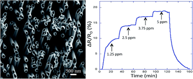
Fig. 4a shows the sensor response under different gaseous NH3 concentrations below 5 ppm, for the 40 nm diameter PPy nanofibers network in 50% humid air condition. It can be seen that the PPy network displayed excellent sensitivity since the sensor could detect very low concentration of gaseous NH3. Indeed, a large response of about 10% was obtained in direct contact with 1.25 ppm of NH3 molecules. The strong dependence of the sensitivity with the nanofibers diameter makes it difficult to compare with other reports available on PPy nanofiber-based NH3 sensors. However, the sensitivity values of the 40 nm diameter PPy sample have been found to be better or at least similar to those reported in previous works.5,13,17–20 Fig. 4b shows the changes in resistance over concentration ranges of 1 ppm to 50 ppm of NH3 for the 40 nm diameter PPy nanofibers network in 50% humid air condition. The sensitivity values of the PPy network increased with NH3 ppm levels. However, it can be seen that the change in the response of the prepared PPy sample did not proceed linearly with a higher slope of the sensitivity at low NH3 concentrations, as previously reported for PPy gas sensors.5,17,18 Finally, in our experiments we also found that both the initial resistance of the fabricated PPy sensor and its sensitivity to NH3 remained unchanged as the relative humidity varied from 20% to 100%. Therefore, the as-prepared PPy nanofibers network sensors could be indeed useful for environmental gas sensing applications at room temperature.


image file: c6ra03439j-f4.tif
Fig. 4 (a) Sensitivity response under gaseous NH3 concentrations below 5 ppm, for the 40 nm diameter PPy nanofibers network in 50% humid air condition. (b) The response values for a wider range of gaseous NH3 concentrations, for the latter network in 50% humid air.

4. Conclusions

We reported an easy fabrication approach for producing centimeter-scale networks of self-standing electrically interconnected PPy NTs and NWs. These dense arrays of electronically conductive polymeric nanostructures can successfully act as high performance chemiresistive gaseous NH3 sensors. Their manufacturing was based on the development of simple and low-cost technologies that did not require any sophisticated processing methods such as the lithographic techniques. Concretely, the 3D network-based sensors were made by combining established and reliable techniques such as track-etching technology for the fabrication of interconnected channels in nanoporous polymer templates and a fairly common electrochemical polymerization for the controlled growth of the crossed nanofiber networks.

The as-prepared network structures offered several advantages in terms of large surface-to-volume ratio and/or hollow inner geometry, as well as easy transport of charge carriers, which are all key aspects in the development of gas sensors with high sensitivity and fast response. The sensor based on small diameter PPy nanofibers array (40 nm) gave the highest response in low NH3 concentration. The response to 1.25 ppm NH3 was about 10% at room temperature. The response value for the 40 nm diameter PPy network under gaseous NH3 concentrations in the range of 5–50 ppm was a factor 4–8 larger than for the 230 nm diameter PPy sample due to the enhanced active surface of the sample. Furthermore, the sensors showed almost no humidity sensitivity from 20% to 100% relative humidity.

It is worth pointing out that this synthesis approach is highly versatile as it offers great tunability. Indeed, a wide range of 3D networks of high aspect-ratio nanostructures (i.e. NWs or NTs) of controlled orientation, packing factor, diameter and material composition can be prepared by this method. This work paves the way to provide a cost-effective and technically reliable method for large-scale production of interconnected arrays of high-aspect ratio nanostructures of different compositions that combines a high specific surface with a good electrical connectivity and mechanical robustness for promising applications in many fields, including the energy storage technology and sensors manufacturing.

Acknowledgements

This work was partly supported by the Fédération Wallonie-Bruxelles (ARC 13/18-052, Supracryst). D. Lahem acknowledges financial support from WBGREEN program (Walloon Region of Belgium), in the framework of the CAPTINDOOR project. We also thank Mr Damien Lefevre for his technical assistance during the PPy electropolymerization, as well as Mrs Pascale Lipnik for the TEM investigations.

References

  1. M. Tian, W. Wang, Y. Wei and R. Yang, J. Power Sources, 2012, 211, 46–51 CrossRef CAS.
  2. W. Wang, M. Tian, A. Abdulagatov, S. M. George, Y. C. Lee and R. Yang, Nano Lett., 2012, 12, 655–660 CrossRef CAS PubMed.
  3. S. Xiong, C. Yuan, X. Zhang and Y. Qian, CrystEngComm, 2011, 13, 626–632 RSC.
  4. C. Wei, H. Pang, B. Zhang, Q. Lu, S. Liang and F. Gao, Sci. Rep., 2013, 3, 2193–2197 Search PubMed.
  5. Q. S. Kwon, S. J. Park, H. Yoon and J. Jang, Chem. Commun., 2012, 48, 10526–10528 RSC.
  6. M. Rauber, I. Alber, S. Müller, R. Neumann, O. Picht, C. Roth, A. Schökel, M. E. Toimil-Molares and W. Ensinger, Nano Lett., 2011, 11, 2304–2310 CrossRef CAS PubMed.
  7. M. R. J. Scherer and U. Steiner, Nano Lett., 2013, 13, 3005–3010 CrossRef CAS PubMed.
  8. E. Araujo, A. Encinas, Y. Velázquez-Galván, J. M. Martínez-Huerta, G. Hamoir, E. Ferain and L. Piraux, Nanoscale, 2015, 7, 1485–1490 RSC.
  9. E. J. Crossland, M. Kamperman, M. Nedelcu, C. Ducati, U. Wiesner, D. M. Smilgies, G. E. Toombes, M. A. Hillmyer, S. Ludwigs, U. Steiner and H. J. Snaith, Nano Lett., 2008, 9, 2807–2812 CrossRef PubMed.
  10. D. H. Wang, H. P. Jakobson, R. Ko, J. Tan, R. Z. Fineman, D. H. Yu and Y. F. Lu, Chem. Mater., 2006, 18, 4231–4237 CrossRef CAS.
  11. J. Martin, M. Martin-Gonzalez, J. F. Fernandez and O. Caballero-Calero, Nat. Commun., 2014, 5, 5130–5135 CrossRef CAS PubMed.
  12. E. Ferain and R. Legras, Nucl. Instrum. Methods Phys. Res., Sect. B, 2003, 208, 115–122 CrossRef CAS.
  13. X. Yang, L. Li and F. Yan, Sens. Actuators, B, 2010, 145, 495–500 CrossRef CAS.
  14. T. Patois, J. B. Sanchezb, F. Bergerb, J. Y. Rauchc, P. Fieveta and B. Lakarda, Sens. Actuators, B, 2012, 171, 431–439 CrossRef.
  15. M. A. Chougule, S. G. Pawar, S. L. Patil, B. T. Raut, P. R. Godse, S. Sen and V. B. Patil, IEEE Sens. J., 2011, 11, 2137–2141 CrossRef CAS.
  16. S. C. Hernandez, D. Chaudhuri, W. Chen, N. V. Myung and A. Mulchandani, Electroanalysis, 2007, 19, 2125–2130 CrossRef CAS.
  17. L. Zhang, F. Meng, Y. Chen, J. Liu, Y. Sun, T. Luo, M. Li and J. Liu, Sens. Actuators, B, 2009, 142, 204–209 CrossRef CAS.
  18. Ishpal and A. Kaur, Appl. Phys., 2013, 113, 094504 Search PubMed.
  19. X. Yang and L. Li, Synth. Met., 2010, 160, 1365–1367 CrossRef CAS.
  20. H. Yoon, M. Chang and J. Jang, J. Phys. Chem. B, 2006, 110, 14074–14077 CrossRef CAS PubMed.
  21. C.-W. Lin, B.-J. Hwang and C.-R. Lee, Mater. Chem. Phys., 1999, 58, 114–120 CrossRef CAS.
  22. H. Liu, et al., Nano Lett., 2004, 4, 671–675 CrossRef CAS.
  23. L. Al-Mashat, H. D. Tran, W. Wlodarski, R. B. Kaner and K. Kalantar-Zadeh, IEEE Sens. J., 2008, 8, 365–370 CrossRef CAS.
  24. S. Demoustier-Champagne, E. Ferain, C. Jérôme, R. Jérôme and R. Legras, Eur. Polym. J., 1998, 34, 1767–1774 CrossRef CAS.
  25. J. P. Spatz, B. Lorenz, K. Weishaupt, H. D. Hochheimer, V. Menon, R. Parthasarathy, C. R. Martin, J. Bechtold and P. H. Hor, Phys. Rev. B: Condens. Matter Mater. Phys., 1994, 50, 14888–14892 CrossRef CAS.
  26. S. De Vito and C. Martin, Chem. Mater., 1998, 10, 1738–1741 CrossRef CAS.
  27. O. Reynes and S. Demoustier-Champagne, J. Electrochem. Soc., 2005, 152, D130–D135 CrossRef CAS.
  28. C. Debiemme-Chouvy, Electrochem. Commun., 2009, 11, 298–301 CrossRef CAS.
  29. R. Xiao, S. Cho, R. Liu and S. B. Lee, J. Am. Chem. Soc., 2007, 129, 4483–4489 CrossRef CAS PubMed.
  30. Y. Velázques-Galván, J. M. Martínez-Huerta, J. de la Torre Medina, Y. Danlée, L. Piraux and A. Encinas, J. Phys.: Condens. Matter, 2014, 26, 026001 CrossRef PubMed.
  31. S. Carquigny, J. B. Sanchez, F. Berger, B. Lakard and F. Lallemand, Talanta, 2009, 78, 199–206 CrossRef CAS PubMed.
  32. Y. Zhang, S. Cui, J. Chang, L. E. Ocola and J. Chen, Nanotechnology, 2013, 24, 025503 CrossRef PubMed.
  33. E. C. Dickey, O. K. Varghese, K. G. Ong, D. Gong, M. Paulose and C. A. Grimes, Sensors, 2002, 2, 91–110 CrossRef CAS.
  34. J.-H. Cho, J.-B. Yu, J.-S. Kim, S.-O. Sohn, D.-D. Lee and J.-S. Huh, Sens. Actuators, B, 2005, 108, 389–392 CrossRef CAS.

This journal is © The Royal Society of Chemistry 2016