Mono- and di-nuclear nickel(II) complexes derived from NNO donor ligands: syntheses, crystal structures and magnetic studies of dinuclear analogues

Pravat Ghoraia, Anindita Chakrabortyb, Anangamohan Panjac, Tapan Kumar Mondala and Amrita Saha*a
aDepartment of Chemistry, Jadavpur University, Kolkata-700032, India. E-mail: asaha@chemistry.jdvu.ac.in; amritasahachemju@gmail.com; Tel: +91-33-2457294
bMolecular Materials Lab, Chemistry and Physics of Materials Unit, Jawaharlal Nehru Centre for Advanced Scientific Research, Banglore-560064, India
cPostgraduate Department of Chemistry, Panskura Banamali College, Panskura RS, Purba Medinipur, West Bengal 721 152, India

Received 1st February 2016 , Accepted 31st March 2016

First published on 4th April 2016


Abstract

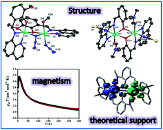
The present report deals with the synthesis and structural characterisation of a mononuclear (1), a di(phenoxido)-bridged dinuclear (2) and a phenoxido/azide bridged dinuclear (3) nickel(II) complexes derived from NNO donor Schiff base ligands. Structural studies reveal that, in all complexes, the nickel(II) ions are hexa-coordinated in a distorted octahedral environment in which tridentate NNO ligand binds the metal centre in the meridional configuration. The variable-temperature (2–300 K) magnetic susceptibility measurements of dinuclear analogues (2 and 3) show that the interaction between the metal centres is moderately ferromagnetic (J = 15.6 cm−1 for 2 and J = 15.3 cm−1 for 3). Broken symmetry density functional calculations of exchange interaction have been performed on complexes 2 and 3 and provide a good numerical estimate of J values (J = 10.31 cm−1 for 2 and J = 17.63 cm−1 for 3) to support the experimental results. Most importantly, compound 2 is only the second example where ferromagnetic coupling is operative in the class of di(phenoxido)-bridged dinickel(II) complexes. The bridging Ni–O–Ni angle being close to the crossover region would provide significant information to get better insight into the magneto-structural correlation in these systems. On the other hand, compound 3 is an important addition to a family of very few hetero-bridged (phenoxido/azide) discrete compounds of nickel(II).


Introduction

There have been extensive studies of the magnetic properties of di- and polynuclear transition metal complexes with exchange-coupled magnetic1,2 centres for the search of single-molecule magnets (SMMs).3–8 The design of such molecule-based magnets relies on the presence of both intra- and inter-molecular coupling. Therefore, the most important systems to explore fundamental aspects are discrete dinuclear exchange-coupled metal complexes because they make possible the evaluation of the pairwise exchange interactions, knowledge of which at least assess qualitatively the magnetic coupling in higher nuclearity systems. In fact, the most of the experimental or theoretical magneto-structural correlations have been established from the studies of dinuclear compounds to understand the basics of the magnetic coupling and to establish proper structure–property relationship.9–17 A rational approach to synthesize discrete exchange-coupled dinuclear metal complexes depends on synthetic strategy that involves the use of particular transition metal ions, blocking ligands and flexidentate bridging ligands with the objective to propagate specific magnetic interactions with interesting structures. The well-known bridging anions like N3, NCS, N(CN)2, CN,C2H3O2, C6H5O, and OH etc., are often used to form such compounds.18 Among them, azido and hydroxo (phenoxide and alkoxide) bridges are the most versatile mediators of magnetic exchange interactions between paramagnetic ions due to their different modes of coordination ability.13,19 Interestingly, while multinuclear Cu(II)-compounds featuring double hetero-bridges μ-hydroxido/alkoxido/phenoxido and μ-X (X = azide, thiocyanate, cyanate, etc.) are quite common in the literature,20 those of hetero-bridged compounds of other 3d metal ions21–23 have been less investigated, and therefore, this area deserves more attention. Among the transition metals, Ni(II) deserves special mention due to its large single-ion zero-field splitting, and as a consequence numerous di- and polynuclear Ni(II) complexes with interesting magnetic properties have been derived.24,25

Schiff-base ligands have been extensively studied as blocking ligands in coordination chemistry mainly due to their ease of synthesis, tremendous structural diversities and immense possibilities towards different applications including molecular magnetism.26 Common strategy to develop di- and polynuclear transition metal complexes is to the use of blocking ligands with lesser donor sites in combination with bridging ligands. Therefore, NNO or NNN donor Schiff base ligands along with various polyatomic bridging anions are excellent combinations to produce various structural architectures, and useful from the magnetic coupling point of view.27,28 Schiff bases derived from salicylaldehydes and 8-aminoquinoline used as blocking ligands are scare in literature.29,30 To the best of our knowledge, only limited numbers of Cu(II)-binuclear complexes of this ligand have been structurally characterized,29,30 while none is reported for nickel. We are interested in developing the coordination chemistry of nickel(II) derived from 8-aminoquinoline and salicyladehyde/o-vanillin to investigate whether it would cause any change in the structure and magnetic properties of the resulting complexes. Accordingly, in this present report, we have synthesized and structurally characterized one mononuclear, one homo- and one hetero-bridged dinuclear Ni(II) complexes, Ni(L1)2 (1), [Ni2(L2)2(SCN)2(DMF)2] (2) and [Ni2(L1)2(N3)2] (3), respectively, where HL1 and HL2 are the tridendentate Schiff base ligands derived from 8-aminoquinoline and o-vanillin/salicylaldehyde, respectively, as shown in Scheme 1. Magnetic characterization has been carried out for dinuclear complexes 2 and 3. We also report here DFT calculations to provide a qualitative theoretical interpretation of overall magnetic behaviour of these dinuclear analogues. Both experimental and theoretical data suggest that the dinuclear nickel(II) analogues exhibit ferromagnetic interaction.


image file: c6ra02982e-s1.tif
Scheme 1 Drawing of the ligands used in this study.

Experimental section

Materials

Materials such as 8-aminoquinoline, o-vanillin and salicylaldehyde were purchased from Sigma-Aldrich, India. The chemicals were of reagent grade and used without further purification. All other chemicals and solvents were of reagent grate and used as received.

Caution! Azide salts of metal complexes with organic ligands are potentially explosive. Only a small amount of material should be prepared and it should be handled with great care.

Synthesis of the Schiff-base ligands

Tridentate Schiff base ligand (HL1) was prepared by the standard method.29 Briefly, 1.0 mmol of 8-aminoquinoline (145 mg) was mixed with 1.0 mmol of o-vanillin (152 mg) in 20 ml of methanol. The resulting solution was heated to reflux for ca. 1 h, and allowed to cool. Ligand HL2 was synthesized following the identical procedure using salicylaldehyde (1.0 mmol, 122 mg) instead of o-vanillin. The dark orange methanol solutions were used directly for complex formation.

Synthesis of Ni(L1)2 (1)

NiCl2·6H2O (237 mg, 1.0 mmol) dissolved in 10 ml of methanol was added to a methanolic solution (10 ml) of the ligand (HL1, 2.0 mmol) with constant stirring for about 3 h. The resulting solution was filtered, and the filtrate was left to stand in the air. X-ray quality red colour single crystals of complex 1 were obtained in several days upon slow evaporation of the filtrate at ambient temperature. Yield: 430 mg (70%). Anal. calcd for C34H26N4O4Ni: C 66.65%, H 4.28%, N 9.15%. Found: C 66.76%, H 4.36%, N 9.01%. IR (cm−1, KBr): ν(C[double bond, length as m-dash]N) 1606 m; ν(C[double bond, length as m-dash]N, py) 1533 m.

Synthesis of [Ni2(L2)2(SCN)2(DMF)2] (2)

NiCl2·6H2O (237 mg, 1.0 mmol) and HL2 (1.0 mmol) were combined in a 20 ml methanol, and to the mixture 5 ml DMF solution of sodium thiocyanate (81 mg, 1.0 mmol) was added with stirring. Finally few drops of triethylamine were added with stirring. The resulting mixture was heated to reflux for 1 h during which time colour of the solution changed to dark reddish brown. The reaction mixture was then filtered and kept at room temperature. Analytically pure dark-brown crystals suitable for X-ray diffraction were obtained from the solution after several days, which was collected by filtration and washed with methanol/ether and air dried. Yield: 670 mg (77%). Anal. calcd for C40H36N8O4S2Ni2: C 55.03%, H 4.16%, N 12.84%. Found: C 54.87%, H 4.27%, N 12.66%. IR (cm−1, KBr): ν(C[double bond, length as m-dash]N) 1608 m; ν(C[double bond, length as m-dash]N, py) 1581 m; ν(SC[N with combining macron]) 2097s.

Synthesis of [Ni2(L1)2(N3)2] (3)

Complex 3 was synthesized from methanol/water solvent mixture following the very similar procedure as described for complex 2 but HL1 and sodium azide were used instead of HL2 and sodium thiocyanate, respectively. Colour: dark brown, yield: 650 mg (82%). Anal. calcd for C35H30N10O5Ni2: C 53.42%, H 3.84%, N 17.81%. Found: C 53.58%, H 3.72%, N 17.62%. IR (cm−1, KBr): ν(C[double bond, length as m-dash]N) 1606 m; ν(C[double bond, length as m-dash]N, py) 1537 m; ν(N3) 2057, 2032s.

Physical measurements

Elemental analyses for C, H and N were carried out using a Perkin-Elmer 240C elemental analyser. Infrared spectra (400–4000 cm−1) were recorded from KBr pellets on a Nicolet Magna IR 750 series-II FTIR spectrophotometer. The powder X-ray diffraction (PXRD) data were registered with a Bruker D8 Advance X-ray diffractometer using Cu-Kα radiation (λ = 1.5418 Å) operating at 40 kV and 40 mA. The XRD patterns were recorded in the 2θ range of 3–50 using Lynxeye detector (1D mode) with a step size of 0.02 and a dwell time of 1 s per step. DC magnetic susceptibility data of polycrystalline powder samples of 2 and 3 were collected on a Vibrating Sample Magnetometer, PPMS (Physical Property Measurement System, Quantum Design, USA) in the temperature range of 2 K to 300 K with an applied field 1000 Oe. Field variation (−5 kOe to 5 kOe) magnetization measurement was carried out at 2 K.

X-ray crystallography

Single crystal X-ray diffraction data of complexes 1–3 were collected on a Bruker SMART APEX-II CCD diffractometer using graphite monochromated Mo/Kα radiation (λ = 0.71073 Å). The unit cells were determined from the setting angles of 36 frames of data. Data processing, structure solution, and refinement were performed using the Bruker Apex-II suite program. All available reflections to 2θmax were harvested and corrected for Lorentz and polarization factors with Bruker SAINT plus.31 Reflections were then corrected for absorption, inter-frame scaling, and other systematic errors with SADABS.31 The structures were solved by the direct methods and refined by means of full matrix least-square technique based on F2 with SHELX-97 software package.32 All the non-hydrogen atoms were refined with anisotropic thermal parameters. All the hydrogen atoms belonging to carbon were placed in their geometrically idealized positions, while hydrogen atom connected to oxygen atom of DMF molecule was located on the difference Fourier map and all of them were constrained to ride on their parent atoms. Crystal data and details of the data collection and refinement for 1–3 are summarized in Table 1.
Table 1 Crystal data and structure refinement of complexes 1–3
Compound 1 2 3
Empirical formula C34H26N4O4Ni C40H36N8O4SNi2 C35H30N10O5Ni2
Formula weight 613.30 874.31 788.11
Temperature (K) 298 (2) 298 (2) 298 (2)
Wavelength (Å) 0.71073 0.71073 0.71073
Crystal system Monoclinic Triclinic Orthorhombic
Space group P21/n P[1 with combining macron] Pbca
a (Å) 12.5834(7) 9.488(3) 18.6631(5)
b (Å) 10.2983(6) 10.734(4) 18.8121(5)
c (Å) 21.547(1) 11.133(4) 19.0739(5)
α (°) 90 73.787(7) 90
β (°) 98.706(3) 68.999(7) 90
γ (°) 90 84.003(7) 90
Volume (Å3) 2760.1(3) 1016.4(6) 6696.7(3)
Z 4 1 8
Dcalc (g cm−3) 1.476 1.428 1.563
Absorption coefficient (mm−1) 0.752 1.080 1.185
F(000) 1272 452 3248
θ Range for data collection (°) 1.77–27.24 1.98–27.50 1.87–25.04
Reflections collected 41[thin space (1/6-em)]174 9071 91[thin space (1/6-em)]058
Independent reflection/Rint 3699/0.0711 2902/0.0583 4660/0.0492
Data/restraints/parameters 6084/0/388 4260/0/253 5912/0/472
Goodness-of-fit on F2 1.022 1.072 1.021
Final R indices [I > 2σ(I)] R1 = 0.0922, wR2 = 0.1316 R1 = 0.1037, wR2 = 0.2515 R1 = 0.0287, wR2 = 0.0642
R indices (all data) R1 = 0.1101, wR2 = 0.1598 R1 = 0.1338, wR2 = 0.2853 R1 = 0.0447, wR2 = 0.0717


Results and discussion

Syntheses and IR spectroscopy

In the synthesis of metal complexes 1–3, we used two different Schiff base ligands HL1 and HL2. HL1 was derived from 1[thin space (1/6-em)]:[thin space (1/6-em)]1 condensation of o-vanillin and 8-aminoquinoline in methanolic solution, whereas ligand HL2 was synthesized using salicylaldehyde instead of o-vanillin. Mononuclear complex 1 was synthesized simply by allowing Schiff base HL1 to react with NiCl2·6H2O in a methanol solution in a 2[thin space (1/6-em)]:[thin space (1/6-em)]1 molar ratio. Complex 2 was obtained by adding a methanolic solution of NiCl2·6H2O to a methanolic solution of Schiff base ligand HL2 followed by DMF solution of NaSCN in a 1[thin space (1/6-em)]:[thin space (1/6-em)]1[thin space (1/6-em)]:[thin space (1/6-em)]1 molar ratio in the presence of few drops of triethylamine. Complex 3 was synthesized adopting very similar procedure as that of 2, by adding a methanolic solution of NiCl2·6H2O to a methanolic solution of Schiff base ligand HL1, followed by aqueous solution of NaN3 in a 1[thin space (1/6-em)]:[thin space (1/6-em)]1[thin space (1/6-em)]:[thin space (1/6-em)]1 molar ratio. A comparison of the powder XRD patterns of 2 and 3 (Fig. S1 and S2) with that of the simulated powder XRD patterns of the single crystals clearly show the purity of the bulk samples which are in accord with the elemental analyses results.

In the IR spectra of complexes 1, 2 and 3, a strong and sharp band due to azomethine ν(C[double bond, length as m-dash]N) appears at 1606, 1608 and 1606 cm−1, respectively. In the spectrum of complex 2, a strong absorption band is observed at 2098 cm−1, which characterizes ν(C[triple bond, length as m-dash]N) vibration of thiocyanate anions.28,33 Complex 3 shows two sharp absorption bands, at 2057and 2032 cm−1, consistent with the presence of both end-on bridging and terminal azide in the structure.34

Description of the crystal structures

Complex 1 crystallizes in the monoclinic space group P21/n. The perspective view of the mononuclear nickel(II) complex is shown in Fig. 1. Selected bond distances and angles with their estimated standard deviations are listed in Table 2. Two HL1 ligands bind the metal ion in a N2O fashion using a phenolate oxygen atom, imine nitrogen atom and amine nitrogen atom, forming one five and one six membered chelate rings, and each of them occupies one meridional position of the octahedral structure of nickel(II). The five-membered chelate bite angles N3–Ni1–N4 [79.92(11)] and N1–Ni1–N2 [79.35(12)°] are close but differ from other two bite angles N4–Ni1–O3 [89.75(10)] and N2–Ni1–O1 [87.86(10)] which are considerably close to the ideal octahedral value (90°). The four coordinating atoms making up the basal plane are two phenoxo-O atoms [O3 and O1] and two quinoline-N atoms [N1 and N3] from the two Schiff bases, while the axial sites are occupied by two imine-N atoms [N2 and N4]. From close inspection of bond distances it has been observed that Ni–N(quinoline) bond distances are relatively longer compared to Ni–O(phenoxo) bond distances. The chelate bite angles and the transoid angles strongly suggest that the complex has a distorted octahedral structure. The solid state structure of 1 is stabilized by edge-to-edge π⋯π stacking, C–H⋯π and nonconventional C–H⋯O hydrogen bonding interactions as shown in Fig. S3.
image file: c6ra02982e-f1.tif
Fig. 1 Crystal structure of 1 showing atom numbering scheme. Hydrogen atoms are omitted for clarity and ellipsoids are drawn at 30% probability.
Table 2 Bond distances (Å) and bond angles (°) of 1–3a
For complex 1
a Symmetry code: a = 1 − x, −y, −z.
Ni1–N1 2.099(3) Ni1–O1 2.056(2)
Ni1–N2 2.055(3) Ni1–O3 2.032(2)
Ni1–N3 2.102(3) Ni1–N4 2.039(3)
N1–Ni1–N2 79.35(12) N3–Ni1–N4 79.92(11)
N1–Ni1–O1 167.16(11) N3–Ni1–O3 169.58(10)
N2–Ni1–N4 173.78(11) N4–Ni1–O3 89.75(10)
N2–Ni1–O1 87.86(10)    

For complex 2
Ni1–N1 2.051(5) Ni1–O2 2.061(4)
Ni1–N2 2.022(5) Ni1–O1a 2.157(4)
Ni1–N3 2.051(6) S1–C17 1.630(7)
Ni1–O1 2.020(4) N3–C17 1.165(9)
N2–Ni1–N1 81.2(2) N2–Ni1–O2 172.92(18)
O1–Ni1–N2 90.40(17) N3–Ni1–O1a 172.93(19)
O1–Ni1–N1 168.52(19) Ni1–O1–Ni1a 98.08(16)

For complex 3
Ni1–N1 2.0436(18) Ni2–N3 2.1333(19)
Ni1–N2 2.0127(19) Ni2–N6 2.0253(19)
Ni1–N3 2.1611(18) Ni2–N7 2.0434(18)
Ni1–O1 1.9784(15) Ni2–N8 2.1197(19)
Ni1–O3 2.0127(15) Ni2–O3 2.0077(14)
Ni1–O4 2.2562(16) Ni2–O5 2.1619(17)
N3–N4 1.207(3) N8–N9 1.189(3)
N4–N5 1.152(3) N9–N10 1.173(3)
O1–Ni1–N2 91.37(7) N6–Ni2–N3 169.28(7)
N2–Ni1–N1 81.36(7) N3–Ni1–O4 154.57(7)
O3–Ni1–O4 74.37(6) O3–Ni2–N7 170.96(7)
N6–Ni2–N7 81.88(8) N8–Ni2–O5 177.92(7)
O3–Ni2–N6 89.47(7) Ni2–O3–Ni1 102.67(6)
N2–Ni1–O3 174.65(7) Ni2–N3–Ni1 93.94(7)
O1–Ni1–N1 172.29(7)    


The crystal structure of 2 consists of a discrete di-(phenoxido)-bridged centro symmetrical unit of formula [Ni2(L2)2(SCN)2(DMF)2], (Fig. 2). Selected bond lengths and angles are presented in Table 2. In this complex, each nickel(II) centre is hexa-coordinated with a distorted octahedral geometry. Nickel(II) centre is bonded to three donor atoms of the deprotonated Schiff base ligand HL2 through the quinone amine nitrogen atom N1, the imine nitrogen atom N2, and the phenoxido oxygen atom O1 as well as to a bridging phenoxido-oxygen atom O1a [a: 1 − x, −y, −z] from the symmetry-related Schiff base ligand. Furthermore, the metal centre is coordinated with a monodentate NCS anion through N3 and a DMF molecule through O2 centre to complete the coordinative requirement for the octahedral geometry. As is usually found in this type of double phenoxido-bridged Ni(II) dimers, the Ni2O2 core is slightly asymmetric, because each Ni(II) ion is closer to the phenoxido oxygen atom of L2 [Ni1–O1 = 2.020(4), Table 2] than that of the phenoxido oxygen atom of the symmetry-related Schiff base ligand [Ni1–O1a = 2.157(4)]. The two Ni atoms are separated by 3.156(1) Å, and the Ni1–O1–Ni1a bridge angle is 98.08(16)°. The tridentate ligand coordinates to the metal ion in the meridional configuration which is usual for this type of rigid ligand. Note that Ni1–O1 and Ni1–N2 are the two shortest bonds, and bonds trans to N1 [Ni1–O1a] are significantly lengthened in 2. The axial positions of the metal centres are occupied by the bridging phenoxo-O atom (O1a) from the tridentate ligand at a distance of 2.157(4) Å, and the thiocyanate nitrogen atom N3 at a distance of 2.051(6) Å. The four donor atoms namely O1, N2, N1 and O2 describe the basal plane around the Ni1 centre, and the displacement of the metal centre from the mean square plane towards the axially coordinated O1a atom is 0.012(3) Å. The equatorial plane is characterized by the bond lengths Ni1–O1 = 2.020(4), Ni1–N2 = 2.022(5), Ni1–N1 = 2.051(5) and Ni1–O2 = 2.061(4) Å. The solid state architecture of 2 is stabilized by edge-to-edge π⋯π stacking, C–H⋯π and nonconventional C–H⋯O hydrogen bonding interactions as shown in Fig. S4.


image file: c6ra02982e-f2.tif
Fig. 2 Molecular structure of 2 showing atom numbering scheme. Hydrogen atoms are omitted for clarity and ellipsoids are drawn at 30% probability.

The crystal structure of [Ni2(L1)21,1-N3)(N3)(CH3OH)] (3) is shown in Fig. 3. The structural analysis reveals that the dinickel(II) compound consists of two deprotonated L1 ligands. Of the two phenoxo-oxygen atoms of two L1, one oxygen atom O3 bridges the two metal centres (Ni1 and Ni2) while the second oxygen O1 acts as a monodentate ligand and is coordinated to Ni1 centre. Again, of the two methoxy oxygen atoms of two L1 ligands, O4 is coordinated to Ni1 while the second oxygen atom O2 is noncoordinated. The quinoline nitrogen atoms (N1, N7) and imine nitrogen atoms (N2, N6) of each of the two L1 are coordinated to each of the two metal centres. In addition to the phenoxo bridge, the metal centres in the dinuclear core are also bridged by the nitrogen atom N3 of an end-on azide ligand. The remaining coordination position of Ni2 is satisfied by the oxygen atom (O5) of a methanol molecule. Thus, both metal centres are hexa-coordinated, but the coordination environments of Ni1 and Ni2 consist of a different set of donor atoms (N3O3 for Ni1 and N4O2 for Ni2). The bond lengths and angles in the coordination environment of the metal centres are listed in Table 2. In case of Ni1 centre, the bond distances involving the bridging and monodentate phenoxo-oxygen atom and the imine nitrogen atom are shorter than other three bond lengths, which follow the order Ni–O(methoxy) > Ni–N(μ1,1-N3) > Ni–N(quinoline). In contrast, the trend of bond distances in Ni2 is slightly different from the Ni1 centre because these are not similarly coordinated. For the Ni2 centre, the bond distances involving the bridging phenoxo-oxygen atom, quinoline nitrogen atom and the imine nitrogen atom are again shorter than other three bond lengths, which follow the order Ni–O(methanol) > Ni–N(μ1,1-N3) > Ni–N(terminal N3). For Ni1, bridging phenoxo-oxygen atom (O3) and the imine nitrogen (N2) occupy the axial positions, while the monodentate phenoxo (O1), methoxy oxygen atom (O4), the bridging azide (N3) and quinoline nitrogen atom (N1) define the basal plane. In the case of Ni2, the terminal azide (N8) and methanol oxygen (O5) occupy the axial positions, while the bridging azide (N3), imine nitrogen atom (N6), quinoline nitrogen atoms (N7), and bridging phenoxo oxygen (O3) define the basal plane. The range of the transoid angles are 154.61(7)–174.65(7) for Ni1 and 169.28(7)–177.92(7) for Ni2. The ranges of the cisoid angles are 101.53(7)–74.37(6) for Ni1 and 81.88(8)–94.45(7) for Ni2 in 3. Clearly, both the transoid and the cisoid angles deviate significantly from ideal values of an octahedral geometry. The molecular packing of the complex is stabilized by nonconventional hydrogen bonding interactions as shown in Fig. S5.


image file: c6ra02982e-f3.tif
Fig. 3 Crystal structure of 3 showing atom numbering scheme. Hydrogen atoms are omitted for clarity and ellipsoids are drawn at 30% probability.

Magnetic properties

The plot of χMT versus T for 2 is displayed in Fig. 4. The room temperature χMT value is 2.07 cm3 mol−1 K. From room temperature to 7 K, there is a gradual increase in χMT value and beyond this maximum, χMT value gradually decreases. An overall ferromagnetic interaction is evident from the nature of χMT versus T plot. The decrease of χMT value beyond 7 K is probably a consequence of the zero-field splitting arising from single-ion anisotropy of Ni(II) ions and possible inter dimer interaction. To understand the nature of magnetic exchange and to determine the value of the coupling constant (J), the χMT data (in the dominant ferromagnetic region 300 K–22 K) were fitted to the isotropic spin Hamiltonian: Ĥ = −1Ŝ2,35 for the Ni(II) dimer (S1 = S2 = 1). The temperature dependence of χM for such a Ni(II) dimeric system can be written as following eqn (1):
 
χM = (22g2/kT) {[exp(J/kT) + 5[thin space (1/6-em)]exp(3J/kT)]/[1 + 3[thin space (1/6-em)]exp(J/kT) + 5[thin space (1/6-em)]exp(3J/kT)]} + (1)

image file: c6ra02982e-f4.tif
Fig. 4 The plots of χMT vs. T for 2. The solid red line indicates the best fit obtained in the temperature range 10 K–300 K (see text).

In this expression, N, β, k and g have their usual meanings and the last term () is the temperature-independent paramagnetism which is assumed to be 200 × 10−6 cm3 mol−1 for two nickel(II) ions. The χMT data (300 K–10 K) were fitted using eqn (1) and the best-fit parameters are J = 15.6 cm−1 with g = 2.01, and R = 4.7 × 10⁻⁴ {R = ∑[(χM)obs − (χM)calc]2/[(χM)obs]2} (Fig. 4). The value of J suggests ferromagnetic interaction, which is also validated by the positive value of Weiss constant θ of 20.4 K obtained from Curie–Weiss fitting of the plot of 1/χM vs. T in the temperature range 300–140 K (Fig. S6). The maximum value of magnetization at 2 K reaches of 3.54 (Fig. 5), which is slightly less than the expected value for ferromagnetically coupled Ni(II) dimers.


image file: c6ra02982e-f5.tif
Fig. 5 The M vs. H curve for 2 at 2 K.

Fig. 6 shows the variable-temperature magnetic susceptibility of 3 measured at 1000 Oe. At 300 K, the χMT value is 2.8 cm3 mol−1 K, typical for two Ni(II) ions with g > 2.00. Upon cooling, χMT value increases and reaches a maximum value of 3.74 cm3 mol−1 at 17 K, and then finally decreases with temperature. Similar to compound 2, a dominant ferromagnetic interaction is suggested by the nature of χMT versus T plot, while the decrease of χMT value beyond the maximum is result of the zero-field splitting as well as the possible inter dimer antiferromagnetic interaction. The best fit parameters using eqn (1) show J = 15.3 cm−1 with g = 2.32, and R = 2.6 × 10⁻⁴ {R = ∑[(χM)obs − (χM)calc]2/[(χM)obs]2} (Fig. 6). The positive value of J suggests the existence of a ferromagnetic coupling within the Ni(II) dimer. The temperature dependence of the reciprocal susceptibility plot above 120 K (Fig. S7) follows the Curie–Weiss law with a Weiss constant θ of 11.25 K, which is also consistent with the ferromagnetic coupling between Ni(II) ions. The M vs. H curve at 2 K reaches a maximum value of 3.8 (Fig. 7), which is very close to the expected value for ferromagnetically coupled Ni(II) dimer.


image file: c6ra02982e-f6.tif
Fig. 6 The plot of χMT vs. T for 3. The solid red line indicates the best fit obtained in the temperature range 22 K–300 K (see text).

image file: c6ra02982e-f7.tif
Fig. 7 The M vs. H curve for 3 at 2 K.

DFT study and magneto-structural correlations

In order to get better insight into the magnetic exchange phenomena in the binuclear nickel(II), phenoxo-O and azido-N bridged complexes 2 and 3, “broken-symmetry” density functional theory (DFT)36 calculations have been carried out using the hybrid B3LYP exchange-correlational functional37 in Gaussian 09 program.38 All elements except nickel have been assigned the 6-31+G(d) basis set. The LanL2DZ basis set with effective core potential has been employed for the nickel atoms.39 The broken-symmetry procedure can be applied to a system of two nickel(II) ions, each containing two unpaired spins. First a self-consistent field (SCF) calculation is carried in quintet state (HS) of the molecule. In the next stage, another SCF calculation is performed taking two spins up on a nickel atom and two spins down on the other nickel atom, which is referred to as the broken-symmetry (BS) solution. Finally, the magnetic exchange coupling constant (J) value is evaluated using the Yamaguchi formula,40 J = (EBSEHS)/(〈S2HS − 〈S2BS), where EHS and EBS are the energies, 〈S2HS and 〈S2BS are the spin-squared operator in the high-spin (HS) and broken symmetry (BS) state. The calculated J value for complex 3 is found to be 17.63 cm−1 and is comparable to the experimental value 15.3 cm−1 (Table 3), while for complex 2 the theoretical value 10.31 cm−1 is found to be lower compare to the experimental value 15.6 cm−1. In both the cases the positive J value indicates, there is ferromagnetic interactions between the nickel centres. The spin density plots in high-spin and broken-symmetry states are shown in Fig. 8 and 9 for complexes 2 and 3, respectively. The spin density plots suggest, in addition to the nickel centres, the spin density is well distributed in bridging azide-N and phenoxo-O atoms and so the magnetic exchange phenomena have been taking place through the bridging atoms in the complexes.
Table 3 Experimental and DFT calculated magnetic exchange coupling constants for 2 and 3
Complexes EHS (a.u.) EBS (a.u.) S2HS 〈S2BS Jtheo (cm−1) Jexp (cm−1)
2 −3419.3024026 −3419.3022145 6.011 2.007 10.31 15.6
3 −2613.420067 −2613.4197459 6.015 2.011 17.63 15.3


image file: c6ra02982e-f8.tif
Fig. 8 Spin density plots of 2 correspond to (a) high spin state and (b) broken symmetry state. The isodensity cutoff value is 0.004 e bohr−3. Blue and green colours represent positive and negative spins respectively.

image file: c6ra02982e-f9.tif
Fig. 9 Spin density plots of 3 correspond to (a) high spin state and (b) broken symmetry state. The isodensity cutoff value is 0.004 e bohr−3. Blue and green colours represent positive and negative spins respectively.

As already described in the structural section, 2 is a Ni(II) dimer with a di-phenoxo bridging unit. Experimental and theoretical magneto-structural correlations carried out by several groups have clearly shown that the major factor controlling the exchange coupling in planar alkoxido– and phenoxido–Ni(μ-O)2Ni complexes is the bridging Ni–O–Ni angle (θ).24 According to these magneto-structural correlations, a ferromagnetic coupling is expected when Ni–O–Ni angle is close to 90°. As Ni–O–Ni angle deviates from 90°, the ferromagnetic coupling decreases and above the critical Ni–O–Ni angle of ∼96 to 98°, the coupling becomes antiferromagnetic.24 The bridging Ni–O–Ni angle of 98.089(16)° in 2 suggests that magnetic coupling between two Ni(II) centres could be either weak antiferromagnetic of even ferromagnetic as the angle falls in the crossover region. Moreover, it has also been seen from the theoretical studies that the out-of-plane displacement of the phenyl carbon atom directly linked to the phenoxo oxygen atom from the Ni2O2 plane (τ) is of as much importance as bridging angle θ in determining the sign and direction of the J value. It has been shown from the theoretical studies that the AF interaction decreases with increase in τ value.24 Although the bridging angle falls in the crossover region but the larger τ angle of 39.5° in 2 prevent an adequate pathway for super exchange (weakening antiferromagnetic coupling), and thus it is reasonable to think that the ferromagnetic interaction between the Ni(II) ions observed for 2 is not unexpected. It should be noted that, to the best of our knowledge, 2 is the second example of a structurally and magnetically characterized bis(μ-alkoxido or phenoxido) bridged dinuclear nickel(II) complex which shows ferromagnetic coupling between two metal centres. Careful inspection of the literature results shows that all the reported di-(alkoxido or phenoxido)-bridged dinuclear nickel(II) complexes show strong antiferromagnetic interactions except in one case where the ferromagnetic coupling is observed in which bridging angle was found to be 95.6°.41 As can be seen from the magneto-structural parameters for the reported di-(alkoxido or phenoxido)-bridged dinuclear nickel(II) complexes in Table 4,41–53 in most of the antiferromagnetically coupled systems, bridging angles are found to be above 99°. Interestingly, there is one Ni(II) dimer presenting a very similar bridge to that of compound 2 with Ni–O–Ni bond angle of 98.28° but in that case antiferromagnetic coupling is observed.42 Magneto-structural correlation data clearly suggest how slight variation of bridging angle affects magnitude of the magnetic coupling. In addition to this difference in bridging angle, several other structural dissimilarities perhaps play the significant role in the opposite sign of the magnetic coupling in these complexes: (i) 8-aminoquinoline part of the ligand imposes geometrical restraint in the present case but the ligand used in the previous study was flexible enough, (ii) structural rigidity enforces the deprotonated ligand L2 to bind with the metal center in meridional fashion while it is facially coordinated in the previous complex and (iii) finally, a major difference is that the coordination environment in the correlated complex consists of a N2O4 donor set in which donor atoms from tridentate ligands exclusively occupy the axial positions, while it is N3O3 in complex 2 and one of the apical position is occupied by exogenous SCN ligand.42 All these above facts strongly suggest that as a large number of structural parameters such as bond angles, bond lengths, out of plane displacement, dihedral angles and the remaining coordination of planes need to be considered to account for the variation of the exchange interaction in this type of complexes. The bridging angle of the most of the reported compounds in these systems is above 99°, and thus a large number of examples on such systems covering a wide range of bridging angles (especially at the region of crossover angle) are required to develop proper magneto-structural correlation.

Table 4 Magnetic and structural parameters of the double phenoxido or alkoxido-bridged octahedral dinuclear nickel(II) complexes
Compound θ (°)a Jexp (cm−1) M⋯M (Å) Ref.
a Average bridging angle.
[Ni2(L)(CH3CO2)2]·10H2O 95.6 10.1 3.005 41
2 98.08 15.3 3.156 This work
[Ni2(L)2(NO2)2]·CH2Cl2·C2H5OH·2H2Oa 98.28 −5.26 3.053 42
[Ni(L)(OOCCF3)(CH3OH)]2 98.7 −19.2 3.077 43
[Ni(L)(OOCCF3)(H2O)]2 98.92 −19.5 3.066 43
[Ni2(L) (N3)2(H2O)2]·CH3CN 99.15 −18.8 3.085 44
[Ni2(Hemp)(H2O)4](F3CCOO)2·2H2O 99.16 −24.6 3.071 45
[{Ni(Hsalhyph)Cl(H2O)}2]·2DMF 99.2 −14.8 3.085 46
[Ni2L(NCS)2(H2O)2]·2DMFa 99.2 −21.3 3.105 47
Ni2(L)2(NO3)2] 99.31 −20.34 3.124 48
[Ni2L(H2O)4](ClO4)2·4NH2CONH2 99.5 −17.0 3.100 47
[Ni2(L)2(NO3)2] 99.75 −24.27 3.119 49
Ni2(L)2(NO2)2] 100.0 −25.25 3.156 48
[Ni2(Hemp)(H2O)4](ClCH2COO)2·2H2O 100.15 −29.6 3.101 45
[Ni2L2(OAc)2] 100.58 −36.18 3.176 50
[(NiL)2]·CH3OH·4H2O 100.6 −66.4 3.1538 51
[Ni2L(MeOH)2(ClO4)2]·2NHEt3ClO4a 101.3 −29.5 3.135 47
[Ni2(μ-L)(acac)2(H2O)]·CH3CN 101.5 −12.6 3.203 52
[Ni2(sym-hmp)2](BPh4)2·3.5DMF·0.5(2-PrOH) 105.43 −69.7 3.237 53


Unlike to di-(phenoxo)-bridged complexes, μ-phenoxo–μ1,1-azide bridged complexes possess dissimilar bridges, and thus there are many parameters which can affect the magnitude of J. Here, the exchange interaction propagated mainly through the phenoxo and end-on azido bridges and the structural parameters related to these bridges should be taken into account to understand the magnetic behaviour of a compound having a μ-phenoxo–μ1,1-azide bridging moiety. As we mentioned earlier, when the Ni–phenoxido–Ni bridge angle is greater than 96–98°, antiferromagnetic behaviour is expected. In contrast, the interaction is predicted to be ferromagnetic for the end-on (EO) azide-bridged Ni(II) complexes, with J increasing upon increasing the bridging angle, yielding a maximum at around 104°. In addition to the bridging angle, it has also been established theoretically that the extent of ferromagnetic interaction in EO azide-bridged Ni(II) complexes decreases linearly with the Ni–N bond lengths. These three governing structural parameters of the μ-alkoxido/phenoxido–μ-azide nickel(II) systems along with their exchange integrals are listed in Table 5.15,55–57 Of them, it is clear from the magneto-structural correlation table that the parameters relevant to the azide bridging are the key factor governing the sign and strength of the magnetic interaction between two metal centres. In fact, all the reported μ-alkoxido/phenoxido–μ-azide nickel(II) compounds exhibit ferromagnetic interaction. All these structural parameters of 3 are very similar to previously reported μ-alkoxido/phenoxido–μ-azide nickel(II) systems, and thus nature and magnitude of interactions observed in 3 is reasonable.

Table 5 Magnetic and structural parameters of μ-phenoxo–μ1,1-azide nickel(II) compounds
Compound Jexp (cm−1) Ni–O–Ni (deg) Ni–N–Ni (deg) Average Ni–O (Å) Average Ni–N (Å) Asymmetry in Ni–N (Å) Ref.
3 15.6 102.68 93.92 2.01 2.145 0.03 This work
[Ni2(L)21,1-N3)(N3)(H2O)]·CH3CH2OH 16.6 103.0 98.0 2.035 2.11 0.06 54
[Ni2(L)21,1-N3)(CH3CN)(H2O)](ClO4)·H2O·CH3CN 16.9 104.7 97.0 2.005 2.115 0.01 54
[Ni2(HL)3(μ1,1-N3)]·3H2O 5.0 106.9 96.3 1.99 2.15 0.12 55
[Ni(L)(μ1,1-N3)Ni(L1) (N3)(OH2)]·H2O 25.6 106.7 96.5 1.985 2.14 0.02 15
[Ni2(L)(N3)(H2O)]·CH3OH·H2O 2.85 102.3 95.6 2.02 2.125 0.03 56
[Ni2(HL)21,1-N3)(o-vanillin)]·H2O 6.69     2.006 2.125 0.05 57


Conclusion

We have synthesized and structurally characterized one mononuclear (1) and one di(phenoxido)-bridged dinuclear (2) and one μ-phenoxido/μ-azide bridged dinuclear (3) nickel(II) complexes derived from NNO donor Schiff base ligands. Structural studies reveal that the metal centres in all complexes are hexa-coordinated in a distorted octahedral geometry in which the rigid tridentate ligands span around the metal centre in the meridional position. Both complexes exhibit ferromagnetic interaction at lower temperatures. Interestingly, the observed J values for both compounds 2 and 3 are well matched with J values obtained from broken symmetry density functional calculations. It is important to note that the di(phenoxido)-bridged dinickel(II) complex (2) is only the second example where ferromagnetic coupling is operative in-between two nickel(II) centres. The bridging Ni–O–Ni angle being close to the crossover region would provide significant information to get better insight into the magneto-structural correlation in these systems. As there are very few hetero-bridged discrete compounds of dinickel(II), compound 3 is an important addition in the family of such systems.

Acknowledgements

A. S. gratefully acknowledges the financial support of this work by the DST, India (Sanction no. SB/EMEQ-159/2013 dated 10/10/2013). A. P. also gratefully acknowledges the financial support of this work by the CSIR, New Delhi, India (Sanction no. 01(2834)/15/EMR-II dated 02/06/15). Crystallography was performed at the DST-FIST, India, funded single crystal diffractometer facility at the Department of Chemistry, Jadavpur University, India. Dedicated to Professor Sreebrata Goswami on the occasion of his 60th birthday.

References

  1. (a) O. Kahn, Angew. Chem., Int. Ed., 1985, 24, 834 CrossRef; (b) J. S. Miller and A. J. Epstein, Angew. Chem., Int. Ed., 1994, 33, 385 CrossRef.
  2. J. S. Miller and M. Drillon, Magnetism: Molecules to Materials, Wiley-VCH, Weinheim, 2001 Search PubMed.
  3. (a) E. K. Brechin, C. Boskovic, W. Wernsdorfer, J. Yoo, A. Yamaguchi, E. C. Sanudo, T. R. Concolino, A. L. Rheingold, H. Ishimoto, D. N. Hendrickson and G. Christou, J. Am. Chem. Soc., 2002, 124, 9710 CrossRef CAS PubMed; (b) Single-Molecule Magnets and Related Phenomena, ed. R. Winpenny, Springer, Berlin, 2006 Search PubMed.
  4. J. R. Friedman, M. P. Sarachik, J. Tejada and R. Ziolo, Phys. Rev. Lett., 1996, 76, 3830 CrossRef CAS PubMed.
  5. (a) D. Gatteschi and R. J. Sessoli, Angew. Chem., Int. Ed., 2003, 42, 268 CrossRef CAS PubMed; (b) D. Gatteschi and R. J. Sessoli, J. Magn. Magn. Mater., 2004, 272–76, 1030 CrossRef.
  6. R. Bagai, W. Wernsdorfer, K. A. Abboud and G. Christou, J. Am. Chem. Soc., 2007, 129, 12918 CrossRef CAS PubMed.
  7. C. J. Milios, A. Vinslava, P. A. Wood, S. Parsons, W. Wernsdorfer, G. Christou, S. P. Perlepes and E. K. Brechin, J. Am. Chem. Soc., 2007, 129, 8 CrossRef CAS PubMed.
  8. T. C. Stamatatos, K. A. Abboud, W. Wernsdorfer and G. Christou, Angew. Chem., Int. Ed., 2007, 46, 884 CrossRef CAS PubMed.
  9. E. Ruiz, J. Cirera, J. Cano, S. Alvarez, C. Loose and J. Kortus, Chem. Commun., 2008, 52 RSC.
  10. R. Koner, H. H. Lin, H. H. Wei and S. Mohanta, Inorg. Chem., 2005, 44, 3524 CrossRef CAS PubMed.
  11. A. A. Lozano, M. Sáez, J. Pérez, L. García, L. Lezama, T. Rojo, G. López, G. García and M. D. Santana, Dalton Trans., 2006, 3906 RSC.
  12. (a) J. Ribas, A. Escuer, M. Monfort, R. Vicente, R. Cortés, L. Lezama and T. Rojo, Coord. Chem. Rev., 1999, 193–195, 1027 CrossRef CAS; (b) M. Verdaguer, A. Bleuzen, V. Marvaud, J. Vaissermann, M. Seuleiman, C. Desplanches, A. Scuiller, C. Train, R. Garde, G. Galley, C. Lomenech, I. Rosenman, P. Veillet, C.Cartier and F. Villain, Coord. Chem. Rev., 1999, 190–192, 1023 CrossRef CAS.
  13. T. C. Stamatatos, D. Foguet-Albiol, S. C. Lee, C. C. Stoumpos, C. P. Raptopoulou, A. Terzis, W. Wernsdorfer, S. O. Hill, S. P. Perlepes and G. Christou, J. Am. Chem. Soc., 2007, 129, 9484 CrossRef CAS PubMed.
  14. (a) T. Sato, W. Mori, Y. Xie, N. Kanehisa, Y. Kai, M. Fuji, S. Goto, E. Nagai and Y. Nakao, Inorg. Chim. Acta, 2006, 359, 2271 CrossRef CAS; (b) A. K. Boudalis, J.-M. Clemente-Juan, F. Dahan and J.-P. Tuchagues, Inorg. Chem., 2004, 43, 1574 CrossRef CAS PubMed.
  15. S. K. Dey, N. Mondal, M. S. E. Fallah, R. Vicente, A. Escuer, X. Solans, M. Font-Barda, T. Matsushita, V. Gramlich and S. Mitra, Inorg. Chem., 2004, 43, 2427 CrossRef CAS PubMed.
  16. A. Roth, A. Buchholz, M. Rudolph, E. Sch6tze, E. Kothe and W. Plass, Chem.–Eur. J., 2008, 14, 1571 CrossRef CAS PubMed.
  17. (a) D.-Y. Wu, W. Huang, W.-J. Hua, Y. Song, C.-Y. Duan, S.-H. Li and Q.-J. Meng, Dalton Trans., 2007, 1838 RSC; (b) A. N. Georgopoulou, C. P. Raptopoulou, V. P. Rafael Ballesteros, B. Abarca and A. K. Boudalis, Inorg. Chem., 2009, 48, 3167 CrossRef CAS PubMed.
  18. (a) A. Escuer and G. Aromi, Eur. J. Inorg. Chem., 2006, 4721 CrossRef CAS; (b) M. Ferbinteanu, H. Miyasaka, W. Wernsdorfer, K. Nakata, K. Sugiura, M. Yamashita, C. Coulon and R. Clérac, J. Am. Chem. Soc., 2005, 127, 3090 CrossRef CAS PubMed; (c) S. S. Massoud, F. A. Mautner, R. Vicente, A. A. Gallo and E. Ducasse, Eur. J. Inorg. Chem., 2007, 1091 CrossRef CAS; (d) A. J. Tasiopoulos, A. Vinslava, W. Wernsdorfer, K. A. Abboudand and G. Christou, Angew. Chem., Int. Ed., 2004, 43, 2117 CrossRef CAS PubMed; (e) A. Panja, P. Guionneau, I.-R. Jeon, S. M. Holmes, R. Clérac and C. Mathonière, Inorg. Chem., 2012, 51, 12350 CrossRef CAS PubMed.
  19. (a) M. F. Charlot, O. Kahn, M. Chaillet and C. Larrieu, J. Am. Chem. Soc., 1986, 108, 2574 CrossRef CAS; (b) L. K. Thompson and S. S. Tandon, Comments Inorg. Chem., 1996, 18, 125 CrossRef CAS.
  20. Y.-C. Chou, S.-F. Huang, R. Koner, G.-H. Lee, Y. Wang, S. Mohanta and H.-H. Wei, Inorg. Chem., 2004, 43, 2759 CrossRef CAS PubMed.
  21. (a) G. Ambrosi, P. Dapporto, M. Formica, V. Fusi, L. Giorgi, A. Guerri, M. Micheloni, P. Paoli, R. Pontellini and P. Rossi, Dalton Trans., 2004, 3468 RSC; (b) J. M. Clemente-Juan, C. Mackiewicz, M. Verelst, F. Dahan, A. Bousseksou, Y. Sanakisand and J.-P. Tuchagues, Inorg. Chem., 2002, 41, 1478 CrossRef CAS PubMed.
  22. S. Hazra, R. Koner, P. Lemoine, E. C. Sañudo and S. Mohanta, Eur. J. Inorg. Chem., 2009, 3458 CrossRef CAS.
  23. (a) G. Aromí, S. Parsons, W. Wernsdorfer, E. K. Brechin and E. J. L. McInnes, Chem. Commun., 2005, 5038 RSC; (b) T. C. Stamatatos, K. A. Abboud, S. P. Perlepes and G. Christou, Dalton Trans., 2007, 3861 RSC; (c) M.-L. Tong, M. Monfort, J. M. C. Juan, X.-M. Chen, X.-H. Bu, M. Ohba and S. Kitagawa, Chem. Commun., 2005, 233 RSC; (d) A. N. Georgopoulou, C. P. Raptopoulou, V. Psycharis, R. Ballesteros, B. Abarca and A. K. Boudalis, Inorg. Chem., 2009, 48, 3167 CrossRef CAS PubMed.
  24. J. Ruiz, A. J. Mota, A. Rodríguez-Diéguez, I. Oyarzabal, J. M. Secob and E. Colacio, Dalton Trans., 2012, 41, 14265 RSC.
  25. (a) L. Botana, J. Ruiz, A. J. Mota, A. Rodríguez-Diéguez, J. M. Seco, I. Oyarzabal and E. Colacio, Dalton Trans., 2014, 43, 13509 RSC; (b) M. Pait, A. Bauzá, A. Frontera, E. Colacio and D. Ray, Inorg. Chem., 2015, 54, 4709 CrossRef CAS PubMed; (c) R. Boča, Coord. Chem. Rev., 2004, 248, 757 CrossRef; (d) R. Herchel, R. Boča, J. Krzystek, A. Ozarowski, M. Durán and J. van Slageren, J. Am. Chem. Soc., 2007, 129, 10306–10307 CrossRef CAS PubMed.
  26. (a) K. Liu, W. Shi and P. Cheng, Coord. Chem. Rev., 2015, 289–290, 74 CrossRef CAS; (b) A. Escuer, J. Esteban, S. P. Perlepes and T. C. Stamatatos, Coord. Chem. Rev., 2014, 275, 87 CrossRef CAS.
  27. C. Adhikary and S. Koner, Coord. Chem. Rev., 2010, 254, 2933 CrossRef CAS.
  28. R. Biswas, S. Mukherjee, P. Kar and A. Ghosh, Inorg. Chem., 2012, 51, 8150 CrossRef CAS PubMed.
  29. (a) H. Xu, C. He, Y. X. Sui, X. M. Ren, L. M. Guo, Y. G. Zhang, S. Nishihara and Y. Hosokoshi, Polyhedron, 2007, 26, 4463 CrossRef CAS; (b) S. Nayak, P. Gamez, B. Kozlevčar, A. Pevec, O. Roubeau, S. Dehnen and J. Reedijk, Polyhedron, 2010, 29, 2291 CrossRef CAS.
  30. M. Shyamal, T. K. Mandal, A. Panja and A. Saha, RSC Adv., 2014, 4, 53520 RSC.
  31. G. M. Sheldrick, SAINT, Version 6.02, SADABS, Version 2.03, Bruker AXS Inc, Madison, Wisconsin, 2002 Search PubMed.
  32. G. M. Sheldrick, SHELXL-97, Crystal Structure Refinement Program, University of Gottingen, 1997 Search PubMed.
  33. P. Kar, R. Biswas, M. G. B. Drew, A. Frontera and A. Ghosh, Inorg. Chem., 2012, 51, 1837 CrossRef CAS PubMed.
  34. F. A. Mautner, S. Hanna, R. Cortes, L. Lezama, M. G. Barandika and T. Rojo, Inorg. Chem., 1999, 38, 4647 CrossRef CAS PubMed.
  35. L. Toma, L. M. Toma, R. Lescouëzec, D. Armentano, G. De Munno, M. Andruh, J. Cano, F. Lloret and M. Julve, Dalton Trans., 2005, 1357 RSC.
  36. (a) L. Noodleman, J. Chem. Phys., 1981, 74, 5737 CrossRef CAS; (b) L. Noodleman and D. A. Case, Adv. Inorg. Chem., 1992, 38, 423 CrossRef CAS; (c) L. Noodleman, C. Y. Peng, D. A. Case and J. M. Mouesca, Coord. Chem. Rev., 1995, 144, 199 CrossRef CAS.
  37. (a) A. D. Becke, J. Chem. Phys., 1993, 98, 5648 CrossRef CAS; (b) C. Lee, W. Yang and R. G. Parr, Phys. Rev. B: Condens. Matter, 1988, 37, 785–789 CrossRef CAS.
  38. M. J. Frisch, G. W. Trucks, H. B. Schlegel, G. E. Scuseria, M. A. Robb, J. R. Cheeseman, G. Scalmani, V. Barone, B. Mennucci, G. A. Petersson, H. Nakatsuji, M. Caricato, X. Li, H. P. Hratchian, A. F. Izmaylov, J. Bloino, G. Zheng, J. L. Sonnenberg, M. Hada, M. Ehara, K. Toyota, R. Fukuda, J. Hasegawa, M. Ishida, T. Nakajima, Y. Honda, O. Kitao, H. Nakai, T. Vreven, J. A. Montgomery, J. J. E. Peralta, F. Ogliaro, M. Bearpark, J. J. Heyd, E. Brothers, K. N. Kudin, V. N. Staroverov, R. Kobayashi, J. Normand, K. Raghavachari, A. Rendell, J. C. Burant, S. S. Iyengar, J. Tomasi, M. Cossi, N. Rega, J. M. Millam, M. Klene, J. E. Knox, J. B. Cross, V. Bakken, C. Adamo, J. Jaramillo, R. Gomperts, R. E. Stratmann, O. Yazyev, A. J. Austin, R. Cammi, C. Pomelli, J. W. Ochterski, R. L. Martin, K. Morokuma, V. G. Zakrzewski, G. A. Voth, P. Salvador, J. J. Dannenberg, S. Dapprich, A. D. Daniels, Ö. Farkas, J. B. Foresman, J. V. Ortiz, J. Cioslowski and D. J. Fox, GAUSSIAN 09 (Revision D.01), Gaussian, Inc., Wallingford CT, 2009 Search PubMed.
  39. (a) P. J. Hay and W. R. Wadt, J. Chem. Phys., 1985, 82, 270 CrossRef CAS; (b) W. R. Wadt and P. J. Hay, J. Chem. Phys., 1985, 82, 284 CrossRef CAS; (c) P. J. Hay and W. R. Wadt, J. Chem. Phys., 1985, 82, 299 CrossRef CAS.
  40. (a) K. Yamaguchi, H. Fukui and T. Fueno, Chem. Lett., 1986, 625–628 CrossRef CAS; (b) M. Nishino, S. Yamanaka, Y. Yoshioka and K. Yamaguchi, J. Phys. Chem. A, 1997, 101, 705–712 CrossRef CAS.
  41. Y. Aratake, M. Ohba, H. Sakiyama, M. Tadokoro, N. Matsumoto and H. Okawa, Inorg. Chim. Acta, 1993, 212, 183 CrossRef CAS.
  42. S. Naiya, H. S. Wang, M. G. B. Drew, Y. Song and A. Ghosh, Dalton Trans., 2011, 40, 2744 RSC.
  43. D. Sadhukhan, A. Ray, G. Pilet, C. Rizzoli, G. M. Rosair, C. J. Gómez-García, S. Signorella, S. Bellú and S. Mitra, Inorg. Chem., 2011, 50, 8326 CrossRef CAS PubMed.
  44. S. Hazra, S. Mondal, M. Fleck, S. Sasmal, E. C.Sañudo and S. Mohanta, Polyhedron, 2011, 30, 1906 CrossRef CAS.
  45. M. Pait, A. Sarkar, E. Colacio and D. Ray, Proc. Natl. Acad. Sci., India, Sect. A, 2014, 84, 189 CrossRef CAS.
  46. A. Burkhardt, A. Buchholz, H. Görls and W. Plass, Z. Anorg. Allg. Chem., 2013, 639, 2516 CrossRef CAS.
  47. (a) K. K. Nanda, R. Das, L. K. Thompson, K. Venkatsubramanian, P. Paul and K. Nag, Inorg. Chem., 1994, 33, 1188 CrossRef CAS; (b) K. K. Nanda, L. K. Thompson, J. N. Bridson and K. Nag, J. Chem. Soc., Chem. Commun., 1994, 1337 RSC.
  48. R. Biswas, S. Giri, S. K. Saha and A. Ghosh, Eur. J. Inorg. Chem., 2012, 2916 CrossRef CAS.
  49. P. Mukherjee, M. G. B. Drew, C. J. Gómez-García and A. Ghosh, Inorg. Chem., 2009, 48, 5848 CrossRef CAS PubMed.
  50. P. Mukherjee, M. G. B. Drew, C. J. Gómez-García and A. Ghosh, Inorg. Chem., 2009, 48, 4817 CrossRef CAS PubMed.
  51. M. Dieng, O. Diouf, M. Gaye, A. S. Sall, P. Pérez-Lourido, L. Valencia, A. Caneschi and L. Sorace, Inorg. Chim. Acta, 2013, 394, 741 CrossRef CAS.
  52. I. Oyarzabal, J. Ruiz, A. J. Mota, A. Rodríguez-Diéguez, J. M. Seco and E. Colacio, Dalton Trans., 2015, 44, 6825 RSC.
  53. H. Sakiyama, K. Tone, M. Yamasaki and M. Mikuriya, Inorg. Chim. Acta, 2011, 365, 183 CrossRef CAS.
  54. S. Sasmal, S. Hazra, P. Kundu, S. Dutta, G. Rajaraman, E. C. Sanudo and S. Mohanta, Inorg. Chem., 2011, 50, 7257 CrossRef CAS PubMed.
  55. R. Koner, S. Hazra, M. Fleck, A. Jana, C. R. Lucas and S. Mohanta, Eur. J. Inorg. Chem., 2009, 4982 CrossRef CAS.
  56. A. Banerjee, R. Singh, D. Chopra, E. Colacio and K. K. Rajak, Dalton Trans., 2008, 6539 RSC.
  57. X. Qin, Y. Ji, Y. Gao, L. Yan, S. Ding, Y. Wang and Z. Liu, Z. Anorg. Allg. Chem., 2014, 640, 462 CrossRef CAS.

Footnote

Electronic supplementary information (ESI) available: File contains Fig. S1–S7. CCDC 1449944–1449946 contain the supplementary crystallographic data for complexes 1 and 2, respectively. For ESI and crystallographic data in CIF or other electronic format see DOI: 10.1039/c6ra02982e

This journal is © The Royal Society of Chemistry 2016