Insights into the macroporosity of freeze-cast hierarchical geopolymers

Elettra Papaa, Valentina Medria, Patricia Benito*b, Angelo Vaccarib, Simone Buganib, Jakub Jaroszewiczc and Elena Landia
aNational Research Council of Italy, Institute of Science and Technology for Ceramics (CNR-ISTEC), Via Granarolo 64, 48018 Faenza, Italy
b“Toso Montanari” Department of Industrial Chemistry, Alma Mater Studiorum, University of Bologna, Viale Risorgimento 4, 40136 Bologna, Italy. E-mail: patricia.benito3@unibo.it
cFaculty of Materials Science and Engineering, Warsaw University of Technology, 141 Woloska Str., 02-507 Warsaw, Poland

Received 25th January 2016 , Accepted 25th February 2016

First published on 26th February 2016


Abstract

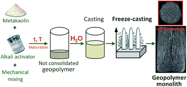
Geopolymer monoliths with controlled lamellar macroporosity and total porosity ranging from 60% to 70% were prepared by ice-templating a partially geopolymerized slurry. Both the maturation treatment of the starting mixture and the water specifically added for freeze-casting were tailored to modify both the geopolymerization and viscosity of the slurry, and, consequently, its freezing behavior, in order to optimize the final lamellar architecture. Following a room temperature maturation treatment, a 50% water content added for freezing developed thick lamellae and wide pores. A lower water content (30%) and curing at 80 °C after maturation at room temperature (for both 50% and 30% H2O) was conducive to a narrow lamellar pore width distribution in the 30–130 μm range. However, the consumption of water due to geopolymerization in samples cured at 80 °C led to a decreased length and thickness of the lamellae. Lastly, the interparticle meso- and macropores (0.003 to 1 μm) within the geopolymer lamellae were only slightly modified by the maturation treatment.


1. Introduction

Geopolymers are amorphous alkaline aluminosilicate binders with a 3D framework of randomly cross-linked silica and alumina tetrahedral units and charge-balancing hydrated alkali cations (i.e. K+, Na+) located in their cavities.1 Geopolymers are easily prepared at near ambient temperature starting from an aluminosilicate solid (fly ash, slag, or metakaolin) and an aqueous solution of an alkali activator (sodium or potassium hydroxide and/or silicate). In short, the geopolymer is obtained through a dissolution–precipitation process. First the silica-alumina source dissolves at high pH, then the silica and alumina tetrahedra condense, thus forming oligomers that transform into a gel that finally sets. This consolidation is chemical rather than thermal.2,3 Geopolymers are considered to be the “green” alternative to Portland Cement,4 and may find applications as fire-resistant and thermal insulating elements,5,6 as well as for encapsulating hazardous7 and nuclear8 waste.

The properties of geopolymer materials, and therefore their applications, depend on the aluminosilicate source, Si/Al ratio, aluminum and silicon availability, alkali activator, temperature, curing time, and water content.2 Water modifies the dissolution/precipitation mechanism; moreover, since water is not chemically bound to the structure but enclosed in its cavities, it alters the inter-particle bonding to form the gel and triggers the structural reorganization process; in other words, it acts as a pore former.9–11

The textural properties of geopolymers, and in particular those of metakaolin-based geopolymers, have been studied widely,12–18 since the pore network determines the mechanical properties12 and durability.19 On the other hand, the tailoring of the geopolymer pore structure expands their applications as thermal insulators,20,21 filters,22 and catalysts23,24 (being the amorphous counterpart of zeolites).

In recent years, much research has been focused on developing geopolymers with hierarchical porosity. Geopolymer particles can be arranged in three-dimensional structures to produce materials with a pore size from a few tenths of a nanometer to several millimeters, and a total pore volume from 30% to 90%.25 Foaming is the most common approach for achieving this.26 Geopolymer foams are prepared by adding a foaming or a blowing agent to the geopolymer slurry, and exploiting any entrapped or generated evolving gases during the synthesis. For instance, H2 bubbles evolve due to the redox reaction of metallic species such as Al27 and Si powders28,29 in the alkaline solution. On the other hand, organic templates were used to develop the macroporosity. Surfactants were added30 or formed in situ31 to the geopolymer gel to obtain a total porosity of up to 80%. Medpelli et al.,32 on the other hand, used a triglyceride oil for a reactive emulsion template; spherical macropores (10–50 μm) were produced in the geopolymer matrix via the oil droplet template in the emulsion. Geopolymer foams containing pores (10–200 μm) were also prepared with alkoxysilanes as a hydrophobic template.33 Lastly, polylactic acid (PLA) fibers acted as pore formers;34 porous geopolymers with controlled pore size, aspect ratio, and orientation were obtained by kneading the PLA fibers into the geopolymer paste, followed by the extrusion and elimination of the template.

We recently reported on a new approach for obtaining lamellar geopolymer monoliths with a hierarchical porosity, i.e. the freeze-casting of the geopolymer slurry.35 This is an environmentally friendly process for producing monoliths made by mesoporous geopolymer matrices, while avoiding the use of organic templates. The unidirectional freezing of the geopolymer aqueous suspension, followed by ice sublimation under reduced pressure, produced a solid with unidirectional channel-like (lamellar) pores that were the replicas of ice crystals.

It is well known that the properties of freeze-cast materials depend on solidification conditions, solvent, solid content,36 particle size,37 particle–particle interactions,38,39 and the use of additives.40 Since the solidification is often directional, pores exhibit an anisotropic morphology in the solidification plane; that is, porous channels run from the bottom to the top of samples.41 When dealing with geopolymers, the freezing process is more challenging. Unlike ceramics ice-templating – which uses a colloidal inert ceramic suspension – geopolymer freezing is performed with a reactive slurry. Not only do the macropores develop, but geopolymerization also takes place during ice-templating,35 the latter being completed during drying. Therefore, the viscosity of the slurry and the particle–particle interaction are modified during the process.

In our previous work,35 starting from an aqueous slurry of metakaolin and potassium di-silicate with molar ratios SiO2[thin space (1/6-em)]:[thin space (1/6-em)]Al2O3 = 4 and H2O/K2O = 13.5, geopolymerization was triggered through a maturation step, without reaching complete consolidation. Different amounts of water (20, 50, 70 vol%) were then added to the geopolymer paste to induce lamellar ice growth by unidirectional freezing. Hierarchical geopolymers with mesoporous matrix and lamellar macroporosity were obtained: geopolymer nanoparticles were arranged in lamellae, while the size of the meso- and macropores depended on the water content added after the maturation step. Lamellar monoliths (height 25 mm) were produced with an optimized solid loading of 36 wt% and 50 vol% of additional water, while further water addition induced some cracks on lamellar surfaces.

The aim of this work was to further tailor the macroporosity of freeze-cast monoliths. We modified the amount and morphology of the lamellar macroporosity in geopolymer monoliths by increasing the water content in the starting geopolymer slurry, and adjusting maturation steps and related viscosity before the freezing process.

2. Experimental

2.1. Sample preparation

Metakaolin grade M1200S was purchased from Imerys. The composition of metakaolin has been reported elsewhere.42 Activating alkali solutions were prepared by dissolving KOH pellets (purity >85% from Sigma-Aldrich) in distilled water and by adding fumed silica powder (99.8% from Sigma-Aldrich) under magnetic stirring as described in a previous work.25 Two potassium di-silicate solutions were prepared with a molar ratio SiO2[thin space (1/6-em)]:[thin space (1/6-em)]K2O = 2 and H2O[thin space (1/6-em)]:[thin space (1/6-em)]K2O = 13.5 and 23, respectively. The two slurries (coded G13 and G23), with a theoretical Si/Al molar ratio equal to 2, were prepared by the mechanical mixing of metakaolin and the potassium di-silicate solution for 20 min at 100 rpm.

After preparation, the slurries G13 and G23 underwent a maturation step at room temperature (r.t.) for 4 and 24 hours, respectively (treatments coded T1 and T2), to reach viscosity values of the same order of magnitude. Another maturation step (T3) was tested on slurry G23: after 24 h at r.t. the mixture was left for 1 h at 80 °C in a heater.

After maturation, distilled water was added to the slurries (30% and 50 vol%) and mechanically mixed for 8 min. Mixtures were cast in cylindrical rubber molds (Ø = 15 mm), pre-cooled on the freeze dryer shaft set at −40 °C (Edwards Mod.MFD01, Crawley, UK), to produce monoliths reaching a height of 25 mm. A flow-chart of the freeze-casting process is shown in Fig. 1 and slurry codes and the treatments applied have been listed in Table 1. The water addition is reported as vol% over the theoretical volume of the geopolymer solid matrix plus the added water. Solid loadings refer to the wt% of the metakaolin in the starting slurries.


image file: c6ra02232d-f1.tif
Fig. 1 Flow-chart of the freeze-casting of the geopolymers.
Table 1 Maturation step, water addition, solid loading, and viscosity at 100 s−1 of the geopolymer slurries for ice-templating. Chemical, morphological and textural characteristics of the geopolymer monoliths: Si/Al and K/Al molar ratios measured by XRF; bulk density and total porosity (eqn (1)); lamellar pore width and lamellae thickness referred to the microstructure of the monoliths' top surfaces (Fig. 5); total pore volume measured by mercury porosimetry; specific surface area (SBET) and pore volume (Vp) obtained through N2 adsorption/desorption measurements
Code Slurries Monoliths
Maturation step Added H2O (vol%) Solid loading (wt%) Viscosity at 100 s−1 (Pa s) Si/Ala K/Ala Bulk density (g cm−3) Total porosity (%) Lamellar pore widthb (μm) Lamella thicknessb (μm) Accessible porosityc (%) SBET (m2 g−1) Vpd (cm3 g−1)
a Measured by XRF.b Measured by SEM image analysis.c Measured by MIP.d Measured by N2 adsorption/desorption.
G13-T1-50 (ref. 35) T1 4 h r.t. 50 36 0.02 1.87 0.36 0.8 64 6–60 10–110 26.61 39.4 0.115
G23-T2-30 T2 24 h r.t. 30 35 0.03 1.47 0.26 0.9 62 10–240 30–85 53.56 38.1 0.144
G23-T2-50 50 27 0.01 1.50 0.27 0.7 71 10–360 5–500 41.74 33.4 0.123
G23-T3-30 T3 24 h r.t.+ 1 h 80 °C 30 35 n.d 1.60 0.33 0.8 65 50–120 50–250 60.35 38.6 0.121
G23-T3-50 50 27 0.75 1.66 0.33 0.7 73 70–100 30–250 54.27 35.8 0.112


Cast slurries were frozen at −40 °C and the solidified liquid phase was sublimated in the freeze dryer at P = 10 Pa in 24 h. Final monoliths were rinsed in distilled water to remove any residue of unreacted potassium silicate and then dried for 2 h in a heater at 100 °C. At least five samples of each slurry were produced for the reproducibility assessment of the ice-templating process.

2.2. Characterization techniques

The viscosity of the slurries (as prepared, after maturation steps T1, T2 and T3, and after water addition for ice-templating) was characterized by a controlled-stress rotational rheometer (Bohlin C-VOR 120, Malvern, UK) equipped with a parallel plate sensor with diameter 20 mm (PP20) or 60 mm (PP60), while forcing the gap to 0.5 mm. Flow curves were determined by increasing the shear rate from 0 to 100 s−1 and then by decreasing from 100 to 0 s−1.

Si/Al and K/Al molar ratios in final consolidated samples were determined by X-ray fluorescence (XRF) analysis. Measurements were performed in a PANalytical Axios Advanced WD-XRF (wavelength dispersive X-ray fluorescence) spectrometer equipped with X-ray tube (Rh target), working at 4 kW. The pellets to be analyzed (diameter 13 mm) were prepared by mixing 0.400 g of the sample with 0.100 g wax (binder) at 100 kN for 120 s.

The percent values of sample total porosity were calculated according to the eqn (1):

 
Total porosity (%) = [1 − (bulk density/true density)] × 100 (1)

The bulk density of samples was determined by weight-to-volume ratio. The volume was geometrically measured by using a caliper (accuracy ±0.05 mm). The true density, i.e. mass/volume of the solid determined by helium pycnometry (Multivolume pycnometer 1305 by Micrometrics), was 2.25 g cm−3 and 2.33 g cm−3, respectively for the G13 and the G23 matrices.25,35

Three-dimensional monolithic structures were studied by μ-Computer Tomography. Samples were scanned using the Skyscan Micro-CT system model 1172 (Skyscan Bruker, Kontich, Belgium). The SkyScan 1172 scanner was operated at 100 kV and 100 μA, with exposure time set to 240 ms. A 180° rotation scan was performed around the vertical axis and with a rotation step of 0.2°. During scanning, a 0.5 mm aluminum filter was used to improve the image quality. The necessary field of view (FOV) for the sample was determined, and resulted in an optimal image pixel size of 9.8 μm. The projected images were reconstructed in 2000 × 2000 pixel-sized cross-sectional images by using a modified Feldkamp cone-beam reconstruction algorithm43 (NRecon, v.1.6.9 software; Skyscan Bruker). Slices were converted into an 8 bit BMP output format while the values within the dynamic range were mapped into gray levels 0–255. This output format was suitable for further processing, while the structural information, such as volume fraction and structure orientation, was obtained from binary images using a commercially available software CTan, v. 1.14.4; Skyscan Bruker. The solid/pore threshold was set both by trying different values and by visually inspecting the appearance of cross sections.44 A ±2 threshold variation results in a variation of less than 1% in the structural parameters, such as porosity.

The morphological and microstructural features of freeze-cast geopolymers were examined by Environmental Scanning Electron Microscopy (E-SEM FEI Quanta 200, FEI Company) and by SEM-FEG (Zeiss).

The pore size distribution in the range 0.0058–100 μm was analyzed by mercury porosimetry (surface tension = 0.48 N m−1 and contact angle = 140°, Thermo Finnigan Pascal 140 and Thermo Finnigan Pascal 240).

The measurements of specific surface area, pore volume, and pore size distribution in the 2–500 nm range were carried out using a Micromeritics ASAP 2020 instrument by N2 adsorption/desorption at −196 °C. Samples were previously degassed under a vacuum, heated up to 250 °C, and maintained at a pressure below 15 μm Hg for 60 min. The specific surface area was calculated by the Brunauer–Emmet–Teller (BET) method. The total pore volume was obtained at p/p0 = 0.995. Pore size distributions were obtained by the BJH method using the desorption branch. Powders in the form of ground and 600 μm sieved samples were analyzed. The measurement error is related to the accuracy of Hg intrusion porosimetry, MIP, (<4%) and N2 adsorption/desorption techniques (<1%).

3. Results and discussion

3.1. Rheology

Viscosity is particularly important in freeze-casting since, for a given particle size, it determines the critical freezing-front rate at which particle trapping occurs inside the growing ice crystals.45

The logarithmic plots of the viscosity vs. shear rate are shown in Fig. 2a and b for both the G13 and G23 compositions, before and after maturation steps and water addition for freeze-casting. Geopolymer slurries showed a pseudo-plastic behavior, while the viscosity was largely dependent on the H2O/K2O molar ratio.29 Indeed, Favier et al.46 reported that colloidal interactions among metakaolin particles are negligible while hydrodynamic effects control the rheological behavior, namely the viscosity of the aqueous alkaline activator.


image file: c6ra02232d-f2.tif
Fig. 2 Logarithmic plots of the viscosity vs. shear rate of the geopolymer slurries for both the compositions G13 (a) and G23 (b) before and after maturation/curing steps and water addition for freeze-casting. The data error is ±5%.

As shown in Fig. 2b, after maturation step T2 (room temperature for 24 h) the geopolymerization progress produced an increased viscosity. The long maturation time (24 h) for G23 was set to reach a viscosity of the same order of magnitude as G13 and G13-T1. Obviously, the water addition for ice templating decreased the viscosity, but slurries G13-T1 + H2O and G23-T2 + H2O again had similar viscosities (Table 1).

After the curing treatment T3 on G23, the resulting paste showed high plasticity, whereas the viscosity could not be measured. It follows that, after water addition, the viscosity of G23-T3 + H2O was one order of magnitude higher than that of the slurries G13-T1 + H2O and G23-T2 + H2O (Table 1), with a curve very similar to that of G23-T2 (Fig. 2b).

3.2. Chemical composition

The chemical composition of ice-templated monoliths was studied by XRF, Table 1. The Si/Al ratio was equal to 1.99 and 1.93 in 97% geopolymerized G13 and G23 matrices, respectively.25,35 Contrary to G13-T1-50, the Si/Al ratio for G23 monoliths was well below that expected. The water addition had an almost negligible effect on the composition, while the maturation treatment increased the Si/Al ratio. As the maturation was extended for 1 h at 80 °C, the silicon content increased slightly, and the Si/Al ratio varied from 1.5 to 1.6. The K/Al ratio in freeze-cast samples was also lower than that expected (0.80),35 its values being lower in G23-T2-30 and G23-T2-50 monoliths. It follows that both Si/Al and K/Al molar ratios increased with T3 maturation, thus indicating an increase in geopolymerization, while the Si/Al ratio was also affected slightly by the amount of water added and the resulting freezing rates.

As reported in a previous paper,35 the potassium silicate aqueous solution continuously modified its chemical composition during geopolymerization (maturation step), in agreement with the increase in viscosity, as mentioned above. After water addition and freezing at −40 °C, the K2O aqueous solution started to crystallize into ice, while SiO2 was either partially segregated by lamellar ice crystals or embedded into the geopolymer matrix, depending on the freezing rates. The soluble K2O was removed by water rinsing after sublimation.

3.3. Macro to mesoporosity

Bulk densities and total porosities are shown in Table 1. Total porosity mainly depended on the initial solid loading, but it was also slightly affected by maturation steps, as will be discussed in the following paragraph. The porous network of samples from the millimeter to nano scale was studied by a combination of micro-CT, SEM, Hg intrusion, and N2 adsorption/desorption.
3.3.1. Computed tomography. The evolution of the casting in the vertical direction of the prepared monoliths was analyzed by micro-CT (μ-CT) images. Some virtual slices of the monoliths, parallel and perpendicular to the ice growth direction, were studied (Fig. 3). For the latter, cross sections were analyzed in three different regions situated 11 mm apart. An estimate of total porosity and pore size distribution (accounting for pore size over 9.8 μm) were also obtained by analyzing the reconstructed images (Fig. 4).
image file: c6ra02232d-f3.tif
Fig. 3 μ-CT slices of the monoliths visualized in the “XZ” (longitudinal sections; the freezing direction (Z) is from the sample bottom-zone 1 toward the sample top-zone 3) and “XY” planes (transversal sections, A–C).

image file: c6ra02232d-f4.tif
Fig. 4 Pore structure thickness (StTh) distributions obtained from the analysis of the computed tomography data. The total porosity values (%) were included in the images.

The structural gradient in the vertical direction, characteristic of ice-templated samples,47 was observed in all monoliths (Fig. 3). Broadly speaking, three regions could be identified, and are labeled as zones 1, 2, and 3 in Fig. 3. A compact solid with small pores developed at the bottom of the cylinder in contact with the cold mold (zone 1). A high cooling rate (supercooled region) produced small ice crystals that engulfed the geopolymer particles. Moving away from the bottom to the top, the temperature decreased and the growth behavior of the ice crystals changed to a steady-state; crystals aligned steadily in the temperature gradient direction and geopolymer particles concentrated and assembled in lamellae (zones 2 and 3). In the transition region, zone 2, a mixture of random and lamellar pores coexisted, while in zone 3, some better defined lamellar pores developed. Thicker lamellae and larger pores were observed at the top of the cylinders, since the solidification rate decreased.48 In zone 3, lamellar pores were both straight and tilted in the vertical direction, since the columnar ice front rejected the particles in directions which were tangential to the solid/liquid interface displacement direction,47 an event which appeared to be more pronounced in the vicinity of the mold wall. Some cracks were observed in zone 2 for G23-T3-30 and in zone 3 for G23-T3-50 propagating perpendicularly to the ice front. Cracks were formed during freezing due to residual stresses resulting from the ice volume expansion in a more geopolymerized slurry.

Some differences in both the length of zones and the size and number of pores were identified. Both the amount of water and the maturation treatment modified the freezing dynamics in the solidification process and, as a result, the porosity, since the viscosity and freezing temperature were altered.

The initial compact zone 1 was larger in samples with low water content and, regardless of water content, in T3 samples (Fig. 3). The formation of a denser slurry under such preparation conditions led to difficulty in the rejection of particles by the ice front. Moreover, upon increasing the solid content, the thermodynamic solidification temperature was lowered by particle–particle interactions, and the critical supercooling value for the ice formation was reached sooner.38 Transversal slices (Fig. 3 slices A) revealed that for the G23-T2-50 and G23-T2-30 samples the inner part of the cylinder, where the freezing rate was slower, contained some lamellar pores. A random network of fine pores of undefined shape (at the resolution of the available images) developed in the sample G23-T3-50. Total porosities calculated in these slices were 58% and 47% for G23-T2-50 and G23-T3-50, while they decreased to 40% and 20% for G23-T2-30 and G23-T3-30 (Fig. 4).

In the middle and top slices (slices B and C in Fig. 3), pores of different width and length (actually representing the cross sections of the longitudinal channel-like pores) were arranged in domains. In general, but especially in the top slices, thicker lamellae developed with the increase in water content, while the maturation treatment shortened them and, consequently, the size of the domains. Pore size distributions and total porosities are shown in Fig. 4. The broadness of the width distribution and the average pore width increased in the top slices. G23-T2-50 had the broadest distribution and highest total porosity values. Specifically, in the middle and top slices, pore width ranges were 30–170 and 30–300 μm, while porosity values were 57% and 65%, respectively. Large pores merged, giving rise to interconnected pores. G23-T2-30, G23-T3-30, and G23-T3-50 developed quite similar pore width distributions (i.e. 30–130 μm, top slice) and porosity values (approx. 50%). The main differences among these three samples depended on the length and arrangement of the pores, namely on the long-range pore order.

A lower amount of water and a soft maturation treatment were conducive to a better arrangement of ice crystals, and therefore of the pores. This occurred mainly in the top slices and outer part of the cylinder, where the temperature was lower. The ice crystals started growing at the contact of the mold, because it was pre-cooled. Ice crystals grew diagonally to the mold's lateral surface in G23-T2-30 and G23-T3-50; whereas in G23-T3-30 it was possible to observe that pores were concentrically arranged and parallel to the mold outside the cylinder whereas they were randomly organized inside.

3.3.2. Scanning electron microscopy. SEM images were taken on the monoliths' top surfaces in order to study the morphology of lamellae at higher magnifications than in μ-CT images (Fig. 5). In addition to the above-mentioned differences in the lamellar organization and dimensions and, consequently, in the main pores at the macroscopic level (Fig. 3 and 4), further differences can be evidenced at the micro- and submicroscopic level by SEM (Fig. 5). In μ-CT images at very low magnification, G23-T2-50 showed well developed wider pores and thicker lamellae than other samples, which had randomly oriented shorter and thinner lamellae. SEM images confirmed that the top surfaces of freeze-cast samples were formed by lamellae with the same orientation and arranged in macro-domains.
image file: c6ra02232d-f5.tif
Fig. 5 SEM micrographs of fracture surfaces of samples G23-T2-30, G23-T2-50, G23-T3-30, and G23-T3-50.

The size of macro-domains depended on the samples, as did the thickness and width of the lamellae and pores (Fig. 5). In particular, sample G23-T2-50 showed a second order of lamellar arrangements; specifically, the solid portions observed at low magnification by μ-CT images were made by very thin lamellae and pores aligned in parallel. Therefore, in this sample, a sort of lamellar micro-domains inside the macro-domain was clearly observed, unlike other samples at the same magnification. It should be noted that some lamellae were cracked in G23-T3-30 and G23-T3-50, as previously observed in the μ-CT images.

Lamellae were built up by bound geopolymer nanoparticles. These particles were more abundant, homogeneous in size, and smoother in T3 than in T2 monoliths, thus confirming both a more homogeneous gelation process and a higher degree of geopolymerization. Some jagged dendritic-like features, running in the solidification direction, were also observed in T2 samples, as previously detected in the G13-T1-50 sample;35 in some cases, they formed protuberances and bridges between adjacent lamellae.

The morphological characteristics of the monoliths' top surface (namely the lamellar pore width, which is the short axis of pores cut perpendicular to the freezing direction,49 and lamella thickness) are listed in Table 1. The differences in the ice crystal growth could be related to the geopolymerization degree and, therefore, to the differences in the viscosity of slurries. It has been reported that the lamellar pore width decreased when the freezing rate increased.41,50 This effect was clear in monoliths subjected to the maturation treatment T2: the lower the solid loading, the lower the freezing rate. The pore width size range increased when the solid loading decreased (Table 1), while the mean value was estimated to be 40 μm in G13-T2-50, 70 μm in G23-T2-30, and about 100 μm in G23-T2-50. The maturation treatment T3 caused water consumption due to both improved geopolymerization and water evaporation, while the higher viscosity increased the freezing rate,45 thus resulting in a narrow pore width size range (Table 1) and mean values of about 70–80 μm in G23-T3 monoliths. The lamellar pore width is known to decrease both with an increasing freezing rate and decreasing solid loading,41,50 resulting in a great variability in lamella thickness, and in particular in a wider size range with lamellar micro-domains inside macro-domains for G23-T2-50. The formation of micro- and macro-domains was reported with mutually miscible solvents, due to the growing of populations of crystals at different times.51,52 Similarly, a high dilution and the consequent slow freezing favored the de-mixing of potassium silicate solution: the water component, enriched in soluble K2O, starts to crystallize into ice, while the remaining liquid portion is over-saturated with respect to SiO2.35 Thus the K2O/SiO2 ratio locally varies, leading to different temperatures and rates of solidification.

3.3.3. Hg intrusion and N2 adsorption/desorption. The pores in the geopolymer matrix, making up the lamellae, were evaluated by Hg intrusion and N2 adsorption/desorption.

A trimodal pore size distribution was obtained by Hg intrusion for the all the samples (in Fig. 6a results from G23-T2-50 and G23-T3-50 are shown as representative), with pores in the 0.01–0.1, 0.1–10, and 10–100 μm ranges. The shapes of the pore size distributions were not largely modified by either the water content or the treatment. These values may differ from the results obtained by μ-CT due to either the intrinsic spatial resolution of μ-CT or the bottleneck effect of MIP.53


image file: c6ra02232d-f6.tif
Fig. 6 Pore size distributions measured by Hg intrusion porosimetry (a) and N2 adsorption/desorption (b) of samples G23-T2-50 and G23-T3-50.

The N2 adsorption/desorption isotherms were of type II (Fig. 7), with no plateau at higher relative pressures, thus indicating that when the relative pressure was increased, the volume adsorption continued.54 The hysteresis loops attributed to the N2 capillary condensation into interconnected mesopores were a mixture of types H2 and H3. Thus samples could be described as macroporous, with mesopores due to the aggregation of geopolymer particles. The changes in shape of the hysteresis loops were related to changes in the pore size distributions.


image file: c6ra02232d-f7.tif
Fig. 7 N2 isotherms of samples G23-T2-50, G23-T3-50, G23-T2-30 and G23-T3-30.

BJH pore size distributions were monomodal in the 0.003–0.2 μm range (in Fig. 6b the curves of G23-T2-50 and G23-T3-50 are shown as representative). Irrespective of the water content, in T3 samples there was a shift toward smaller pore sizes than in T2 samples, with maxima centered at approximately 0.006 and 0.01 μm, respectively. The change in the geopolymerization degree, and consequently in the organization of particles in the slurry, may explain the pore size distributions within the lamellae. However, the role of the Si/Al ratio cannot be ruled out.12,55 Lastly, it should be noted that the data obtained by Hg intrusion and N2 gas adsorption were consistent in the 0.01–0.1 μm range.

Despite differences in the geopolymerization degree, BET surface area values (Table 1) remained constant regardless of the treatment, while they increased slightly for samples with a lower water content. Conversely, the total pore volume (Table 1) decreased with the curing treatment, regardless of the water content.

The small differences observed in the geopolymer network indicated that, even though there were large differences in lamellar macropores, the assembly of the particles was not greatly modified during freeze-casting.

4. Conclusions

Monoliths made with lamellae of geopolymer particles with tailored hierarchical porosity can be prepared by the freeze-casting of the geopolymer slurry. The lamellae arranged in domains, and the lamellar macroporosity both depended on the maturation treatment of the geopolymer slurry and on the water content added for freeze-casting. They both modified the water/solid ratio in the slurry and, consequently, the viscosity and freezing behavior. In particular, the structural gradient along the monolith, the thickness, length, and orientation of the lamellae, and the pore width were all altered.

The addition of a high water content to the geopolymer slurry (low solid content) subjected to a room temperature maturation treatment decreased the viscosity and freezing rate. Consequently, the length of the initial compact zone of the monolith close to the freezing mold was shorter, whereas the thickness of both the lamellae and pores, which developed when ice crystals aligned, increased.

The opposite behavior was obtained after a further maturation of the slurry at 80 °C, because an improved geopolymerization consumed some water. However, in this situation, not only was a narrow pore width distribution created with thinner geopolymer layers in between, but the size of the lamellar domains also decreased.

Despite the changes in macroporosity, the features of the geopolymer particles building the lamellae were not altered much during freeze-casting. Indeed, the interparticle mesopores and macropores and specific surface area values were rather constant in all the samples, and only slightly modified by the maturation treatment.

References

  1. J. Davidovits, Geopolymers Chemistry and Applications, ed. J. Davidovits, Institut Geopolymere, Saint-Quentin, France, 2008 Search PubMed.
  2. P. Duxson, A. Fernández-Jiménez, J. L. Provis, G. C. Lukey, A. Palomo and J. S. J. van Deventer, J. Mater. Sci., 2007, 42, 2917–2933 CrossRef CAS.
  3. A. Mellado, C. Catalán, N. Bouzón, M. V. Borrachero, J. M. Monzó and J. Payá, RSC Adv., 2014, 4, 23846–23852 RSC.
  4. C. Shi, A. Fernández Jiménez and A. Palomo, Cem. Concr. Res., 2011, 41, 750–763 CrossRef CAS.
  5. V. Vaou and D. Panias, Miner. Eng., 2010, 23, 1146–1151 CrossRef CAS.
  6. J. Temuujin, A. Minjigmaa, W. Rickard, M. Lee, I. Williams and A. Van Riessen, J. Hazard. Mater., 2010, 180, 748–752 CrossRef CAS PubMed.
  7. J. Zhang, J. L. Provis, D. Feng and J. S. J. van Deventer, J. Hazard. Mater., 2008, 157, 587–598 CrossRef CAS PubMed.
  8. E. R. Vance and D. Perera, Geopolymers for nuclear waste immobilisation, in Geopolymers: Structure, Processing, Properties and Industrial Applications, ed. J. L. Provis and J. S. J. van Deventer, CRC Press, 2009, pp. 403–422 Search PubMed.
  9. F. Frizon and C. Joussot Dubien, Patent publication no. US2010 0222204 A1, 2010.
  10. E. Prud'homme, P. Michaud, E. Joussein, J. M. Clacens and S. Rossignol, J. Non-Cryst. Solids, 2011, 357, 1270–1278 CrossRef.
  11. P. Steins, A. Poulesquen, O. Diat and F. Frizon, Langmuir, 2012, 28, 8502–8510 CrossRef CAS PubMed.
  12. P. Duxson, J. L. Provis, G. C. Lukey, S. W. Mallicoat, W. M. Kriven and J. S. J. van Deventer, Colloids Surf., A, 2005, 269, 47–58 CrossRef CAS.
  13. R. R. Lloyd, J. L. Provis, K. J. Smeaton and J. S. J. van Deventer, Microporous Mesoporous Mater., 2009, 126, 32–39 CrossRef CAS.
  14. C. F. Maitland, C. E. Buckley, B. H. O'Connor, P. D. Butler and R. D. Hart, J. Appl. Crystallogr., 2011, 44, 697–707 CrossRef CAS.
  15. J. L. Provis, R. J. Myers, C. E. White, V. Rose and J. S. J. van Deventer, Cem. Concr. Res., 2012, 42, 855–864 CrossRef CAS.
  16. S. Lee, H.-T. Jou, A. van Riessen, W. D. A. Rickard, C.-M. Chon and N.-H. Kang, Construct. Build. Mater., 2014, 52, 221–226 CrossRef.
  17. P. Steins, A. Poulesquen, F. Frizon, O. Diat, J. Jestin, J. Causse, D. Lambertin and S. Rossignol, J. Appl. Crystallogr., 2014, 47, 316–324 CAS.
  18. J. Melar, G. Renaudin, F. Leroux, A. Hardy-Dessources, J.-M. Nedelec, C. Taviot-Gueho, E. Petit, P. Steins, A. Poulesquen and F. Frizon, J. Phys. Chem. C, 2015, 119, 17619–17632 CAS.
  19. Y. Ma, J. Hu and G. Ye, Fuel, 2013, 104, 771–780 CrossRef CAS.
  20. E. Kamseu, Z. N. M. NGouloure, B. N. Alib, S. Zekeng, U. C. Melo, S. Rossignol and C. Leonelli, Energ. Build., 2015, 88, 45–56 CrossRef.
  21. Z. N. M. NGouloure, B. Nait-Ali, S. Zekeng, E. Kamseu, U. C. Melo, D. Smith and C. Leonelli, Journal of Building Engineering, 2015, 3, 58–69 CrossRef.
  22. M.-X. Xu, Y. He, C.-Q. Wang, X.-F. He, X.-Q. He, J. Liu and X.-M. Cui, Appl. Clay Sci., 2015, 115, 254–259 CrossRef CAS.
  23. S. Candamano, P. Frontera, A. Macario, F. Crea, J. B. Nagy and P. L. Antonucci, Chem. Eng. Res. Des., 2015, 96, 78–86 CrossRef CAS.
  24. S. Sharma, D. Medpelli, S. Chen and D.-K. Seo, RSC Adv., 2015, 5, 65454–65461 RSC.
  25. E. Landi, V. Medri, E. Papa, J. Dedecek, P. Klein, P. Benito and A. Vaccari, Appl. Clay Sci., 2013, 73, 56–64 CrossRef CAS.
  26. Z. Zhang, J. L. Provis, A. Reid and H. Wang, Construct. Build. Mater., 2014, 56, 113–127 CrossRef.
  27. J. L. Bell and W. M. Kriven, Preparation of Ceramic Foams from Metakaolin-Based Geopolymer Gels, in Developments in Strategic Materials: Ceramic Engineering and Science Proceedings, ed. H. T. Lin, K. Koumoto, W. M. Kriven, E. Garcia, I. E. Reimanis and D. P. Norton, 2009, vol. 29, pp. 97–111 Search PubMed.
  28. E. Prud'homme, P. Michaud, E. Joussein, C. Peyratout, A. Smith, S. Arrii-Clacens, J. M. Clacens and S. Rossignol, J. Eur. Ceram. Soc., 2010, 30, 1641–1648 CrossRef.
  29. V. Medri, E. Papa, J. Dedecek, H. Jirglov, P. Benito, A. Vaccari and E. Landi, Ceram. Int., 2013, 39, 7657–7668 CrossRef CAS.
  30. M. Strozi Cilla, P. Colombo and M. R. Morelli, Ceram. Int., 2014, 40, 5723–5730 CrossRef CAS.
  31. M. Strozi Cilla, M. R. Morelli and P. Colombo, J. Eur. Ceram. Soc., 2014, 34, 3133–3137 CrossRef.
  32. D. Medpelli, J.-M. Seo and D.-K. Seo, J. Am. Ceram. Soc., 2014, 97, 70–73 CrossRef CAS.
  33. B. E. Glad and W. M. Kriven, J. Am. Ceram. Soc., 2015, 98, 2052–2059 CrossRef CAS.
  34. K. Okada, A. Imase, T. Isobe and A. Nakajima, J. Eur. Ceram. Soc., 2011, 31, 461–467 CrossRef CAS.
  35. E. Papa, V. Medri, P. Benito, A. Vaccari, S. Bugani, J. Jaroszewicz, W. Swieszkowski and E. Landi, Microporous Mesoporous Mater., 2015, 215, 206–214 CrossRef CAS.
  36. S. Deville, Adv. Eng. Mater., 2008, 10, 155–169 CrossRef CAS.
  37. S. Deville, E. Maire, A. Lasalle, A. Bogner, C. Gauthier, J. Leloup and C. Guizard, J. Am. Ceram. Soc., 2010, 93, 2507–2510 CrossRef CAS.
  38. T. Waschkies, R. Oberacker and M. J. Hoffmann, Acta Mater., 2011, 59, 5135–5145 CrossRef CAS.
  39. A. Lasalle, C. Guizard, E. Maire, J. Adrien and S. Deville, Acta Mater., 2012, 60, 4594–4603 CrossRef CAS.
  40. B. Delattre, H. Bai, R. O. Ritchie, J. De Coninck and A. P. Tomsia, ACS Appl. Mater. Interfaces, 2014, 6, 159–166 CAS.
  41. S. Deville, E. Saiz and A. P. Tomsia, Acta Mater., 2007, 55, 1965–1974 CrossRef CAS.
  42. V. Medri, S. Fabbri, J. Dedecek, Z. Sobalik, Z. Tvaruzkova and A. Vaccari, Appl. Clay Sci., 2010, 50, 538–545 CrossRef CAS.
  43. L. A. Feldkamp, L. C. Davis and J. W. Kress, J. Opt. Soc. Am., 1984, 1, 612–619 CrossRef.
  44. S. Bugani, M. Camaiti, L. Morselli, E. Van de Casteele and K. Janssens, Anal. Bioanal. Chem., 2008, 391, 1343–1350 CrossRef CAS PubMed.
  45. U. G. Wegst, M. Schecter, A. E. Donius and P. M. Hunger, Philos. Trans. R. Soc., A, 2010, 368, 2099–2121 CrossRef CAS PubMed.
  46. A. Favier, J. Hot, G. Habert, N. Roussel and J. B. d'Espinose de Lacaillerie, Soft Matter, 2014, 10, 1134–1141 RSC.
  47. S. Deville, E. Maire, A. Lasalle, A. Bogner, C. Gauthier, J. Leloup and C. Guizard, J. Am. Ceram. Soc., 2009, 92, 2489–2496 CrossRef CAS.
  48. T. Waschkies, R. Oberacker and M. J. Hoffmann, J. Am. Ceram. Soc., 2009, 92, S79–S89 CrossRef CAS.
  49. P. M. Hunger, A. E. Donius and U. G. K. Wegst, Acta Biomater., 2013, 9, 6338–6348 CrossRef CAS PubMed.
  50. E. Landi, D. Sciti, C. Melandri and V. Medri, J. Eur. Ceram. Soc., 2013, 33, 1599–1607 CrossRef CAS.
  51. C. Guizard, J. Leloup and S. Deville, J. Am. Ceram. Soc., 2005, 97, 2020–2023 CrossRef.
  52. Y. Tang, S. Qiu, C. Wu, Q. Miao and K. Zhao, J. Eur. Ceram. Soc., 2016, 36, 1513–1518 CrossRef CAS.
  53. S. Bugani, M. Camaiti, L. Morselli, E. Van de Casteele and K. Janssens, X-Ray Spectrom., 2007, 36, 316–320 CrossRef CAS.
  54. K. S. W. Sing, D. H. Everett, R. A. W. Haul, L. Moscou, R. A. Pierotti, J. Rouquerol and T. Siemieniewska, Pure Appl. Chem., 1985, 57, 603–619 CrossRef CAS.
  55. P. Duxson, G. C. Lukey and J. S. van Deventer, J. Non-Cryst. Solids, 2006, 352, 5541–5555 CrossRef CAS.

This journal is © The Royal Society of Chemistry 2016
Click here to see how this site uses Cookies. View our privacy policy here.