DOI:
10.1039/C6RA01269H
(Paper)
RSC Adv., 2016,
6, 28194-28199
Highly sensitive and selective “naked eye” sensing of Cu(II) by a novel amido–imine based receptor: a spectrophotometric and DFT study with practical application†
Received
15th January 2016
, Accepted 6th March 2016
First published on 8th March 2016
Abstract
We report the synthesis of a novel compound (E)-bis-N′-((1H-pyrrol-2-yl)methylene)-pyridine-2,6-carbohydrazide and its sensing ability to detect copper(II) ion in aqueous medium by a sharp color change from yellow to brown, the sensing limit being 4.0 × 10⁻⁹ M. Theoretical modeling of the compound and its copper complex was performed. Practical utility was explored by successful paper strip response.
Introduction
Among the various transition metals important for sustaining life processes, copper holds special importance, owing to a very interesting “two-faced” property. On one hand, Cu2+ plays an indispensable role in proper functioning of mammalian metabolism due to its presence in various enzymes such as amine oxidases, ceruloplasmin, hephaestin, lysyl oxidase and superoxide dismutase (along with zinc)1 along with its various industrial2 and agricultural3 uses. On the other hand, excess copper intake opens up an array of physiological disorders as well as causes diseases of serious concern. Whereas nausea, vomiting, tissue damage are some of the minor effects, gastrointestinal catarrh, kidney damage, hypoglycemia and dyslexia are effects of deep concern.4 Neurodegenerative diseases which pose a serious threat such as Alzheimer's disease, Menkes disease and Wilson's disease are supposed to be aggravated by excess copper due to its participation in the formation of reactive oxygen species (ROS).5 As a major amount of copper accumulates in the body through drinking water, detection of copper in water by simple techniques poses an important and enticing challenge in research.
With the advent of sophisticated techniques, various sensors have been reported for detection of copper such as fluorescence ‘turn-on/off’ sensors, quantum dot-based assays and electrochemical sensors.6–26 But sensing by the above mentioned techniques requires use of sophisticated instruments, hence is expensive and time consuming. Moreover, in most cases the sensing experiments were carried out using metal solutions in water and organic solvent mixture7 and hence are of greater academic than practical interest. Thus, there is still need for selective, sensitive copper sensors in aqueous medium. In the present work, we report a simply synthesized Schiff base receptor (E)-bis-N′-((1H-pyrrol-2-yl)methylene)-pyridine-2,6-carbohydrazide by the condensation of pyridine 2,6 dicarboxyhydrazide and pyrrole-2-aldehyde for visual sensing of copper in water/methanol mixed solvent accompanied by a sharp color change. The sensing limit of our reported sensor (∼4.0 × 10⁻⁹ (M)) is much lower compared to other reports utilizing ‘naked eye’ change/change in emission as the tool for detection.8 Furthermore, test strips coated with the receptor molecule showed a change in color when aqueous copper solution was dropped onto them.
Experimental
Reagents and apparatus
Pyridine-2,6-dicarboxylic acid, pyrrole-2-aldehyde, hydrazine hydrate and metal perchlorate salts were purchased from Sigma-Aldrich and were used without further purification. 1H-NMR and 13C-NMR were recorded on Bruker Advanced Supercon 300 MHz and chemical shifts are expressed in ppm using TMS as internal standard. IR spectrum was recorded on Perkin Elmer spectrum-100 and UV-vis titrations and related experiments were conducted in Hitachi U-3501 Spectrophotometer. UV-vis titration with copper was carried out with 1.0 × 10⁻⁴ (M) aqueous solution of copper. Mass spectra were recorded on Waters Xevo G2-S Q TOF mass spectrometer.
Synthesis of (E)-bis-N′-((1H-pyrrol-2-yl)methylene)-pyridine-2,6-carbohydrazide
The synthesis of receptor (E)-bis-N′-((1H-pyrrol-2-yl)methylene)-pyridine-2,6-carbohydrazide (1) was achieved in three steps (Scheme 1).
 |
| | Scheme 1 Synthesis of compound 1. | |
Synthesis of ethyl pyridine-2,6-dicarboxylate
It was prepared according to reported procedure and used without further characterization.9
Synthesis of pyridine-2,6-dicarboxyhydrazide
It was prepared using procedure reported elsewhere and used in the final step without further characterization.10
Synthesis of (E)-bis-N′-((1H-pyrrol-2-yl)methylene)-pyridine-2,6-carbohydrazide (1)
0.1 g of pyridine-2,6-dicarboxyhydrazide (∼0.5 millimoles) was taken in a 50 mL round bottomed flask and dissolved in 20 mL dry methanol. 0.095 g of pyrrole-2-aldehyde (∼1.0 millimole) was added in portions to the aforesaid solution of pyridine-2,6-dicarboxyhydrazide. The solution was stirred under reflux for 4 hours on a water bath. The resulting solution was cooled and allowed to stand for overnight when shining greenish yellow crystals of 1 separated out. The product was filtered under suction, dried in vaccum under reduced pressure and was characterized by 1H-NMR, 13C-NMR and IR spectroscopy. Yield: 0.139 g (79.65%); IR (KBr, cm−1): 3358.80 (pyrrole –NH), 3212.65 (amide –NH), 2923.49, 2852.66, 1649.14 (imine), 1615.47 (amide –C![[double bond, length as m-dash]](https://www.rsc.org/images/entities/char_e001.gif) O), 1587.19, 1531.57, 1117.66, 1032.48, 733.74, 680.18 (ESI, Fig. S1†); 1H-NMR (300 MHz, DMSO-d6, 290 K, TMS): 12.029 (s, 2H, –NH), 11.635 (s, 2H, –CONH), 8.515 (s, 2H, –CHN), 8.293–8.315 (m, 3H, Ar-H), 6.996 (s, 2H, Ar-H), 6.589 (s, 2H, Ar-H), 6.180 (s, 2H, Ar-H) (ESI, Fig. S2†); 13C-NMR (300 MHz, DMSO-d6, 290 K, TMS): 159.45, 148.90, 143.28, 140.32, 127.53, 125.52, 123.55, 114.61, 110.00 (ESI, Fig. S3†).
O), 1587.19, 1531.57, 1117.66, 1032.48, 733.74, 680.18 (ESI, Fig. S1†); 1H-NMR (300 MHz, DMSO-d6, 290 K, TMS): 12.029 (s, 2H, –NH), 11.635 (s, 2H, –CONH), 8.515 (s, 2H, –CHN), 8.293–8.315 (m, 3H, Ar-H), 6.996 (s, 2H, Ar-H), 6.589 (s, 2H, Ar-H), 6.180 (s, 2H, Ar-H) (ESI, Fig. S2†); 13C-NMR (300 MHz, DMSO-d6, 290 K, TMS): 159.45, 148.90, 143.28, 140.32, 127.53, 125.52, 123.55, 114.61, 110.00 (ESI, Fig. S3†).
Results and discussions
Receptor 1 has been synthesized by a simple condensation reaction between pyridine-2,6-dicarboxyhydrazide and pyrrole-2-aldehyde. It has been characterized by 1H NMR, 13C NMR and IR spectroscopy. The presence of a singlet at 12.02 corresponds to the pyrrole –NH proton, the signal for the same is obtained at 3358.80 cm−1 in IR spectrum.26 The singlet at 11.635 corresponded to 3212.65 cm−1 in IR spectrum.26 The singlet at 8.515 appeared for imine proton and the signal for the same functional group is obtained at 1649.14 cm−1 in the IR spectrum.26
Visual sensing of Cu(II)
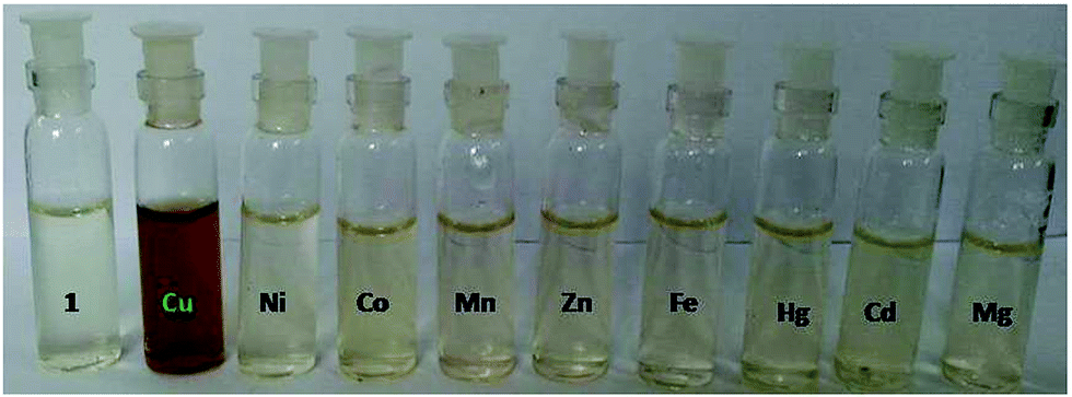
For checking the optical response of receptor 1 towards metal ions, the receptor was dissolved in aqueous methanol mixture (methanol![[thin space (1/6-em)]](https://www.rsc.org/images/entities/char_2009.gif) :water 3:7) and perchlorate salts of transition metals dissolved in deionised water were added (2 equiv.) individually to it. To our delight, the receptor instantaneously showed a sharp change from light yellow to brown by addition of Cu(II) only (Fig. 1), indicating a likely interaction between 1 and copper. The sharp color change can be attributed to a possible intramolecular charge transfer due to complexation with copper.11,12 Under similar conditions, no addition of other metals, including those commonly available in water, showed any significant color change (ESI, Fig. S4†). The selective color change for Cu2+ is a probable outcome of judicious balance between N and O donor centre, for which Cu2+ shows a particular affinity as well as the matching of ionic radius of Cu2+ with the cavity size of receptor 1.
:water 3:7) and perchlorate salts of transition metals dissolved in deionised water were added (2 equiv.) individually to it. To our delight, the receptor instantaneously showed a sharp change from light yellow to brown by addition of Cu(II) only (Fig. 1), indicating a likely interaction between 1 and copper. The sharp color change can be attributed to a possible intramolecular charge transfer due to complexation with copper.11,12 Under similar conditions, no addition of other metals, including those commonly available in water, showed any significant color change (ESI, Fig. S4†). The selective color change for Cu2+ is a probable outcome of judicious balance between N and O donor centre, for which Cu2+ shows a particular affinity as well as the matching of ionic radius of Cu2+ with the cavity size of receptor 1.
 |
| | Fig. 1 Color change of receptor 1 upon addition of 10 equivalents of metal solutions (∼10−4 (M)). | |
UV-vis experiments
To further check the selectivity of receptor 1 towards Cu(II) ion, 10 equivalent of 0.1 mM aqueous solution of transition metals in the form of perchlorate salts were added to 10 μM solution of receptor 1 in aqueous methanol (methanol:water 3:7, v/v) and absorption spectra were recorded using UV-vis spectrophotometer. The selectivity of sensor 1 towards Cu(II) was satisfyingly ensured with the generation of a new red shifted peak at ∼392 nm with two shoulders at ∼375 and ∼416 nm respectively, whereas receptor 1 furnished a strong peak at ∼330 nm (π → π* transition), followed by a weak shoulder at 260 nm respectively. However, addition of other metals (10 equivalents, 0.1 mM aqueous solution) produced too small a change in the absorbance to take into account (ESI, Fig. S5†) and thus the response for Cu(II) was much greater than any other metal ions (Fig. 2). The interaction of 1 with Cu(II), though already indicated from optical color change, could now be rationalized to be the result of complexation between 1 and Cu(II) due to the aforesaid spectral change and the selectivity of compound 1 towards copper can be firmly concluded. In order to investigate the nature of complexation between 1 and Cu(II) a titration experiment was performed using a stock aqueous solution of copper of concentration 1.0 × 10⁻⁴ (M). As already mentioned, the receptor 1 (dissolved in 30% aq. methanol) exhibited two peaks at ∼260 nm and ∼330 nm. Upon addition of 1 equivalent of Cu(II) a broad peak was generated at ∼405 nm along with decrease in the intensity of the peak at ∼330 nm (ESI, Fig. S6†). However, further addition of Cu(II) up to 12 equivalents i.e., until saturation resulted in the resolution of the aforesaid broad peak into one sharp peak at ∼392 nm accompanied by two shoulders at ∼375 nm and ∼416 nm (Fig. 3). Thus, the gradual increase in absorbance value of the newly generated red shifted peak at the ∼392 nm wavelength occurs at the expanse of decrease in absorbance of the receptor's own peak.
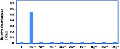 |
| | Fig. 2 UV-vis response upon addition of 10 equivalents of Cu2+, bar graph showing response of Cu2+ to receptor 1 (10 equivalents of other metals also added). | |
 |
| | Fig. 3 UV-visible titration of 10 μM solution of receptor 1 with Cu2+. | |
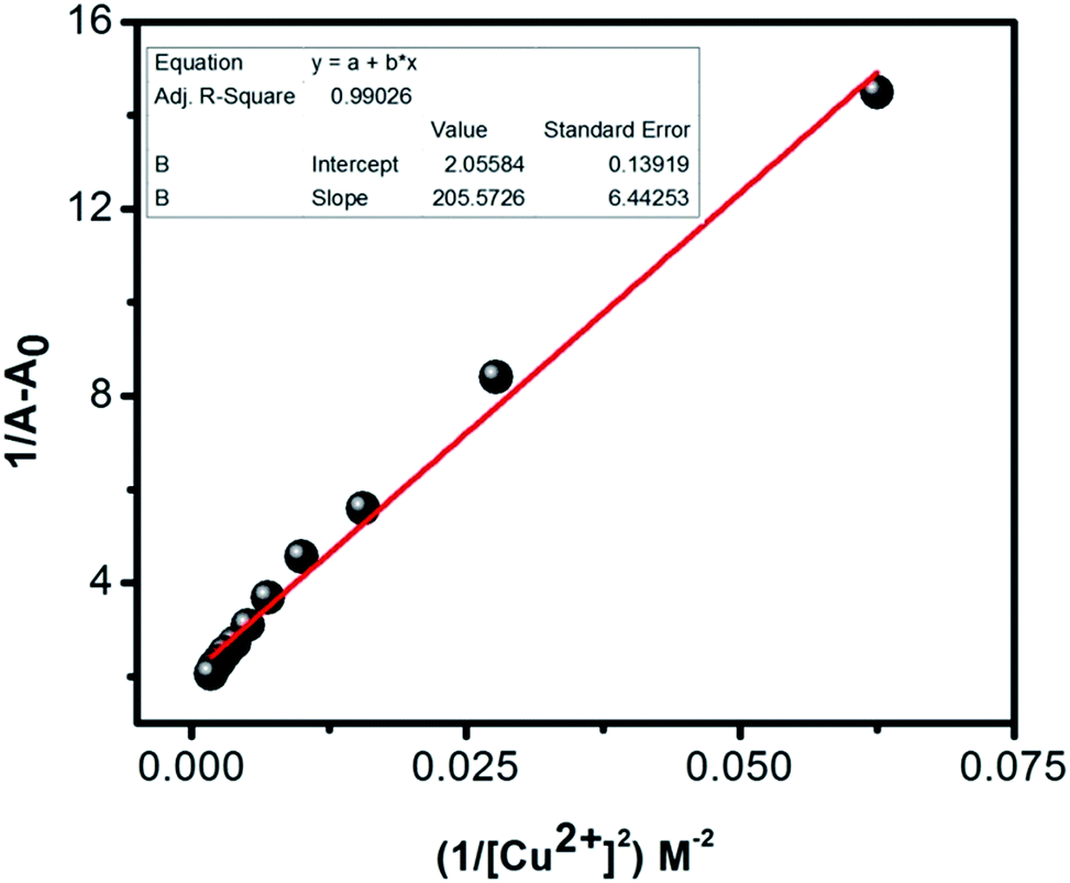
Stoichiometry of complexation and formation constant
With the aim of finding out the stoichiometry and binding constant of the generated complex, the Benesi–Hildebrand (B–H) method was employed, the data sets of absorbance and concentration used in UV-vis titration being reused. The B–H equation is: 1/(A − A0) = 1/(bΔε[G]0[H]0Ka) + 1/(bΔε[H]0), where A and A0 stand for the absorbance values of the host–guest complex and the host (receptor 1 in this case), Δε represents the difference in molar extinction coefficients of the host–guest complex and host whereas b is a constant. The plot of 1/(A − A0) vs. [G]0 (the guest concentration) shows a linearity when the host guest interaction is 1:1 in nature. However, for non-linear interactions (say 1:n) the physical quantity in the abscissa is to be raised to the appropriate order ([G]0n) to obtain linearity. The absorbance of the peak at ∼392 nm was considered for construction of the B–H plot. The plot of 1/(A − A0) showed a linearity (with the fitting parameter being 0.99026) when plotted against the reciprocal of the concentration of Cu2+ squared (Cu2+ being the ‘guest’ in this case), hinting at a 1:2 binding between 1 and Cu2+. The value of binding constant (Ka), which is the ratio of intercept to slope, turned out to be (6.25 ± 1.35) × 1012 M−2 (Fig. 4). So the binding of this ligand with Cu(II) ion is really very strong.
 |
| | Fig. 4 B–H plot for UV-vis titration of 1 with Cu2+ (1.0 × 10⁻⁴ M). | |
Detection limit
The detection limit of receptor 1 for Cu(II) ion was calculated from the calibration curve constructed from the UV-vis titration data set according to IUPAC definition13 (ESI, Fig. S7, Table S1†). A linear fit of absorbance at 392 nm vs. Cu(II) ion concentration plot obtained from the UV-titration curve is used for detection limit measurement. Receptor 1 showed an exceptional detection limit of 4.0 × 10⁻⁹ M for Cu(II). As the detection limit for Cu(II) is comparable to that obtained by sensitive and costly measurement techniques such as fluorescence spectroscopy as also by colorimetry,8 compound 1 stands out among other available chemosensors owing to ‘naked eye’ color change, mild solvent system (methanol:water, 3:7), rapid response and use of cheap instrumental assistance in determining trace amount of copper in water.
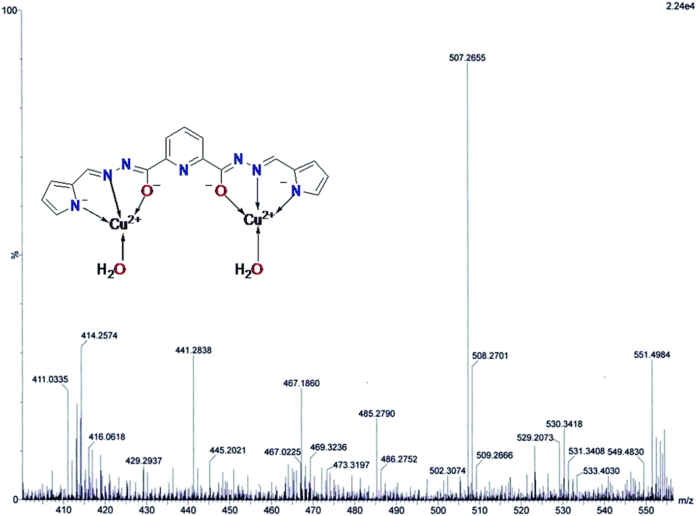
Mass spectral and IR analysis
As mentioned previously, B–H plot indicated 1:2 complex between 1 and Cu(II). To unravel the complexation phenomenon, the complex of receptor 1 and Cu(II) was prepared by refluxing stoichiometric amount of the two complexing partners in methanol, followed by cooling, when a brown powdery mass was obtained. It was filtered under suction, dried in vaccum overnight and then a very dilute solution of the formed complex in methanol was subjected to ESI-MS spectroscopy. A signal at m/z 507.2655 (calculated 506.98) was obtained (Fig. 5). The observed value supports the formation of a 1:2 complex between 1 and Cu(II). Next up, with the task of investigating the participation of the functional groups in 1 responsible for complexation, the IR spectrum of the prepared complex was recorded (ESI Fig. S8†). The shift in the position of imine moiety's peak in the aforementioned spectrum (from a sharp band at ∼1649 cm−1 to a mere hinge at ∼1616 cm−1) clearly proves its involvement in the complexation as also a broad band corresponding to water (∼3434.97 cm−1) was obtained, along with disappearance of the pyrrole –NH signal at ∼3358 cm−1. Finally, to merge our findings from IR and mass spectrometry to visualize the complex, it can be concluded that the pyrrole moieties lost one proton each and the imine moieties as also the tautomerised amide functionality in 1 were involved in the complexation, the co-ordination number being satisfied by two water molecules (Fig. 5).
 |
| | Fig. 5 Mass spectrum of complex of 1 with Cu2+ along with the proposed structure. | |
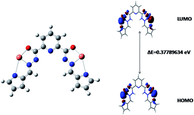
DFT calculations
The urge of delving into the electronic structure of receptor 1 and its copper complex along with the purpose of supporting our experimental findings prompted us to carry on theoretical modeling for receptor 1 (Fig. 6) as well as its complex (Fig. 7) with copper(II) by the aid of Gaussian 09W using B3LYP hybrid functional (6-311++g** basis set).14 The optimized structure of complex explains its remarkable stability over receptor 1 as evident from the lower HOMO–LUMO energy gap of the complex compared to receptor 1 (Fig. 6 and 7). The alteration in the bond distances of major functional groups responsible for complexation agrees with our experimental findings (ESI, Table S2†). A quick analysis of the data provided in ESI, Table S2† furnishes the following conclusions: (I) the increment of CO bond length indicates its involvement in complexation with Cu2+. (II) The tautomerism of the amidic proton which renders a partial double bond character to the amidic C–N bond is evident from decrease in the bond length value of C–N bond. (III) The lengthening of the imine bond length establishes its donation to the metal centre, Cu2+ in the present case. Thus, the hypothetical structure of the complex can be well justified from our theoretical findings.
 |
| | Fig. 6 DFT (B3LYP/6-311++g**) optimized structure of receptor 1 showing energy difference between HOMO and LUMO. | |
 |
| | Fig. 7 B3LYP optimized structure of complex of 1 with Cu2+ and difference in HOMO–LUMO energy. | |
Practical application
Encouraged by the high selectivity and sharp color change of receptor 1 toward Cu(II) in aqueous medium, we attempted to explore its potential to detect copper in the solid state. Keeping in mind that of late, several reported Schiff base receptors furnished satisfactory results for paper strip test15 we attempted to carry out paper strip test for copper with compound 1. Test paper coated with receptor 1 was treated with copper solution (perchlorate salt, ∼10−4 M) and much to our satisfaction, readily changed color from yellow to light brown (Fig. 8), thereby revealing the practical utility of compound 1.
 |
| | Fig. 8 Change in color of paper strip coated with 1 upon addition of Cu2+ (∼10−4 M, 2 equiv.). | |
Conclusion
To conclude, we reported the synthesis of a copper selective amido–imine based chemosensor 1 and studied its interaction with copper by spectrophotometric methods and employed mass spectrometry to explore the mode of complexation. Theoretical modeling was also conducted using G09W. The sensing limit of receptor 1 was found to be ∼4.0 × 10⁻⁹ (M) for copper in water, which is well lower than the WHO recommended amount of copper in water (30 μM). Furthermore, 1 responds to paper strip test for Cu2+ by a color change from yellow to light brown. Thus, our reported chemosensor is of significance due to easy synthetic outline, high selectivity and sensitivity and extremely low limit of detection. The practical applicability of our reported chemosensor 1 was demonstrated by successful test kit experiment.
Acknowledgements
AB and SG would like to acknowledge CSIR and UGC for granting their respective fellowships. NG would like to acknowledge UPE and CRNN, University of Calcutta for financial support. The authors would like to give a heartfelt thanks to Prof Dilip K. Maiti for his invaluable suggestions.
References
- G. W. Gokel, W. M. Leevy and M. E. Weber, Crown ethers: sensors for ions and molecular scaffolds for materials and biological models, Chem. Rev., 2004, 104, 2723–2750 CrossRef CAS PubMed.
- G. W. Stratton, in Review in Environmental Toxicology, ed. E. Hodgson, Elsevier, Amsterdam, 1987, pp. 85–94 Search PubMed.
- N. N. Greenwood and A. Earnshow, Chemistry of the elements, Pergamon Press, New York, 1984, p. 628 Search PubMed.
- I. H. Scheinberg and A. G. Morell, in Inorganic Biochemistry, ed. G. L. Eichhorn, Elsevier, Amsterdam, 1973, pp. 306–343 Search PubMed.
- D. Strausak, J. F. B. Mercer, H. H. Dieter, W. Stremmel and G. Multhaup, Copper in disorders with neurological symptoms: Alzheimer's, Menkes, and Wilson diseases, Brain Res. Bull., 2001, 55, 175–185 CrossRef CAS PubMed.
-
(a) T. Kato, S. Nakamur and M. Mirita, Determination of nickel, copper, zinc, silver, cadmium and lead in seawater by isotope dilution inductively coupled plasma mass spectrometry, Anal. Sci., 1990, 6, 623–626 CrossRef CAS;
(b) S. Hong, T. Kang, J. Moon, S. Oh and J. Yi, Surface plasmon resonance analysis of aqueous copper ions with amino-terminated self-assembled monolayers, Colloids Surf., A, 2007, 292, 264–270 CrossRef CAS;
(c) B.-C. Yin, P. Zuo, H. Huo, X.-H. Zhong and B.-C. Ye, DNAzyme self-assembled gold nanoparticles for determination of metal ions using fluorescence anisotropy assay, Anal. Biochem., 2010, 401, 47–52 CrossRef CAS PubMed;
(d) K.-M. Gattas-Asfura and R. M. Leblanc, Peptide-coated CdS quantum dots for the optical detection of copper(II) and silver(I), Chem. Commun., 2003, 21, 2684–2685 RSC;
(e) M. Kupper and J. W. Schultze, A new copper ion selective microelectrode for electrochemical applications, J. Electroanal. Chem., 1997, 427, 129–135 CrossRef;
(f) W. Lin, L. Yuan, W. Tan, J. Feng and L. Long, Construction of fluorescent probes via protection/deprotection of functional groups: a ratiometric fluorescent probe for Cu2+, Chem.–Eur. J., 2009, 15, 1030–1035 CrossRef CAS PubMed.
-
(a) P. Kaur, S. Kaur, K. Singh, P. R. Sharma and T. Kaur, Indole-based chemosensor for Hg2+ and Cu2+ ions: applications in molecular switches and live cell imaging, Dalton Trans., 2011, 40, 10818–10821 RSC;
(b) D. Maity and T. Govindaraju, Highly selective visible and near-IR sensing of Cu2+ based on thiourea–salicylaldehyde coordination in aqueous media, Chem.–Eur. J., 2011, 17, 1410–1414 CrossRef CAS PubMed;
(c) P. Kaur, M. Kaur and K. Singh, Ferrocene based chemosensor for Cu2+ – a dual channel signaling system, Talanta, 2011, 85, 1050–1055 CrossRef CAS PubMed;
(d) S. Dalapati, S. Jana, M. A. Alam and N. Guchhait, Multifunctional fluorescent probe selective for Cu(II) and Fe(III) with dual-mode of binding approach, Sens. Actuators, B, 2011, 160, 1106–1111 CrossRef CAS;
(e) S. H. Mashraqui, T. Khan, S. Sundaram and S. Ghadigaonkar, Phenothiazinepyridyl chalcone: an easily accessible colorimetric and fluorimetric ‘on–off’ dual sensing probe for Cu2+, Tetrahedron Lett., 2008, 49, 3739–3743 CrossRef CAS;
(f) D. Zhang, M. Wang, M. Chai, X. Chen, Y. Ye and Y. Zhao, Three highly sensitive and selective colorimetric and off–on fluorescent chemosensors for Cu2+ in aqueous solution, Sens. Actuators, B, 2011, 168, 200–206 CrossRef;
(g) X. Ma, Z. Tan, G. Wei, D. Wei and Y. Du, Solvent controlled sugar–rhodamine fluorescence sensor for Cu2+ detection, Analyst, 2012, 137, 1436–1439 RSC;
(h) C. Yu, L. Chen, J. Zhang, J. Li, P. Liu, W. Wang and B. Yan, “Off–on” based fluorescent chemosensor for Cu2+ in aqueous media and living cells, Talanta, 2011, 85, 1627–1633 CrossRef CAS PubMed.
-
(a) T. G. Jo, Y. J. Na, J. J. Lee, M. M. Lee, S. Y. Lee and C. Kim, A diaminomaleonitrile based selective colorimetric chemosensor for copper(II) and fluoride ions, New J. Chem., 2015, 39, 2580 RSC;
(b) S. Goswami, S. Maity, A. K. Das and A. C. Maity, Single chemosensor for highly selective colorimetric and fluorometric dual sensing of Cu(II) as well as ‘NIRF’ to acetate ion, Tetrahedron Lett., 2013, 54, 6631–6634 CrossRef CAS;
(c) H. Zhou, J. Wang, Y. Chen, W. Xi, Z. Zheng, D. Xu, Y. Cao, G. Liu, W. Zhu, J. Wu and Y. Tian, New diaminomaleonitrile derivatives containing aza-crown ether: selective, sensitive and colorimetric chemosensors for Cu(II), Dyes Pigm., 2013, 98, 1–10 CrossRef CAS;
(d) M. X. Liu, T. B. Wei, Q. Lin and Y. M. Zhang, A novel 5-mercapto triazole Schiff base as a selective chromogenic chemosensor for Cu2+, Spectrochim. Acta, Part A, 2011, 79, 1837–1842 CrossRef CAS PubMed;
(e) A. Kumar, V. Kumar, U. Diwan and K. K. Upadhyay, Highly sensitive and selective naked-eye detection of Cu2+ in aqueous medium by a ninhydrin–quinoxaline derivative, Sens. Actuators, B, 2013, 176, 420–427 CrossRef CAS;
(f) U. Diwan, A. Kumar, V. Kumar, K. K. Upadhyaya and P. K. Roychowdhury, A water compatible turn ‘on’ optical probe for Cu2+ based on a fluorescein–sugar conjugate, Sens. Actuators, B, 2014, 196, 345–351 CrossRef CAS.
- A. Bremer, C. M. Ruff, D. Girnt, U. Müllich, J. Rothe, P. W. Roesky, P. J. Panak, A. Karpov, T. J. J. Müllich, M. A. Denecke and A. Geist, 2,6-Bis(5-(2,2-dimethylpropyl)-1H-pyrazol-3-yl)pyridine as a ligand for efficient actinide(III)/lanthanide(III) separation, Inorg. Chem., 2012, 51, 5199–5207 CrossRef CAS PubMed.
- R. M. Duke, T. McCabe, W. Schmitt and T. Gunnlaugsson, Recognition and Sensing of Biologically Relevant Anions in Alcohol and Mixed Alcohol–Aqueous Solutions Using Charge Neutral Cleft Like Glycol-Derived Pyridyl–Amidothiourea Receptors, J. Org. Chem., 2012, 77, 3115–3126 CrossRef CAS PubMed.
- A. Kumar, V. Kumar, U. Diwan and K. K. Upadhyay, Highly sensitive and selective naked-eye detection of Cu2+ in aqueous medium by a ninhydrin–quinoxaline derivative, Sens. Actuators, B, 2013, 176, 420–427 CrossRef CAS.
- V. K. Gupta, A. K. Singha, M. R. Ganjalic, P. Norouzic, F. Faridbodc and N. Mergua, Comparative study of colorimetric sensors based on newly synthesized Schiff bases, Sens. Actuators, B, 2013, 182, 642–651 CrossRef CAS.
- IUPAC, Spectrochim. Acta, Part B, 1978, 33, 241–245 CrossRef.
- M. J. Frisch, et al., GAUSSIAN 03, Revision D.01, Gaussian, Inc., Wallingford, CT, 2004 Search PubMed.
-
(a) V. K. Gupta, A. K. Singha, M. R. Ganjalic, P. Norouzic, F. Faridbodc and N. Mergua, Comparative study of colorimetric sensors based on newly synthesized Schiff bases, Sens. Actuators, B, 2013, 182, 642–651 CrossRef CAS;
(b) N. Mergua and V. K. Gupta, A novel colorimetric detection probe for copper(II) ions based on a Schiff base, Sens. Actuators, B, 2015, 210, 408–417 CrossRef;
(c) D. Maity and T. Govindaraju, Highly Selective Colorimetric Chemosensor for Co2+, Inorg. Chem., 2011, 50, 11282–11284 CrossRef CAS PubMed.
- D. Maity and T. Govindaraju, Highly selective visible and near-IR sensing of Cu2+ based on thiourea–salicylaldehyde coordination in aqueous media, Chem.–Eur. J., 2011, 17, 1410–1414 CrossRef CAS PubMed.
- S. Prabhu, S. Saravanamoorthy, M. Ashok and S. Velmathi, Colorimetric and fluorescent sensing of
multi metal ions and anions by salicylaldimine based receptors, J. Lumin., 2012, 132, 979–986 CrossRef CAS.
- Y. J. Zhang, X. P. He, M. Hu, Z. Li, X. X. Shi and G. R. Chen, Highly optically selective and electrochemically active chemosensor for copper(II) based on triazole-linked glucosyl anthraquinone, Dyes Pigm., 2011, 88, 391–395 CrossRef CAS.
- A. Kumar, V. Kumar, U. Diwan and K. K. Upadhyay, Highly sensitive and selective naked-eye detection of Cu2+ in aqueous medium by a ninhydrin–quinoxaline derivative, Sens. Actuators, B, 2013, 176, 420–427 CrossRef CAS.
- H. H. Wang, L. Xue, Z.-J. Fang, G. P. Li and H. Jiang, A colorimetric and fluorescent chemosensor for copper ions in aqueous media and its application in living cells, New J. Chem., 2010, 34, 1239–1242 RSC.
- N. Mergu and V. K. Gupta, A novel colorimetric detection probe for copper(II) ions based on a Schiff base, Sens. Actuators, B, 2015, 210, 408–417 CrossRef CAS.
- M. X. Liu, T. B. Wei, Q. Lin and Y. M. Zhang, A novel 5-mercapto triazole Schiff base as a selective chromogenic chemosensor for Cu2+, Spectrochim. Acta, Part A, 2011, 79, 1837–1842 CrossRef CAS PubMed.
- K. B. Kim, H. Kim, E. J. Song, S. Kim, I. Noh and C. Kim, A cap-type Schiff base acting as a fluorescence sensor for zinc(II) and a colorimetric sensor for iron(II), copper(II), and zinc(II) in aqueous media, Dalton Trans., 2013, 42, 16569 RSC.
- N. Narayanaswamy and T. Govindaraju, Aldazine-based colorimetric sensors for Cu2+ and Fe3+, Sens. Actuators, B, 2012, 161, 304–310 CrossRef CAS.
- R. Sheng, P. Wang, Y. Gao, Y. Wu, W. Liu, J. Ma, H. Li and S. Wu, Colorimetric Test Kit for Cu2+ Detection, Org. Lett., 2008, 10, 21 CrossRef PubMed.
- X. Baoa, J. Yua and Y. Zhoub, Selective colorimetric sensing for F− by a cleft-shaped anion receptor containing amide and hydroxyl as recognition units, Sens. Actuators, B, 2009, 140, 467–472 CrossRef.
Footnote |
| † Electronic supplementary information (ESI) available. See DOI: 10.1039/c6ra01269h |
|
| This journal is © The Royal Society of Chemistry 2016 |
Click here to see how this site uses Cookies. View our privacy policy here.