
Fluorescent yeast containing intracellularly biosynthesized CdSe QDs as a sensitive probe for simple determination of copper(II) in water and plasma†
Yilong Suab,
Qing-Qing Duab,
Xincheng Quab,
Dongyu Wanab,
Yan-Hua Liuab,
Chao Wangc,
Zheng-Yu Yan*ab and
Sheng-Mei Wu*ab
aDepartment of Analytical Chemistry, China Pharmaceutical University, 24 Tongjia Lane, Gulou District, Nanjing 210009, China. E-mail: yanzhengyujiang@126.com; wushengmei80@163.com; Tel: +86-025-86185150
bKey Laboratory of Drug Quality Control and Pharmacovigilance, Ministry of Education, 24 Tongjia Lane, Gulou District, Nanjing 210009, China
cCollege of Bioscience and Technology, China Pharmaceutical University, 210009 Nanjing, China
First published on 2nd February 2016
Abstract
A simple strategy using CdSe QD-containing yeast as a fluorescent probe was developed for tracing copper(II) (Cu2+) in water and plasma. Nanocrystal CdSe QDs were attentively biosynthesized by Saccharomyces cerevisiae in vivo and were well reserved in the cytoplasm. The yeast membrane, with selective permeability, acted as a semipermeable filter to protect the intrafungal QDs from external interference, for example, from certain ions, peptides, proteins and insoluble solids. Moreover, the as-produced CdSe QDs were capped with cell-derived peptides and proteins, ruling out additional intracellular interference from the cytoplasm, including from some ions, lipids and nucleic acids. With the combination of the dual selective effects of both the yeast membrane and the capping proteins of the QDs, this methodology offered a rapid and reliable method for Cu2+ detection in water and plasma with detection limits as low as 1 μmol L−1 and 2 μmol L−1, respectively. In addition, the presence of the chelating reagent CN− could selectively chelate Cu2+ ions into Cu(CN)n(n−2)−a, resulting in the fluorescence recovery of the QDs, which further reinforced the specificity of Cu2+ detection. These results suggest that this technique via biogenic QD-containing microbes provided great potential for body fluid Cu2+ determination with superior selectivity, reliability and simplicity.
Introduction
Copper is essential to human beings as a trace dietary mineral because it is a crucial component of bone, a key constituent of copper proteins for cell respiration and an important factor in the formation of connective tissue.1 Copper deficiency can easily result in anemia-like symptoms. Conversely, Wilson’s disease causes an accumulation of copper in body tissues, which may lead to severe brain and liver damage if untreated.1 Some neurodegenerative diseases (such as Alzheimer’s disease2) are also linked to excessive copper. According to the World Health Organization’s guidance, copper in drinking water is considered to have a significant impact on health.3 Therefore, as a dietary need but a toxic element, close monitoring of the daily intake of copper is required. So far, atomic absorption spectrophotometry,4 electrochemical methods5 and fluorometric assays6–8 have been applied in the detection of trace copper amounts in biological systems, including drinking water and body fluids. However, some conventional methods suffer from various disadvantages, such as time-consuming sample preparations, complicated steps for ion separation and costly instruments.Nowadays, QD-based systems offer an alternative with the virtues of low detection limits, high sensitivity and relatively convenient and economical protocols. Chen et al.9 firstly used water-soluble luminescent CdS quantum dots (QDs) capped with L-cysteine and thioglycerol to detect zinc and copper ions in aqueous solution. The detection limits were 0.8 μM for Zn2+ and 0.1 μM for Cu2+ in water. Mohamed et al.10 employed CdTe and CdZnSe, which were jacketed with glutathione, for Pb2+ detection with a detection limit as low as 40 nM in water. Chan et al.11 enveloped CdSe QDs with 16-mercaptohexadecanoic acid (16-MHA) as a probe for ultrasensitive Cu2+ detection. The technique offered fast and dependable detection of Cu2+ with a detection limit of 5 nM in water, but was much more roughly applied in Dulbecco’s Modified Eagle’s Medium (DMEM D-5671) solutions. With small particle diameters and a high specific surface area, semiconductor QDs possess a much higher surface/internal atom ratio than conventional macroscopic materials. Thus, slight changes in the QDs’ physical and chemical circumstance might deeply impact on their fluorescent luminance, making them have a much great extinction coefficient towards some substances and subsequently a fantastic potential in quantitative determination based on fluorescence quenching.9,10 Compared to the simple system of an aqueous solution, there is significant concern over how to overcome interferences to achieve determination with high selectivity and reliability in complex body fluid.
Having been well reviewed by I. Costas-Mora,12 the selectivity in fluorescent quantitative analysis using QDs mainly comes from four pathways, (1) capping ligands,9,13–16 (2) fluorescence resonance energy transfer,17,18 (3) chemiluminescence, electrochemiluminescence and photoelectrochemical properties,19 and (4) fluorescence-quenching reversible systems.16 Among these pathways, capping ligands outside the nanocrystals which play a critical role in the interaction between the QDs and the analytes are the leading pathway to the selectivity and the sensitivity of QD-based systems. The displacement of ligands with both the analyte and interferents on the QDs’ surface will cause the instability of the QDs, leading to their fluorescence quenching and/or a shift in the fluorescence maximum emission wavelength.
Cell membranes of yeast, with great potential in protecting inner QDs from external interference, were used as a reliable primary selective filter for the fluorescent quantitative analysis of certain ions in a complicated matrix. CdSe QDs generated by biosynthesis in yeast were well conserved inside the cells, and were physically separated from the extracellular surroundings by both the cell membrane and the cell wall of yeast, as we have reported.20 From fundamental knowledge of biology, the cell membrane can regulate what enters the cell, thus controlling the transport of certain inorganic ions and some small water-soluble organic molecules via specific transmembrane proteins for survival.21 The selective permeability of the yeast membrane, which is an essential feature of living cells, works as a selective filter against external interference, as shown in Fig. 1. Yeast may transfer a limited number of macromolecules and even large particles across their membranes by active transport, pinocytosis or phagocytosis (if at all). However, this is time and energy consuming, and thus this can be largely restricted by rapid determination.21
 | ||
| Fig. 1 CdSe QDs were attentively biosynthesized in yeast, and these yeasts can be used as a fluorescent probe for tracing Cu2+ in water and in plasma. The yeast cell membrane with highly selective permeability worked as a semipermeable filter to prevent external interference that might interrupt or obstruct the specific detection of Cu2+. Capped with cell-derived peptides and proteins, the fluorescent yeast probes further ruled out additional intracellular interference from the cytoplasm. | ||
Herein, CdSe QD-containing yeast was explored as a fluorescent yeast probe for the selective detection of copper(II) in water and more complicated matrix plasma. Firstly, the CdSe QDs were biomanufactured by Saccharomyces cerevisiae (ATCC 9763) in vivo by a method we previously reported,20 and the product of biosynthesis was characterized.20 Then, the fluorescence quenching of CdSe QD-containing yeast by Cu2+ ions was quantitatively measured to plot calibration curves as a mathematic model. Thirdly, different kinds of other common metal ions in plasma and water were intentionally added to the suspension of fluorescent yeast probes under the same conditions, in order to investigate the selectivity and sensitivity of the yeast fluorescent probes for the determination of Cu2+. CN− was successfully employed for the recovery of the QDs’ fluorescence that was quenched by Cu2+, which further confirmed the selectivity of detection. Finally, the concentration of Cu2+ in plasma was measured using this method. In conclusion, these fluorescent probes were successfully used for the tracing of Cu2+ ions with a detection limit as low as 1 μmol L−1 in water and 2 μmol L−1 in plasma. To the best of our knowledge, this paper reports for the first time a route for selective ion determination by the use of QDs which are biosynthesized inside living cells.
Results and discussion
1. Quantitative relationship between fluorescence quenching of yeast probes and Cu2+ concentrations in water
The CdSe QDs were biomanufactured and characterized as in previously published work20 reported by our group. A standard addition method was applied to manufacture analytical samples of Cu2+ with different final concentrations to fit the fluorescence intensity of the yeast probes with the working Cu2+ concentrations. As shown in Fig. 2, with the increment of Cu2+ concentration from 0 μM to 500 μM, the fluorescence intensities of the yeast probes were quenched dramatically, indicating the feasibility of Cu2+ detection in water by this method. | ||
| Fig. 2 Emission spectra of CdSe-containing yeast probes upon exposure to different concentrations of Cu2+ in water. The concentrations of Cu2+ were 0, 1, 2, 4, 6, 8, 10, 20, 30, 50, 100, 200, 300 and 500 μM (along the arrow direction), respectively. | ||
According to previous research,9,10,22 fluorescence quenching can be analyzed by the Stern–Volmer equation:
| I0/I = 1 + KSVC |
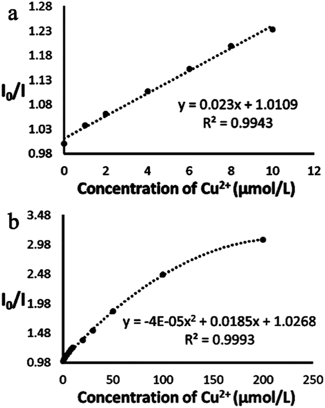
The quantitative relationship between the value of I0/I and the concentration of Cu2+ is shown in Fig. 3. When the concentration of Cu2+ varied from 1 to 10 μmol L−1, the quantitative relationship was a linear fit (y = 0.023x + 1.0109, R2 = 0.9943, where y represents I0/I and x represents the copper concentration, Fig. 3a). Moreover, the fluorescence quenching of the yeast probes was saturated at higher Cu2+ concentrations. When the concentration of Cu2+ varied from 1 to 200 μmol L−1 over a wider concentration range, the relationship had a better binomial fit (y = −0.00004x2 + 0.0185x + 1.0268, R2 = 0.9993, where y represents I0/I and x represents the copper concentration, Fig. 3b). Ultimately, the limit of detection (LOD) was 1 μM, with a distinguished fluorescence quenching of more than 3σ. Meanwhile, three tap water samples were collected and no copper was detected by the method (<1 μmol L−1), which meant the three water samples complied with tap water standards. These simple and low cost experiments put forward the possibility of rapid and sensitive copper ion measurements by green biogenic QDs.
 | ||
| Fig. 3 The correlation of the value of I0/I (565 nm) with the concentration of Cu2+ in water. (a) A linear correlation is observed when the concentration of Cu2+ varied from 1 to 10 μmol L−1, (b) but a binomial correlation can be seen when the concentration of Cu2+ varied from 1 to 200 μmol L−1. | ||
2. Selectivity of yeast fluorescent probes for Cu2+ ion detection
To explore the selective ability of the yeast fluorescent probes’ detection of Cu2+ and their tolerance to different kinds of metal ion solutions, 18 kinds of ions, I− (K+), Ba2+ (Cl−), NH4+ (COO−), Ac− (NH4+), Cd2+ (Cl−), Zn2+ (SO42−), Mg2+ (Cl−), Na+ (Cl−), Ca2+ (Cl−), K+ (Cl−), Al3+ (Cl−), Ag+ (NO3−), Ni+ (SO42−), Cu2+ (Cl−), Pb2+ (NO3−), Hg2+ (Ac−), Fe2+ (SO42−) and Fe3+ (Cl−) were introduced on purpose into the system respectively (Fig. 4 and 5). To magnify the interference of the ions, in this study, ion concentrations were designed much higher than that of blood plasma and even than that of sea water (the extreme example of water, containing Cl− (0.546 mol L−1), Na+ (0.469 mol L−1), Mg2+ (0.0530 mol L−1), SO42− (0.0282 mol L−1), Ca2+ (0.0100 mol L−1), K+ (0.0100 mol L−1)).23 Normally, blood plasma also contains a lot of electrolytes, such as Na+ (142 mOsm L−1), Cl− (108 mOsm L−1), K+ (4.2 mOsm L−1), phosphate (2 mOsm L−1), Ca2+ (1.3 mOsm L−1), Mg2+ (0.8 mOsm L−1) and SO42− (0.5 mOsm L−1), which may interfere with detection.24 Herein, the concentrations of Mg2+ (Cl−), Na+ (Cl−), Ca2+ (Cl−) and K+ (Cl−) were chosen to be 1 mol L−1 while the other six ions were 50 mM (Fig. 4). Meanwhile, Al3+ (Cl−), Ag+ (NO3−), Ni+ (SO42−), Cu2+ (Cl−), Pb2+ (NO3−), Hg2+ (Ac−), Fe2+ (SO42−) and Fe3+ (Cl−) were tested, where the concentrations of the ions were 10 mM (Fig. 5).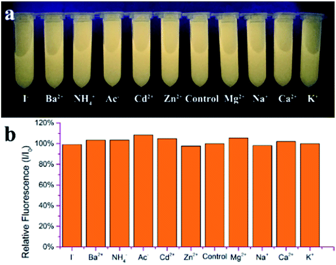 | ||
| Fig. 4 Fluorescence intensity of yeast probes in the presence of various metal ions: I− (K+), Ba2+ (Cl−), NH4+ (COO−), Ac− (NH4+), Cd2+ (Cl−), Zn2+ (SO42−), control, Mg2+ (Cl−), Na+ (Cl−), Ca2+ (Cl−) and K+ (Cl−). (a) Photograph under UV light excitation. (b) Relative fluorescence (I/I0) achieved in the absence and presence of each metal ion, respectively. | ||
 | ||
| Fig. 5 Fluorescence intensity of yeast probes in the presence of various metal ions: Al3+ (Cl−), Ag+ (NO3−), Ni+ (SO42−), Cu2+ (Cl−), control, Pb2+ (NO3−), Hg2+ (Ac−), Fe2+ (SO42−) and Fe3+ (Cl−) in 5 min. (a) Photograph under UV light excitation. (b) Relative fluorescence (I/I0) achieved in the absence and presence of each metal ion, respectively. | ||
With a much higher concentration than in sea water (1 mol L−1 of Mg2+, Na+, Ca2+, K+ respectively and 50 mmol L−1 of I−, Ba2+, NH4+, Ac−, Cd2+, Zn2+ respectively, Fig. 4), the above-mentioned ions did not cause any significant changes in the fluorescence intensity of the biosynthetic QDs, which suggested that the selectivity profile for Cu2+ detection was remarkably wide. It was not surprising for the biogenic probes to exclude many common ion interferents, since their fluorescent CdSe QDs were manufactured inside the yeast. The cytosol also contains large amounts of water-soluble ions, such as Na+ and K+, which hadn’t disturbed the assembly of the CdSe QDs at all, indicating that the resistance of CdSe to these physiological ions was reasonable.
Liang et al.25 reported that bovine serum albumin (BSA) was able to be absorbed on the CdSe QDs, which acted in a manner similar to tri-n-octylphosphine oxide (TOPO), greatly improving their fluorescence intensity and stability. Most importantly, these protein-absorbed QDs removed interference from several metal ions (including Na+, K+, NH4+, Ca2+, Mg2+, Mn2+, Co2+, Zn2+, Fe3+ and Cd2+ with a final concentration of 1 mmol L−1) for Ag+ determination. Besides, Goswami et al.26 developed a model protein BSA capped HgS QD as well, which proved to have the ability to rule out interference from several metal ions (including K+, Na+, Ca2+, Cd2+ and Zn2+ with a final concentration of 50 ppm) for both Cu2+ and Hg2+ determination. This research together inspired the thinking that proteins covered on the nanoparticles’ surface highly enhanced the biocompatibility and fluorescence constancy of the QDs themselves.
According to the probes’ fluorescence quenching results in Fig. 5, 10 mmol L−1 of Al3+, Ag+, Ni+, Pb2+, Fe2+and Fe3+ did also not make any significant impact on the yeast probes. However, the fluorescent yeast probes were quenched when either Cu2+ or Hg2+ ions were introduced (Fig. 5a), indicating that a protocol for selectively detecting Cu2+ was required. It was found that CN− could competitively reduce and chelate Cu2+ ions into Cu(CN)n(n−2)− (ref. 27–29) which had no quenching ability, consequently resuming the fluorescence of QDs that had been quenched by copper(II). For further reinforcement of the selectivity and reliability of the determination method, CN− was applied to check the fluorescence quenching caused by Cu2+ and Hg2+.
As shown in Fig. 6, when CN− was introduced, the fluorescence of the yeast probes quenched by Cu2+ was recovered immediately (Fig. 6 spectra 3 and 4, tube 3 and 4), whereas no change for the Hg2+ quenched samples was observed (Fig. 6 tube 6 and 7, as well as the ESI, Fig. SI 2†), which was in accordance with the report.29 With the supplement of 100 μL sodium cyanide (1 mmol L−1), the concentrations of the fluorescent probes were a little diluted, leading to the slight decrement in fluorescence intensities of the tested sample (Fig. 6 spectra 2, 4 and 5) compared with that of the control fluorescence probes (Fig. 6 spectra 1). According to the reports,28 the pH of the buffer solution influenced the recovery of fluorescence, and the optimum pH for the recovery of fluorescence was near 7. Normally, QDs are easily quenchable in strongly acid or alkaline solutions, and thus it is necessary to keep the pH of the buffer constant at near neutral. Thus, PBS buffer (0.01 mol L−1) with pH 7.4 was used, as beside the above, this pH is also near the pH of the yeast medium and human plasma. 1 mmol L−1 sodium cyanide was overdosed to ensure the complete recovery of fluorescence for concentrations of Cu2+ below 80 μmol L−1 at least (see the ESI, Fig. SI 1†) and had little effect on the fluorescence of the QDs. In addition, as sodium cyanide is hyper toxic and under strict control by the police, any chemical which can selectively chelate Cu2+ and recover the fluorescence, such as curcumin,30 is probably acceptable.
 | ||
| Fig. 6 Fluorescence quenching of yeast probes by Cu2+ and Hg2+ respectively and the fluorescence recovery of the copper treated yeast probes by CN− and their corresponding photographs under UV excitation. (1) Control: fluorescent yeast probes in water, (2) fluorescent yeast probes in water with 100 μL sodium cyanide (1 mmol L−1), (3) fluorescent yeast probes in 20 μmol L−1 Cu2+, (4) copper (20 μmol L−1) treated yeast probes supplemented with 100 μL sodium cyanide (1 mmol L−1), (5) fluorescent probes with 100 μL water, (6) fluorescent probes with 20 μmol L−1 Hg2+, (7) Hg2+ (20 μmol L−1) treated yeast probes supplemented with 100 μL sodium cyanide (1 mmol L−1). | ||
According to ref. 12, 31 and 32, the strong decrement in the QDs fluorescence intensity caused by Cu(II) and Hg(II) was due to the adsorption and formation of new ion based substances with lower solubility than the original QDs, thereby causing their precipitation or agglomeration. For example, Lai et al.31 reported that CdS QDs could be used as a fluorescence probe for copper(II) ion determination, because copper(II) ions were able to quench the CdS QDs by the formation of CuS which had a much lower solubility than CdS. Thereby, Cu2+ or Cu+ ions displaced the exterior Cd2+, producing CuS or Cu2S particles on the surface and forcing the QDs into non-radiative energy transfer when excited. As a result, the fluorescence of QDs was quenched efficiently. Mercury ions displayed a similar phenomena, except that HgS was much more insoluble than both CuS and CdS. These also happened when copper and mercury ions were introduced into the CdSe QDs solution.11
On the other hand, Kurnia et al.33 revealed that when CN− was introduced, Cu(II) was easily reduced by CN− and became Cu(I). This process was assisted by the very strong complexation of Cu(I) by CN− and the sparing solubility of CuCN (s), including the high stability of the Cu(CN)32− species as well as Cu(CN)43−. Meanwhile, Shang et al.28 proved that binding copper to CN− was much stronger than to CdTe QDs. Therefore, in the mixture of CN− and CdTe, copper would prefer to interact with CN− rather than the QDs which led to the recovery of fluorescence. On the other hand, mercuric cyanide was highly soluble in water, and HgTe has much lower solubility than Hg(CN)2, leading to the failure of their dissociation. These phenomena were further backed up by Pei et al.,29 who discovered that 0.4 mM CN− could selectively mask Cu2+ (2–20 μM) and improve the determination of Hg2+ (20 μM). Therefore, CN− was capable of recovering the QD florescence quenched by Cu(II), but not by Hg(II).
After all, with the joint effects of the yeast membrane and protein capping layer of CdSe, all of the above 17 kinds of ions, except Hg2+, could not reduce the fluorescence luminance of the biosynthetic CdSe QDs even if their concentrations were 10 μmol L−1 or more, which exceeds their concentrations in both sea water and plasma. The chelating reagent of CN− was capable of selectively reacting with Cu2+ ions but not Hg2+, which further ensured the selectivity of the measurement.
3. Measurement of Cu2+ in plasma
To assess its practicality of measuring Cu2+ ions in complex body fluid, copper was determined by the calibration curve from 3.1. According to the Reference Interval for Clinical Biochemical Test Items of Chinese, the concentration of plasma copper is in the range of 11.0–22.0 μmol L−1. Therefore, yeast fluorescent probes were firstly used to detect Cu2+ in a plasma sample (a blend of five samples of human plasma obtained from the hospital). At the same time, a standard addition method was applied to determine that there is a quantitative relationship between the fluorescence quenching of yeast probes and spiked Cu2+ concentrations in plasma, by which yeast fluorescent probes were mixed with plasma spiked with different concentrations of Cu2+. Finally, sodium cyanide was added to the mixtures above to double check that the fluorescence quenching was merely caused by Cu2+ (Fig. 7). | ||
| Fig. 7 Fluorescence spectra of Cu2+ measurement in plasma and some of their corresponding photographs under UV excitation. Fluorescent yeast probes in PBS (1), plasma (2), plasma with 30 μmol L−1 Cu2+ (3), PBS with 100 μL sodium cyanide (1 mmol L−1) (4), plasma with 100 μL sodium cyanide (1 mmol L−1) (5), plasma with 30 μmol L−1 Cu2+ and 100 μL sodium cyanide (1 mmol L−1) (6), and PBS with 100 μL water (7). | ||
As shown in Fig. 7, the fluorescent probes were quenched by plasma and plasma spiked with Cu(II) (Fig. 7, spectra 2 and 3). After the usage of the copper(II) chelating agent, the fluorescence intensities in both plasma and the plasma spiked with Cu(II) (Fig. 7, spectra 5 and 6) were raised to the same intensity, just a little lower than that in PBS. The recovered fluorescence intensity was equal to that of the yeast probes in PBS diluted by 100 μL sodium cyanide or water (Fig. 7, spectra 4 and 7). This indicates that the different degree of fluorescent quenching by plasma or the plasma spiked with Cu(II) was only caused by copper ions. Based on the degree of the QDs’ quenching, the copper concentration in the plasma (Fig. 7, spectra 2) was calculated to be about 12.0 μmol L−1 by the calibration curve from 3.1.
The following fluorescence quenching and recovery experiments of the yeast probes in plasma spiked with Cu2+ and CN− were taken in order to indicate the quantitative relationship between the fluorescence quenching of the yeast probes and the Cu2+ concentrations spiked in the plasma. After being spiked with Cu2+ with concentrations from 2 μmol L−1 to 600 μmol L−1, the fluorescence intensities of different copper(II) treated yeast probes were collected and calculated respectively. Along with the increase of Cu2+ in the plasma samples, the fluorescence intensities of the yeast probes steadily decreased (Fig. 8). The linear correlations of the I0/I value with the concentration of spiked Cu2+ are present in Fig. 9. A correlation coefficient of R2 = 0.9985 was achieved when the concentration of copper(II) was in the range of 2–60 μmol L−1, and a binomial correlation was obtained when the concentration of spiked Cu2+ varied widely from 2 to 600 μmol L−1 with a correlation coefficient of R2 = 0.9968. The fluorescence yeast probes were stable in the presence of blood plasma, indicating that the combination of the dual selective effects of both the yeast membrane and the capping proteins of the CdSe can protect the integrity of the QDs. A low σ and a 2 μM LOD was obtained which had a distinguished fluorescence quenching of more than 3σ. CN− was also added and mixed to confirm the specificity of the yeast probes (the fluorescence was recovered in all samples with concentrations of Cu2+ below 80 μmol L−1, data not shown).
 | ||
| Fig. 8 Emission spectra of yeast probes upon exposure to different concentrations of spiked Cu2+ in plasma. The concentration of spiked Cu2+ was 0, 2, 4, 6, 8, 10, 20, 40, 60, 80, 100, 200, 400 and 600 μmol L−1 from the top to the bottom, respectively. | ||
 | ||
| Fig. 9 The correlation of the value of I0/I (562 nm) with the concentration of spiked Cu2+ in plasma. (a) Their relationship was a linear fit when the concentration of Cu2+ varied from 2 to 60 μmol L−1 and (b) a binomial fit when the concentration of Cu2+ varied from 2 to 600 μmol L−1. | ||
After all, this strategy by using fluorescent yeast probes as a whole can not only enhance fluorescence stability of the QDs, but also improve the selectivity and reliability of determination. However, the sensitivity of this method is to some extent lower than those previously reported, which probably stems from the less efficient yield of CdSe QDs.20,34 Future works are demanded to investigate the foundation of the biosynthesis process to improve the yield of CdSe QDs.
Conclusions
Fluorescent yeast probes which contained intracellularly biosynthesized CdSe were used to trace copper(II) in water and plasma by simple and quick protocols. The selective permeability of the yeast membrane meant it could act as a semipermeable and protective filter to prevent the interference of some external ions in the duration of the tests. Moreover, by enveloping with cell-derived peptides and proteins, the as-generated CdSe further eliminated additional intracellular interferences from the cytoplasm. With the conjugated selective effects of both yeast membrane and the capping proteins of CdSe, this convenient technique offered reliable procedures for the detection of Cu2+ ions with a detection limit as low as 1 μmol L−1 in water and 2 μmol L−1 in plasma.The low LOD was fit for copper determination referred to in the Reference Interval for Clinical Biochemical Test Items of Chinese. Although the sensitivity of the method does not meet a few extreme requirements, our work demonstrated the potential of yeast probes as a selective, reliable and simple sensor for copper(II) monitoring and screening in clinically complicated matrices.
Experimental
1. Biosynthesis and characterization of CdSe QDs20
1% (volume ratio) Saccharomyces cerevisiae (ATCC 9763) was firstly cultured in a sterile test tube (18 mm × 180 mm) with 5 mL yeast extracted peptone dextrose (YPD) medium for 18 h, at 250 rpm min−1, 30 °C. Then 200 μL of this preculture was inoculated into 100 mL YPD, amplified and cultured at the same conditions for another 28 h. After that, 10 mL of the culture was transferred into a 50 mL sterilized flask with 5 mmol L−1 Na2SeO3 and cultivated for another 22 h. Yeast pellets from the above 10 mL culture medium were harvested by centrifugation at 4000 rpm for 4 min, then resuspended in 10 mL of fresh YPD containing 1 mmol L−1 CdCl2. These mixtures were cultivated in the dark for 26 h to obtain the desired CdSe QDs in vivo. The yeast cells were finally obtained by centrifugation at 7000 rpm for 3 min, and washed three times with 0.01 mol L−1 phosphate buffer solution (PBS, pH 7.4). The cell pastes were finally stored at −20 °C before use.Absorption spectra of the fluorescent yeast were measured with a UV-1800 UV-Visible spectrophotometer (Shimadzu, Japan). Fluorescence spectra were obtained on a RF-5301 fluorescence spectrophotometer (Shimadzu Corporation, Japan). All fluorescence spectra measurements were performed under the same conditions: the slit widths for both excitation and emission were set at 5 nm and the fluorescent emission spectra were recorded in the wavelength range of 300–700 nm upon excitation at 350 nm. The response time was 0.1 seconds.
2. Detection of metal ions
Firstly, a calibration curve for measuring Cu2+ ions in water was achieved as follows: an aliquot of 5 mL fluorescent yeast suspension (×2) was added to 5 mL Cu2+ solutions with different final concentrations (0, 1, 2, 4, 6, 8, 10, 20, 30, 50, 100, 200, 300 and 500 μM) respectively. The mixtures were then shaken vigorously to mix, and incubated at room temperature for 2 min and shaken vigorously for seconds before measuring the fluorescence spectra. Secondly, each aliquot of 0.75 mL fluorescent yeast suspension (×2) was added to 0.75 mL three tap water samples and a plasma sample. The mixtures were treated as in the above procedure and their corresponding fluorescence spectra were measured. Thirdly, using the same protocol, isometric fluorescent yeast suspension (2×) was added into the solutions with different kinds of metal ions (the final concentrations of the different ions were 1 mol L−1 of Mg2+ (Cl−), Na+ (Cl−), Ca2+ (Cl−) and K+ (Cl−), 50 mM of I− (K+), Ba2+ (Cl−), NH4+ (COO−), Ac− (NH4+), Cd2+ (Cl−) and Zn2+ (SO42−) and 10 mM of Al3+ (Cl−), Ag+ (NO3−), Ni+ (SO42−), Cu2+ (Cl−), Pb2+ (NO3−), Hg2+ (Ac−), Fe2+ (SO42−) and Fe3+ (Cl−), respectively). The fluorescence spectra of the corresponding mixtures were measured in the same way to check their fluorescence changes. Fourthly, calibration curves for measuring Cu2+ ions in plasma were determined as follows: an aliquot of 0.65 mL fluorescent yeast suspension (3×) was added to 0.75 mL real plasma spiked with 0.1 mL of different amounts of Cu2+ stock solutions to give different final Cu2+ concentrations of 0, 2, 4, 6, 8, 10, 20, 40, 60, 80, 100, 200, 400 and 600 μmol L−1 respectively. The mixtures were then mixed and incubated at room temperature for 2 min and shaken vigorously for seconds before detection of their fluorescence spectra.Acknowledgements
This work was supported by the Jiangsu Science Foundation of Education Committee for Youth Scholars (No. BK20140661) and the College Students Innovation Project for the R&D of Novel Drugs (no. J1030830), and the National College Students’ Innovation Entrepreneurial Training Program.References
- P. A. Balch and S. J. Bell, Prescription for Nutritional Healing, Penguin Group, New York, 2006 Search PubMed.
- S. Yujun, Q. Konggang, X. Can, R. Jinsong and Q. Xiaogang, Chem. Commun., 2010, 46, 6572–6574 RSC.
- OMS, Guidelines for drinking-water quality: second addendum to third edition, vol. 1, Recommendations, 2008 Search PubMed.
- J. E. Allan, Spectrochim. Acta, 1961, 17, 459–466 CrossRef CAS.
- P.-G. Su, L.-G. Lin and P.-H. Lin, Sens. Actuators, B, 2014, 191, 364–370 CrossRef CAS.
- Y. Kubo, M. Yamamoto, M. Ikeda, M. Takeuchi, S. Shinkai, S. Yamaguchi and K. Tamao, Angew. Chem., Int. Ed., 2003, 42, 2036–2040 CrossRef CAS PubMed.
- M. A. Haidekker, T. P. Brady, D. Lichlyter and E. A. Theodorakis, J. Am. Chem. Soc., 2006, 128, 398–399 CrossRef PubMed.
- Y.-J. Gong, X.-B. Zhang, C.-C. Zhang, A.-L. Luo, T. Fu, W. Tan, G.-L. Shen and R.-Q. Yu, Anal. Chem., 2012, 84, 10777–10784 CrossRef CAS PubMed.
- Y. Chen and Z. Rosenzweig, Anal. Chem., 2002, 74, 5132–5138 CrossRef CAS PubMed.
- E. Mohamed Ali, Y. Zheng, H.-H. Yu and J. Y. Ying, Anal. Chem., 2007, 79, 9452–9458 CrossRef PubMed.
- C. Yang-Hsiang, C. Jixin, L. Qingsheng, S. E. Wark, S. D. Hee and J. D. Batteas, Anal. Chem., 2010, 82, 3671–3678 CrossRef PubMed.
- I. Costas-Mora, V. Romero, I. Lavilla and C. Bendicho, TrAC, Trends Anal. Chem., 2014, 57, 64–72 CrossRef CAS.
- Y.-H. Zhang, H.-S. Zhang, X.-F. Guo and H. Wang, Microchem. J., 2008, 89, 142–147 CrossRef CAS.
- Y.-H. Chan, J. Chen, Q. Liu, S. E. Wark, D. H. Son and J. D. Batteas, Anal. Chem., 2010, 82, 3671–3678 CrossRef CAS PubMed.
- Z.-C. Liu, J.-W. Qi, C. Hu, L. Zhang, W. Song, R.-P. Liang and J.-D. Qiu, Anal. Chim. Acta, 2015, 895, 95–103 CrossRef CAS PubMed.
- L. Zi, Y. Huang, Z. Yan and S. Liao, J. Lumin., 2014, 148, 359–363 CrossRef CAS.
- J. Li, F. Mei, W.-Y. Li, X.-W. He and Y.-K. Zhang, Spectrochim. Acta, Part A, 2008, 70, 811–817 CrossRef PubMed.
- J. Fu, L. Wang, H. Chen, L. Bo, C. Zhou and J. Chen, Spectrochim. Acta, Part A, 2010, 77, 625–629 CrossRef PubMed.
- W. Guang-Li, X. Jing-Juan and C. Hong-Yuan, Nanoscale, 2010, 2, 1112–1114 RSC.
- S.-M. Wu, Y. Su, R.-R. Liang, X.-X. Ai, J. Qian, C. Wang, J.-Q. Chen and Z.-Y. Yan, RSC Adv., 2015, 5, 79184–79191 RSC.
- B. Alberts, J. Wilson and T. Hunt, Molecular biology of the cell, 2007 Search PubMed.
- L.-H. Jin and C.-S. Han, Anal. Chem., 2014, 86, 7209–7213 CrossRef CAS PubMed.
- A. G. Dickson and C. Goyet, Handbook of methods for the analysis of the various parameters of the carbon dioxide system in sea water. Version 2, Oak Ridge National Lab, TN, United States, 1994 Search PubMed.
- J. E. Hall, Guyton and Hall Textbook of Medical Physiology, Saunders, 2010 Search PubMed.
- J.-G. Liang, X.-P. Ai, Z.-K. He and D.-W. Pang, Analyst, 2004, 129, 619–622 RSC.
- N. Goswami, A. Giri, S. Kar, M. S. Bootharaju, R. John, P. L. Xavier, T. Pradeep and S. K. Pal, Small, 2012, 8, 3175–3184 CrossRef CAS PubMed.
- A. Touceda-Varela, E. I. Stevenson, J. A. Galve-Gasion, D. T. Dryden and J. C. Mareque-Rivas, Chem. Commun., 2008, 1998–2000 RSC.
- L. Shang, L. Zhang and S. Dong, Analyst, 2009, 134, 107–113 RSC.
- J. Pei, H. Zhu, X. Wang, H. Zhang and X. Yang, Anal. Chim. Acta, 2012, 757, 63–68 CrossRef CAS PubMed.
- D. Bagchi, S. Chaudhuri, S. Sardar, S. Choudhury, N. Polley, P. Lemmens and S. K. Pal, RSC Adv., 2015, 5, 102516–102524 RSC.
- S. Lai, X. Chang and C. Fu, Microchim. Acta, 2009, 165, 39–44 CrossRef CAS.
- Z.-X. Cai, H. Yang, Y. Zhang and X.-P. Yan, Anal. Chim. Acta, 2006, 559, 234–239 CrossRef CAS.
- K. Kurnia, D. E. Giles, P. M. May, P. Singh and G. T. Hefter, Talanta, 1996, 43, 2045–2051 CrossRef CAS PubMed.
- Y. Li, R. Cui, P. Zhang, B.-B. Chen, Z.-Q. Tian, L. Li, B. Hu, D.-W. Pang and Z.-X. Xie, ACS Nano, 2013, 7, 2240–2248 CrossRef CAS PubMed.
Footnote |
| † Electronic supplementary information (ESI) available. See DOI: 10.1039/c5ra26714e |
| This journal is © The Royal Society of Chemistry 2016 |