The effect of rol genes on phytoecdysteroid biosynthesis in Ajuga bracteosa differs between transgenic plants and hairy roots

Waqas Khan Kayania, Javier Palazònb, Rosa M. Cusidòb and Bushra Mirza*a
aDepartment of Biochemistry, Faculty of Biological Sciences, Quaid-i-Azam University, Islamabad 45320, Pakistan. E-mail: wkkayani@gmail.com; bushramirza@qau.edu.pk; Fax: +92-51-90644050; Tel: +92-51-90643007
bPlant Physiology Laboratory, Faculty of Pharmacy, University of Barcelona, Avda Joan XXIII s/n, 08028 Barcelona, Spain. E-mail: javierpalazon@ub.edu; rcusido@ub.edu; Fax: +34-93-402-90-43; Tel: +34-93-402-0267

Received 5th January 2016 , Accepted 11th February 2016

First published on 12th February 2016


Abstract

Phytoecdysteroids are secondary metabolites biosynthesized by plants as a defense strategy against phytophagous insects. These are also used in certain medical preparations to reduce depression and prevent infections. We studied the effect of rol genes on phytoecdysteroid biosynthesis in Ajuga bracteosa by transformation through Agrobacterium tumefaciens strain GV3101 harboring pPCV002-ABC. Transformation was confirmed by PCR. Among seven independently generated transgenic lines, a significant increase of phytoecdysteroids was observed in lines 3 and 4 (6728 and 6759 μg g−1 total phytoecdysteroids, respectively) that was 14.5-fold higher than the control plants. Both these lines showed relatively high expression of the rolC gene verified by semi-quantitative RT-PCR and densitometric analysis. We also obtained transgenic hairy root lines of A. bracteosa by transformation with A. rhizogenes strains LBA-9402, A4 and ARqua1. Semi-quantitative RT-PCR and densitometric molecular imaging of 9 transgenic root lines obtained from LBA 9402 revealed a high rolC expression. Transgenic hairy root lines also exhibited phytoecdysteroid production, particularly lines A4-2 and 9402-01 that showed a yield of 4449 and 4123 μg g−1 dry weight, respectively. Complete transgenic plants developed from hairy roots had more phytoecdysteroid content than the parent hairy root lines, suggesting the presence of a possible sink (leaves). Considering the ecdysteroid negative feedback inhibition, we hypothesize that the unavailability of a suitable sink prevents further biosynthesis of phytoecdysteroids in hairy roots. Moreover, sengosterone was not detected in untransformed plants, pPCV002-ABC-generated transgenic plants or untransformed roots, but its presence in some high ecdysteroid-producing hairy root lines suggests its de novo biosynthesis in roots.
Introduction

Phytoecdysteroids, the first recognized steroidal hormones,1 are triterpenoids and more than 250 members of this group have been identified so far in over 100 terrestrial plant families. In this context, less than 2% of the world flora has been investigated to date, ∼6% of which are reported to produce phytoecdysteroids.1 The most common and biologically active phytoecdysteroid found in plants is 20-hydroxyecdysone (20-HE).2

Phytoecdysteroids are reported to promote growth in sheep and quails after ingestion.3,4 They stimulate carbohydrate metabolism and reduce rat hypoglycemia.5 It was reported in 1968 that ecdysteroids enhance the rate of protein synthesis in mammals,6,7 and since then, some Olympian athletes have been suspected of using ecdysteroid supplements.8 20-HE and turkesterone are reported to increase the mass of liver and muscles in rats.9,10 Today, more than 140 dietary supplements containing ecdysone/20-HE are available. Based on purified ecdysteroids and ecdysteroid-containing plant powders/extracts, 150 preparations are listed, including ergogenic supplements like Syntrabol®, Ecdysten®, Ecdybol®, and MethoxyFactor®, which are believed to be growth-promoting.11 The majority of these supplements are consumed as pseudo-steroidal muscle enhancers by weight lifters and bodybuilders.12

Plants are thought to biosynthesize phytoecdysteroids, which mimic insect molting hormones, for their defense role against phytophagous insects. When plant-parasitic nematodes are treated with 20-HE, they undergo abnormal molting, immobility, reduced invasion, impaired development, and/or ultimate death.13 Phytoecdysteroids of Ajuga iva reduced fecundity, fertility and survival of two insect species, the whitefly Bemisia tabaci and the mite Oligonychus perseae.14 When four Ajuga species were tested against two sucking insect species, considerable larval mortality was observed at the post-embryonic developmental stage due to the action of 20-HE, cyasterone and ajugalactone.15 Phytoecdysteroid content in plants is usually ≥0.1% of their dry weight, with a far higher amount distributed throughout the plant than in arthropods.1 Levels are particularly high in tissues that are crucial for plant survival and they vary during plant development.

Ajuga bracteosa is a medicinal plant known worldwide for its traditional use in folk medicine, and has been recommended for the treatment of many diseases in Greco Arab medicine. A. bracteosa is reported to contain up to 2.4 mg g−1 dry weight of 20-HE.16 Total phytoecdysteroid contents of 2053, 1892 and 95 mg kg−1 have been reported for A. bracteosa, A. reptans and A. chamaepitys, respectively.15

The presence of Agrobacterium rhizogenes rol genes in the plant genome powerfully activates/induces the plant secondary metabolism mediated by uncommon signal transduction pathways.17 rol genes are harbored in the T-DNA of the plasmid Ri of A. rhizogenes. After plant tissues are infected with these bacteria, the rol genes are transferred and integrated in the plant genome to produce hairy root disease.18

There is very little information about the production of ecdysteroids in plants/roots overexpressing A. rhizogenes rol genes. The most efficient reported hairy root clone, Ar-4, was obtained from A. reptans var. atropurpurea by transformation with A. rhizogenes MAFF 03-01724 strain. When Ar-4 was cultured for 45 days, its weight increased 230-fold and the content of 20-HE increased 4-fold compared to the original roots.19 The regenerants of this root line had a higher number of smaller sized leaves, active plant growth, and less ecdysteroid biosynthesis in the roots than in the mother hairy root line.20 Other attributes of the mother hairy root line, i.e. a high capacity for rooting and 20-HE production, were stably manifested in the regenerants.21 The same hairy roots, when transformed with gus genes and regenerated to whole plants, stably expressed GUS activity.22 Hairy roots of another Ajuga species, A. multiflora, produced by infection with the A4 strain of A. rhizogenes, showed 10 times more 20-HE content than wild type roots.23 Hairy roots treated with sodium acetate and mevalonic acid increased phytoecdysteroid production 2-fold.24

In previous studies assessing the effect of the environment on phytoecdysteroid biosynthesis, we found it was triggered by cold stress.16 It is believed that almost all plants retain the capacity to synthesize ecdysteroids, but the expression of the genes involved is turned off.25 None of the Ajuga species have been previously transformed with A. tumefaciens, and, as indicated above, only two species (A. reptans and A. multiflora) have been transformed with A. rhizogenes. As rol genes are well-known inducers of plant secondary metabolism, in this study we established several hairy root lines as well as transformed plants after the inoculation of different kinds of explants of Ajuga bracteosa with diverse A. rhizogenes strains in order to obtain new insights into the role of these bacterial genes in phytoecdysteroid biosynthesis.

Materials and methods

Plant material, growth conditions and pPCV002-ABC transformation

A. tumefaciens strain GV3101 harboring rol A, B and C genes cloned in the T-DNA region of pPCV002-ABC from Ri plasmid of A. rhizogenes strain A4 was used for transformation.18 Ex vitro (field-grown) plants underwent surface sterilization by immersion in sodium hypochlorite (30% v/v) for 20 minutes with continuous sonication, immersion in ethanol (70% v/v) for 1 minute, washing with sterilized distilled water 5 times, and drying by blotting on sterile filter paper. These sterile plants were used to prepare nodal region explants which were cultured in in vitro conditions and whole plants were raised. Explants for transformation with the aforementioned strain were prepared from these in vitro regenerated plants. The transformation procedure from ex vitro grown plants to selection, regeneration and acclimatization was optimized previously (Fig. 1a and b). Conditions of the growth room were: 25 ± 2 °C 16 h of photoperiod, illumination of 45 μE m−2 s−1 or 1000 lux and 60% relative humidity.
image file: c6ra00250a-f1.tif
Fig. 1 Development of intact transgenic plants and transgenic hairy roots. (a) Ex vitro source plant. (b) In vitro raised source plant. (c–h) Independent transgenic lines containing T-DNA of pPCV002-ABC. (i) Dense rooting of the plants containing T-DNA of pPCV002-ABC. (j) Plants raised through somatic embryogenesis of hairy roots (regenerants). (k) Regenerants retaining the plageotropic property in roots. (l) Shoots of regenerants. (m) Thin and long monocot type leaves of regenerants. (n) Typical leaves from transgenic plants containing T-DNA of pPCV002-ABC. (o–q) Leaf discs with proximal end containing petiole. (r and s) Hairy roots on SH medium in ex vivo source of explants.

Plant source and its sterilization for hairy root induction

Both in vitro and ex vitro grown explants were used for hairy root induction. For agrobacterial infection of ex vitro grown plants, surface sterilization was performed as described above and sterilized plants were subjected to explant preparation immediately. Both in vitro and ex vitro-grown young and fresh stem sections, root sections, and leaves with petioles were prepared (1–3 cm).

Bacterial strains and procedure for A. rhizogenes-mediated transformation

A. rhizogenes strains LBA-9402, A4 and ARqua1 were streaked down separately in YEB medium solidified with agar. The petri plates were kept inverted in darkness at 27 °C and individual bacterial colonies were used for infection. The following transformation methods were used:
1. Inoculation of the vascular system zone. Agrobacterial colonies were picked up with surgical blades and syringe needles separately. Bacterial colonies adhering to the surgical blade were gently moved over the fresh cut sections of stem and 2–3 slight cuts were also made in the stem and placed upright in the medium. Sterile surgical blades and needles with agrobacteria adhering to their pointed end were used to prick the abaxial side of leaves and root sections at several sites.
2. Sonication-assisted transformation. Explants were prepared as described above and infected with LB-grown agrobacteria, which were harvested and resuspended in liquid MS medium. The explants were sonicated with this bacterial suspension for 20 min according to a previously optimized protocol.26 Thereafter, they were blotted on sterile filter paper, co-cultivated on 0.5 MS for two days and then transferred to the medium containing Claforan 500 mg L−1.

Media used and transfer of A. rhizogenes-infected explants

A wide range of media were screened to optimize hairy root induction, stabilization and stabilized growth (ESI Tables 1–5). Hairy roots together with the mother explant were routinely transferred to new medium every 5 days, and after 3 weeks, they were transferred weekly until the total elimination of bacteria. Claforan was supplemented to the medium in 500 mg L−1 concentration for 4 weeks (5–6 times transferred to fresh medium) and in the absence of agrobacterial growth its concentration was reduced to half in the next 4 weeks and finally eliminated. In the third phase, hairy roots were excised from the mother explant (2–3 cm long) and transferred to the medium every two weeks, with one root line transferred in one petri plate. Hairy roots were maintained in the growth room (conditions described) in darkness throughout.

Molecular analysis

Genomic DNA from transformed plants, control untransformed plants, hairy roots and control untransformed roots was isolated by the CTAB method.27 RNA, DNA and cDNA quantity and quality was tested by a NanoDrop ND-1000 spectrophotometer (NanoDrop Technologies Wilmington, DE, USA). PCR Master Mix (Life Technologies, Spain) was carried out in a thermocycler (Perkin-Elmer Gene Amp PCR System 9600, USA). The integration of T-DNA of pPCV002-ABC and TL-DNA of pRi was confirmed through PCR using the standard method.28 The rolC gene and actin (housekeeping gene) was used in the expression analysis by semi-quantitative RT-PCR in both A. tumefaciens and A. rhizogenes methods of transformation. Total RNA was isolated with the TRIzol® Plus RNA Purification Kit (Life Technologies, Germany.) according to the manufacturer's instructions. For sqRT-PCR, cDNA was prepared from total RNA with SuperScript II reverse transcriptase (Invitrogen, Carlsbad CA) according to their instructions. The genes, primers, primer sequences and PCR conditions are described in the ESI Table 6. Amplified PCR products were resolved at 1.5 percent (w/v) agarose gel electrophoresis and visualized with a UV-Trans illuminator (Life Technology, USA). The expression of genes was further analyzed by densitometry of the amplicons using Kodak molecular imaging v4.0 software.

Studied ecdysteroids and their extraction

All the plant material was screened for the presence of six ecdysteroid standards: 20-hydroxyecdysone (20-HE), ajugalactone (AJL), sengosterone (SG), cyasterone (CYP), polypodine (PoB) and makisterone A (MKA) (ESI Fig. 1). For the extraction of ecdysteroids, a previously optimized protocol29 was followed with some modifications. In brief, powdered plant material (∼500 mg) was twice extracted with 10 mL of methanol (sonication of 20 min at 25 °C at 50/60 Hz with occasional shaking for 5 min, performed in triplicate). The suspension was centrifuged (3000 rpm, 15 min) and the supernatant was recovered. The residue was extracted again with 85% methanol (in the same way). After centrifugation (3000 rpm, 15 min), the supernatant was recovered and both fractions (100% methanol and 85% methanol) were combined and dried in a fume hood. The resulting pellet was resuspended in 85% methanol, sonicated for 20 min and subjected to partial purification using column cartridges (RP-C18) with 85% methanol. The column cartridges (Strata C18-E, 55 μm, 70 A, Phenomenex, USA) were previously activated/equilibrated with 20 mL of methanol followed by 20 mL of 85% methanol. After the filtrate obtained was dried, the pellet was resuspended in 5 mL of 85% methanol and used in the HPLC injection.

RP-HPLC analysis

Analytical HPLC was performed with some modifications of a previously optimized protocol30 at room temperature and the mobile phases were water (A) and acetonitrile (B). The HPLC system used was an integration of a waters 600 controller quaternary pump, waters 717 plus autosampler injector and DAD waters 2996 detector. An AKADY Chromatographica column C 18 RP with Ultrabase 100 ODS 2, with dimensions 150 × 4.6 cm × 5 μm, was used. The injection volume was maintained at 20 μL with a flow rate of 1 mL min−1. The gradient program started with 20% B, reaching 35% in 10 min, 55% in 20 min, 100% at 21 min, maintained at 100% until 26 min and finally at 20% from 27 to 37 minutes (column washing) (ESI Fig. 2). UV spectra were acquired for all the ecdysteroids at 245 nm except for ajugalactone, which was detected at a maximum at 237 nm. These specific conditions resulted in ecdysteroid elution at 5.54 to 12.93 min of retention time.

Results and discussion

T-DNA of pPCV002-ABC and T-DNA of pRi alter plant morphology

Untransformed plants of A. bracteosa have straight unbranched stems that are soft in texture (Fig. 1a and b). The intact transgenic plants generated through pPCV002-ABC transformation were abnormally dwarfed, as their internodes were short and hard in texture (Fig. 1c–h). They had more lateral branches and assumed a bushy appearance. The leaves were twisted at the margins and formed ridges and furrows where the veins branched (Fig. 1c–h and l). The leaves were curled and wrinkled (Fig. 1c–g and h), and the roots were long and highly branched (Fig. 1h and i). Expression of rol genes in plants alters several developmental processes and affects their architecture principally by affecting plant hormone metabolism.31 Hairy roots have the ability to regenerate whole plants.32 The plants, cultured through somatic embryogenesis of transformed callus cells obtained through A4 or LBA-9402 harboring pRi, differed considerably in several morphological aspects from untransformed and pPCV002-ABC-transformed plants (Fig. 1j). These regenerants lacked the central main stem and instead formed a bunch of 20–30 shoots generating from the same position. The plants were thin and possessed a large number of leaves and roots. The leaves were very narrow and long, and lacked the central midrib and a definite petiole (Fig. 1j). The roots grew quickly in the medium and were mainly plagiotropic (Fig. 1k). Regenerants obtained from A. reptans also had a different morphology from the control plants.20 In a previous study, regenerants obtained from the hairy roots of A. reptans presented a dwarf phenotype with an increase in leaf size, leaf number and root mass.21 rolC-transformed carnation plants induced ≤48% more stem cuttings per mother plant and improved dense rooting compared to untransformed plants.33

Development of hairy roots induced by TL-DNA of pRi

Among the explant types tested for hairy root procurement, only leaf discs with a midrib, veins and a petiole-containing proximal end were found suitable for inoculation with the agrobacteria (Fig. 1o–q). Stem and root sections did not produce any hairy roots. In a previous report, petiole-generated calli were used for A. multiflora transformation with the A. rhizogenes strain A4, which successfully generated hairy roots.23

For hairy root induction, MS, B5 and SH media were screened (first phase media, ESI Table 1). A few of the explants produced a small number of hairy roots on MS and B5 media, but on SH34 medium (pH 7.0) growth was vigorous and a large number of hairy roots was obtained (Fig. 1r and s and ESI Table 2). However, this medium was apparently useful only in enhancing induction, since after ≥10 days, the hairy roots began turning pale yellow, followed by browning and death. Among different media screened for hairy root survival (second phase media, ESI Table 1), MS (half-strength) supplemented with 2 mg L−1 IBA was found to be the most effective in reinitiating growth, colour and ramification (ESI Table 3). After 10–15 days (refreshing this medium twice), supplementation with IBA was stopped, and good quality roots were obtained in high quantity (third phase media, ESI Table 1). One month after emergence, the surviving roots were found to be stable on half-strength MS medium (ESI Table 4). Uozumi and coworkers report that during the growth stage, hairy roots significantly increased their growth when the medium was supplemented with NAA (0.1 mg L−1).35

The response of plants, cultivated either in the field or in in vitro conditions, to the infection of different A. rhizogenes strains was also studied. Surface-sterilized field-grown plants and four-week-old in vitro grown plants were infected with A. rhizogenes strains to induce hairy roots (Fig. 1a and b). Compared with in vitro explants, those with an ex vitro source were more prone to develop the hairy root syndrome (ESI Table 5), allowing a quick induction of numerous hairy roots (Fig. 1r and s). Among the tested A. rhizogenes strains, LBA-9402 and A4 were found to induce hairy roots on a large scale (ESI Table 5). Among the three methods of infection tested, needle-prick, sonication-assisted,26 and pointed cut with a surgical blade, the latter was found to be the best for inducing hairy roots (Fig. 1q).

Molecular confirmation of TDNA integration into the plant genome

PCR analysis of rolA, rolB, rolC, and nptII genes in transgenics of pPCV002-ABC revealed a successful integration of T-DNA in the plant genome (Fig. 2a–d). In the same way, successful amplification of the rolC gene in hairy roots confirmed the TL-DNA of pRi integration in the hairy root genome (Fig. 2e). To ensure that the hairy root cultures were devoid of agrobacteria, they were screened for the presence of virD1, with a negative result (Fig. 2f).
image file: c6ra00250a-f2.tif
Fig. 2 PCR products of pPCV002-ABC transgenic plants (a–d), transgenic hairy roots (e and f), SQ-RT-PCR of pPCV002-ABC transgenic plants (g–i) and SQ-RT-PCR of selected transgenic hairy root lines (j–l). (a) rolA gene (308 bp). (b) rolB gene (779 bp). (c) rolC gene (541 bp). (d) npt-II gene (780 bp). (e) rolC gene (534 bp). (f) virD1 gene (438 bp). (g) Densitometerical analysis of actin and rolC genes from pPCV002-ABC transgenic plants. (h) SQ-RT-PCR of pPCV002-ABC transgenic plants with rolC gene (363 bp). (i) SQ-RT-PCR of pPCV002-ABC transgenic plants with actin gene (160 bp). (j) Densitometerical analysis of actin and rolC genes from selected transgenic hairy roots lines. (k) SQ-RT-PCR of selected transgenic hairy root lines with rolC gene (363 bp). (l) SQ-RT-PCR of selected transgenic hairy root lines with actin gene (160 bp). M: marker (100 bp+ and 1.0 kb), PC: positive control (colony PCR), NC: negative control (untransformed plant material), WT; wild-type untransformed plant. Gel snapshots (a–d) shows amplicons of 1–7 independent transgenic lines of intact plants, A1 and A2 are hairy root lines obtained from infection of Agrobacterium rhizogenes A4 strain, L1-L42 are selected transgenic hairy root lines obtained from the infection of A. rhizogenes strain LBA-9402.

rol genes of pPCV002-ABC and pRi are powerful inducers of phytoecdysteroid biosynthesis in transformed plants

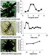
The target phytoecdysteroids were found in a scarce amount in A. bracteosa wild-type plants. We selected seven transgenic lines of pPCV002-ABC, numbered from 1 to 7. They showed a clear increase in total phytoecdysteroid content, e.g. transgenic lines 3 and 4 produced 6728 and 6759 μg g−1 dry weight of total phytoecdysteroids, respectively, which was 14.5 times higher compared to control untransformed in vitro-grown plants (Fig. 3a and b and ESI Table 7a). Semi-quantitative RT-PCR analysis revealed a relatively high expression of the rolC gene in both transgenic lines (Fig. 3h and i) compared to the other studied plant lines, as did densitometric analysis by molecular imaging of corresponding amplicon bands (Fig. 2g).
image file: c6ra00250a-f3.tif
Fig. 3 Phytoecdysteroid contents in different transgenic A. bracteosa samples and evidence of sengosterones' de novo biosynthesis in transgenic hairy root clones. (a) Biosynthesis of phytoecdysteroids in pPCV002-ABC transgenic intact plants. (b) Times increase in detected phytoecdysteroids in pPCV002-ABC transgenic intact plants. (c) Biosynthesis of phytoecdysteroids in selected transgenic hairy roots lines. (d) Times increase in detected phytoecdysteroids in transgenic hairy roots lines. (e) Times increase detected phytoecdysteroids in pPCV002-ABC transgenic intact plants and transgenic hairy roots. (f) Times increase in detected phytoecdysteroids in regenerants. (g) Chromatographs of different transgenic and untransformed plant material regarding biosynthesis of sengosterone. RP-HPLC was conducted in triplicate for each sample. Two factor Complete Randomized Design (CRD) was applied on the data to retrieve analysis of variance (ANOVA). To differentiate in different transgenic material of the same category (intact transformed plants or transgenic hairy roots), it was coupled with Latin Square Design (LSD). Data analyzed with MSTATC 2.0 version. Different letter over the bars (a–c) are statistically significant to each other at P < 0.001 and provided with ±SD.

Out of 59 hairy root lines transformed with TL-DNA of pRi, eleven lines with a high phytoecdysteroid yield were selected. They were obtained after infection with two strains of A. rhizogenes (A4 and LBA-9402) and named: A1, A2, L1, L2, L6, L8, L11, L12, L13, L32 and L42. The hairy roots showed considerably higher levels of phytoecdysteroids compared to the control roots. The highest content, 4449 μg g−1 dry weight, was obtained in A2 (Fig. 3c and ESI Table 7b), followed by 4123 μg g−1 dry weight in L1, which represented a 3.64- and 3.37-fold increase, respectively, compared to control roots (Fig. 3d). The other eight hairy root lines obtained from the LBA-9402 strain produced between 2001 and 2561 μg g−1 dry weight of phytoecdysteroids. The marked differences in phytoecdysteroid production observed in the root lines harboring the T-DNA of these agrobacteria could have arisen from the gene composition of the two different pRi, as well as from the secondary effects of the transformation on root line growth. As T-DNA can be integrated at different sites in the plant genome, the occurrence of genetic changes as a consequence of transformation might provide an explanation in some cases. The hairy root lines obtained from LBA-9402 were analyzed by semi-quantitative RT-PCR, which revealed a notably high rolC expression in the high-yielding root lines (Fig. 2k and l). The densitometric analysis by molecular imaging of corresponding gene bands confirmed a high expression of the rolC gene in L1 and L13 hairy root lines compared with the other studied lines (Fig. 2j). In a previous study, A. reptans was infected by the MAFF 03-01724 strain of A. rhizogenes and among many hairy root lines, Ar-4 (the elite hairy root clone) showed a phytoecdysteroid content 4 times higher than the control roots.19 Among various hairy root lines from another Ajuga species, A. multiflora, obtained by infection with the A. rhizogenes strain A4, one line produced 10 times more 20-HE than the wild-type plant.23

rol genes affected the biosynthesis of AJL far more strongly than that of other phytoecdysteroids

Among 20-hydroxyecdysone (20-HE), ajugalactone (AJL), sengosterone (SG), cyasterone (CYP), polypodine (PoB) and makisterone A (MKA), 20HE, MKA, CYP and AJL were successfully detected at varying levels in the transgenic plants and roots (Fig. 3e). Their production was clearly higher in intact transgenic plants than in hairy roots. While the 20-HE content in hairy roots increased up to 1.2-fold compared to control roots, in intact transgenic plants containing T-DNA of pPCV002-ABC it was 5.6-fold higher compared to untransformed plants. The phytoecdysteroid most enhanced by transformation was AJL, which increased 7.1-fold in hairy roots and 11.0-fold in intact pPCV002-ABC transgenic plants, compared to control roots and plants, respectively.

Induction effect of rol genes on phytoecdysteroid biosynthesis is stronger through pPCV002-ABC than pRi

It is evident from the results that intact transgenic plants yielded more phytoecdysteroids than transgenic hairy roots per g of dry weight. However, these two systems are not comparable, since it is well known that the expression of secondary metabolism depends on several factors, including the part of the plant, the development stage of the considered tissue and culture conditions. Nevertheless, based on these results, we can speculate that transformation through T-DNA of pPCV002-ABC can result in a higher yield of phytoecdysteroids than that of pRi. In a previous study, we found higher amounts of phytoecdysteroids in aerial parts, especially leaves and flowers.16 Moreover, Adler and Grebenok report that during spinach ontogeny 20-HE is transported to the apical regions, that annuals concentrate ecdysteroids in apical regions of the plant, and perennials recycle phytoecdysteroids between their deciduous (leaves) and perennial organs (roots).36 Other studies confirm that phytoecdysteroids are biosynthesized in roots of herbaceous perennials37 and that phytoecdysteroids accumulate in aerial organs of plants e.g., flowers, leaves, stems, and fruit.38 Based on this evidence, we assumed that phytoecdysteroids are synthesized in hairy roots of A. bracteosa and transported to the aerial parts. To explore this hypothesis, the regenerants (intact plants) from somatic embryogenesis of hairy root cells were cultured and analyzed for their phytoecdysteroid content. The results showed that the phytoecdysteroid level in hairy root lines increased up to 1.64-fold compared to untransformed roots, which was further enhanced up to 5.3-fold in their corresponding regenerants. Compared to untransformed roots, phytoecdysteroid levels in regenerants (whole plant) were higher (up to 5.3-fold) than in the corresponding mother hairy root lines (up to 1.64-fold) (Fig. 3f). This differential pattern of phytoecdysteroid biosynthesis suggests the provision of a possible sink (aerial parts, especially leaves) in regenerants. A study on spinach revealed that 20-HE is transported to the apical parts of the plant during growth.36 Our results and previous studies on A. bracteosa suggest that phytoecdysteroids are synthesized in roots and accumulate in the aerial parts. Although the higher phytoecdysteroid content in regenerants than in the corresponding mother hairy root clones remains unaccounted in the current literature, a possible explanation lies in reports of insect ecdysteroid biosynthesis being suppressed by negative-feedback inhibition.39,40 The same might be assumed for plants, leading to the speculation that phytoecdysteroids are synthesized in roots up to a certain amount, after which it is curtailed if no suitable sink (leaves) is available. Previous studies suggest that in hydroponically grown plants, wound-induced accumulation of 20-HE in roots may confer enhanced resistance against subterranean herbivorous insects41 and this could be the result of its de novo biosynthesis in roots.42 As aerial parts of perennial plants are more prone to insect attack, their phytoecdysteroid content needs to be higher to confer resistance.

Hairy roots support de novo biosynthesis of SG

SG was not detected in untransformed plants and roots, nor even in transgenic intact plants of pPCV002-ABC (especially transgenic lines 3 and 4), but it was detected in some transgenic hairy root lines (with a high phytoecdysteroid yield) (Fig. 3g). This does not mean that all the phytoecdysteroids are synthesized in roots, as clarified already by radiolabeled carbon feed experiments.43 We assumed that SG content was below the detection limit in untransformed material. At the same time, its absence in pPCV002-ABC transgenic lines 3 and 4, and its presence in only some of the transgenic hairy roots and regenerants suggests it might be de novo biosynthesized only in roots (Fig. 3g). This hypothesis is further supported by a previous study where tissue-cultured roots of A. reptans produced phytoecdysteroids independently of shoots. However, phytoecdysteroids were not detected in the shoots cultured in the absence of roots.44

Conclusions

Although the precise functions of the rol gene products are not totally known, the genetic changes in the Ajuga bracteosa rol ABC plants and the established hairy root lines apparently not only modified cell differentiation in favor of root formation, but also resulted in normal stimulation of root-specific secondary metabolism. In both cases, this stimulation was positively correlated with the level of expression of the rol C gene, as in both systems the highest expression corresponded to the highest ecdysteroid contents. It is also worth emphasizing that in the absence of effective programs for the cultivation of medicinal plants producing bioactive secondary compounds, most are still collected from the wild.45 This over-collection, together with the disadvantages of traditional cultivation, is leading to a rapid depletion of valuable raw material. Consequently, there is an urgent need to find alternatives to the whole plant in order to meet the market demand for important bioactive products. Biotechnological production systems, such as hairy root cultures, constitute a sustainable and environmentally friendly alternative for the production of secondary compounds with pharmacological interest since they are free of contaminants, unhampered by geographical or political problems, and also guarantee gene containment.

In this work, the establishment of transgenic plants overexpressing rol genes proved to be an effective strategy for increasing secondary metabolite production, since their ecdysteroid levels were higher than in the established hairy root lines. Also of note is the straightforward methodology developed for obtaining and growing Ajuga bracteosa hairy roots with a greatly enhanced biomass and ecdysteroid production. The ecdysteroid levels achieved here may be further increased by optimizing the culture conditions and adding elicitors to the medium.

Competing interest

The authors declare no competing financial interests.

Acknowledgements

GV3101 harboring pPCV002-ABC was kindly provided by Yury Shkryl, Russia. Six standard ecdysteroids were kindly provided by Prof. Josep Coll-Toledano, Department of Biological Chemistry and Molecular Modeling, Spanish National Research Council (SNRC), Barcelona, Spain. We thank the chromatography team at Parc Científic de Barcelona, Spain for RP-HPLC experiments. This project was supported by The Higher Education Commission of Pakistan. Part of this work was financially supported by the Spanish MEC (BIO2014-51861-R) and the Generalitat de Catalunya (2014SGR215).

References

  1. L. Dinan, Phytochemistry, 2001, 57, 325–339 CrossRef CAS PubMed.
  2. L. Dinan, T. Savchenko and P. Whiting, Biochem. Syst. Ecol., 2002, 30, 171–182 CrossRef CAS.
  3. K. Slama, K. Koudela, J. Tenora and A. Maťhová, Experientia, 1996, 52, 702–706 CrossRef CAS PubMed.
  4. D. B. Purser and S. K. Baker, Google Patents, 5922702, 1999.
  5. T. Yoshida, T. Otaka, M. t. Uchiyama and S. Ogawa, Biochem. Pharmacol., 1971, 20, 3263–3268 CrossRef CAS PubMed.
  6. T. Otaka, M. Uchiyama, T. Takemoto and H. Hikino, Chem. Pharm. Bull., 1968, 16, 2426–2429 CrossRef CAS PubMed.
  7. W. Burdette, Science, 1962, 139, 987 Search PubMed.
  8. C. Tsitsimpikou, G. D. Tsamis, P. A. Siskos, M. H. E. Spyridaki and C. G. Georgakopoulos, Rapid Commun. Mass Spectrom., 2001, 15, 1796–1801 CrossRef CAS PubMed.
  9. M. Báthori and Z. Pongrácz, Curr. Med. Chem., 2005, 12, 153–172 CrossRef.
  10. V. Syrov, Nauchnye Doki. Vyss. Shkoly Biol. Nauki., 1983, 11, 16–20 Search PubMed.
  11. R. Lafont and L. Dinan, J. Insect Sci., 2003, 3, 7 CrossRef CAS PubMed.
  12. K. Sláma and R. Lafont, Eur. J. Entomol., 1995, 92, 355 Search PubMed.
  13. I. R. Soriano, I. T. Riley, M. J. Potter and W. S. Bowers, J. Chem. Ecol., 2004, 30, 1885–1899 CrossRef CAS PubMed.
  14. R. Aly, U. Ravid, J. Abu-Nassar, I. Botnick, G. Lebedev, S. Gal, H. Ziadna, G. Achdari, E. Smirov and A. Meir, Pest Manage. Sci., 2011, 67, 1493–1498 CrossRef CAS PubMed.
  15. G. Fekete, L. A. Polgár, M. Báthori, J. Coll and B. Darvas, Pest Manage. Sci., 2004, 60, 1099–1104 CrossRef CAS PubMed.
  16. W. K. Kayani, R. Rani, I. U. Haq and B. Mirza, Steroids, 2014, 87, 2–20 CrossRef PubMed.
  17. V. P. Bulgakov, Biotechnol. Adv., 2008, 26, 318–324 CrossRef CAS PubMed.
  18. A. Spena, T. Schmülling, C. Koncz and J. Schell, EMBO J., 1987, 6, 3891 CAS.
  19. T. Matsumoto and T. Nobukazu, Agric. Biol. Chem., 1991, 55, 1019–1025 CrossRef CAS.
  20. N. Tanaka and T. Matsumoto, Plant Tissue Cult. Lett., 1993, 10, 78–83 CrossRef CAS.
  21. N. Tanaka and T. Matsumoto, Plant Cell Rep., 1993, 13, 87–90 CrossRef CAS PubMed.
  22. N. Uozumi, Y. Ohtake, Y. Nakashimada, Y. Morikawa, N. Tanaka and T. Kobayashi, J. Ferment. Bioeng., 1996, 81, 374–378 CrossRef CAS.
  23. O. T. Kim, M. Manickavasagm, Y. J. Kim, M. R. Jin, K. S. Kim, N. S. Seong and B. Hwang, J. Plant Biol., 2005, 48, 258–262 CrossRef CAS.
  24. D. M. Cheng, G. G. Yousef, M. H. Grace, R. B. Rogers, J. Gorelick-Feldman, I. Raskin and M. A. Lila, Plant Cell, Tissue Organ Cult., 2008, 93, 73–83 CrossRef CAS.
  25. L. Dinan, T. Savchenko and P. Whiting, CMLS, Cell. Mol. Life Sci., 2001, 58, 1121–1132 CrossRef CAS PubMed.
  26. M. I. Georgiev, J. Ludwig-Müller, K. Alipieva and A. Lippert, Plant Cell Rep., 2011, 30, 859–866 CrossRef CAS PubMed.
  27. J. J. Doyle and J. L. Doyle, Focus, 1990, 12, 13–15 Search PubMed.
  28. G. R. Taylor, in PCR. A practical approach, ed. M. J. McPherson, P. Quirka and G. R. Taylor, IRL Press, Oxford, UK, 1991, pp. 1–14 Search PubMed.
  29. A. Castro, J. Coll, Y. A. Tandrón, A. K. Pant and C. S. Mathela, J. Nat. Prod., 2008, 71, 1294–1296 CrossRef CAS PubMed.
  30. J. J. Wu, K. W. Cheng, H. Wang, W. C. Ye, E. T. Li and M. Wang, Phytochem. Anal., 2009, 20, 58–63 CrossRef CAS PubMed.
  31. E. Casanova, M. I. Trillas, L. Moysset and A. Vainstein, Biotechnol. Adv., 2005, 23, 3–39 CrossRef CAS PubMed.
  32. D. Tepfer, Cell, 1984, 37, 959–967 CrossRef CAS PubMed.
  33. A. Zuker, T. Tzfira, G. Scovel, M. Ovadis, E. Shklarman, H. Itzhaki and A. Vainstein, J. Am. Soc. Hortic. Sci., 2001, 126, 13–18 CAS.
  34. R. U. Schenk and A. Hildebrandt, Can. J. Bot., 1972, 50, 199–204 CrossRef CAS.
  35. N. Uozumi, Y. Ohtake, Y. Nakashimada, Y. Morikawa, N. Tanaka and T. Kobayashi, J. Ferment. Bioeng., 1996, 81, 374–378 CrossRef CAS.
  36. J. H. Adler and R. J. Grebenok, Lipids, 1995, 30, 257–262 CrossRef CAS PubMed.
  37. L. Zibareva, Rastit. Resur., 1997, 33, 89–92 CAS.
  38. N. S. Ramazanov, Chem. Nat. Compd., 2005, 41, 361–369 CrossRef CAS.
  39. R. P. Bodnaryk, Arch. Insect Biochem. Physiol., 1986, 3, 53–60 CrossRef CAS.
  40. P. Beydon and R. Lafont, J. Insect Physiol., 1983, 29, 529–533 CrossRef CAS.
  41. E. A. Schmelz, R. J. Grebenok, D. W. Galbraith and W. S. Bowers, J. Chem. Ecol., 1998, 24, 339–360 CrossRef CAS.
  42. E. A. Schmelz, R. J. Grebenok, D. W. Galbraith and W. S. Bowers, J. Chem. Ecol., 1999, 25, 1739–1757 CrossRef CAS.
  43. R. J. Grebenok and J. H. Adler, Phytochemistry, 1991, 30, 2905–2910 CrossRef CAS.
  44. J. Tomás, F. Camps, J. Coll, E. Melé and J. Messeguer, Phytochemistry, 1993, 32, 317–324 CrossRef.
  45. M. Hamayun, S. Afzal and M. A. Khan, Afr. J. Tradit., Complementary Altern. Med., 2006, 3, 57–73 Search PubMed.

Footnote

Electronic supplementary information (ESI) available. See DOI: 10.1039/c6ra00250a

This journal is © The Royal Society of Chemistry 2016
Click here to see how this site uses Cookies. View our privacy policy here.