Conformational perturbation, hydrophobic interactions and oligomeric association are responsible for the enhanced chaperone function of Mycobacterium leprae HSP18 under pre-thermal condition

Sandip Kumar Nandi, Ayon Chakraborty, Alok Kumar Panda and Ashis Biswas*
School of Basic Sciences, Indian Institute of Technology Bhubaneswar, Bhubaneswar-751 013, India. E-mail: abiswas@iitbbs.ac.in; Fax: +91-674-2306202; Tel: +91-674-2576051

Received 4th January 2016 , Accepted 19th June 2016

First published on 20th June 2016


Abstract

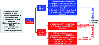
Mycobacterium leprae HSP18 is a small heat shock protein that helps in growth and survival of Mycobacterium leprae pathogen in host species. Recently, we have shown that its chaperone function is enhanced upon heating from 31 to 43 °C which is accompanied by rapid rearrangement of its subunits. We also demonstrated a decrease in its chaperone function when its oligomeric assembly dissociates. However, effect of pre-heating on the structure–function of HSP18 yet remains unexplored. In the present study, we demonstrate that HSP18 undergoes oligomeric association upon pre-heating at 60 °C or above. Surface hydrophobicity and subunit exchange kinetics are also enhanced under similar conditions, which altogether enhances its chaperone function. This study also reveals that perturbation in the secondary structure of HSP18 is completely reversible when heated even at 70 °C. However, alterations in its surface hydrophobicity and quaternary structure do not recover upon pre-heating at 60 °C and above. Interestingly, when pre-heated below 60 °C, conformational (except tertiary conformation) perturbations in HSP18 are completely reversible. Also, its chaperone function remains unaltered when pre-heated below 60 °C. Thus, conformational fluctuations in quaternary structure and subunit exchange dynamics may be the two most important mechanisms by which HSP18 exhibits chaperone function when exposed to such non-permissible thermal stress. Our data reveals that HSP18 is a very robust protein and can recover its native structural integrity as and when the stress disappears. Possibly, this is an important antigen which influences the survival of Mycobacterium leprae pathogen when it encounters thermal stress in an infected host.


1. Introduction

A widespread interest lies in understanding how an organism adopts to any changes in its cellular homeostasis. Several attempts are being made to understand the relevant processes from basic molecular biology approaches to therapeutic applications. Over the years, researchers have revealed that different species have developed several ways to deal with stress. For example, mammalian species adapt to stress at the cellular level by temporary modification in gene expression to acclimatize to environmental changes1 while plant species maintain their cellular homeostasis by opening and closing of guard cells.2 In addition, for mycobacterial species, mycolic acid, an important component of the cell wall, plays an important role in the architecture, impermeability and interaction with environmental stresses.3,4 In addition, all these species have developed a common evolutionary response system against heat stress, in the form of highly conserved stress proteins known as heat shock proteins (HSPs).

HSPs are ubiquitously expressed in all organisms starting from plant, bacteria, yeast though to humans. The term “HSP” is a misnomer, but yet remains an unanticipated discovery of Ritossa who showed that expression of chromosomal puffs of salivary gland cells in Drosophila are induced by heat shock.5 The heat shock response is triggered by various stresses including heat, ethanol, reactive oxygen species and other toxic substances.6 HSPs are mostly known for their exhibition of molecular chaperone function.7 By virtue of this property, both in vivo and in vitro, HSPs can prevent aggregation of various stressed client proteins and also can assist the refolding of different denatured enzymes.6,8 Based on their molecular weights, they are broadly categorized into two sub-classes: large heat shock proteins (such as HSP100, HSP90, HSP70, HSP60, etc.) and small heat shock proteins (sHSPs) (such as, HSP27, HSP20, HSP18, HSP16.9, etc.). Among the large heat shock protein family, HSP60 and HSP70 are the best characterized molecular chaperones. DnaK, a member of the HSP70 family, executes chaperone function in the presence of co-chaperones (DnaJ and GrpE) and ATP.9,10 Similarly, GroEL (a member of the HSP60 family) prevents the aggregation and facilitate folding of unfolded proteins both in vitro and in vivo.10 HSP90 and HSP100 also exhibit molecular chaperone function.11

Another class of ATP independent molecular chaperones whose monomeric molecular weight ranges between 10 and 43 kDa are classified into two categories: (a) small heat shock proteins (sHSPs) containing a conserved “α-crystallin domain” such as αA-crystallin, αB-crystallin, HSP14, HSP16.3, HSP16.5, HSP16.9, HSP20, HSP27 etc.12,13 and (b) other small chaperones devoid of “α-crystallin domain” such as bacterial skp, bacterial spy, HSP33, mitochondrial chaperone, HdeA, HdeB, Cpn10, Cpn21 etc.14–19 sHSPs often exist as an oligomeric assembly with 9–40 subunits.12 They possess a highly conserved “α-crystallin domain” at the centre, flanked by a C-terminal tail and preceded by a highly variable N-terminal region.13 They bind client proteins under chemical and thermal stresses and protect them from misfolding and aggregation.20 Generally, sHSPs can also refold other denatured proteins in an ATP-independent manner and are also known as molecular chaperones.21,22 In vivo, sHSPs encounter several stresses such as thermal stress, redox stress, etc.23 Among these stresses, the impact of thermal stress on the structure and function of different sHSPs has been studied extensively.24–26 Several reports demonstrated that both structure and chaperone function of several sHSPs are modulated upon heating.24,27,28 Apart from direct heating, investigators also studied the structure–function relationship of various sHSPs when pre-exposed to heat i.e. under pre-heating conditions.27,29

Pre-exposure to heat (pre-heating) is one of the most important stresses that all species encounter in the environment. The chaperone function of several molecular chaperones is found to be enhanced upon pre-heat treatment. For example, tubulin, a ubiquitous protein of the eukaryotic cytoskeleton, was found to inhibit DTT induced aggregation of insulin ∼50% more when pre-incubated at 50 °C for 10 min in comparison to the samples which were not pre-incubated.30 In plants, pre-heat treatment of mitochondrial sHSP at 48 °C resulted in enhanced protection of NADH:ubiquinone oxidoreductase (Complex I) of the electron transport chain.31 Lurie et al. reported that pre-heat treatment of mature green tomatoes at 38 °C, protects them from chilling injury.32 Later, Sabehat et al. showed that expression HSP21-encoding tom111 gene in tomatoes under pre-heating conditions is correlated with protection against chilling injury.33 In addition, HSP16.1 which contributes to stress resistance and longevity of Caenorhabditis elegans, when pre-conditioned under mildly elevated temperature, maintains Ca2+ homeostasis under heat stroke and suppresses cell death.34 These results show how pre-heat treatment of a sHSP can act as an effective general protector against multiple necrotic insults. Amongst mammalian sHSPs, α-crystallin was found to exhibit enhanced chaperone activity when pre-heated at 60 °C for 20 min.35 Several authors have also shown that chaperone activity of pre-incubated αA-crystallin was enhanced with an increase in the pre-incubation temperature.27,36 This increased chaperone function of αA-crystallin was also accompanied by perturbation in its secondary structure, enhancement in its oligomeric size and surface hydrophobicity.27,28,36 Amongst mycobacterial sHSPs, HSP16.3 which helps in the growth and survival of Mycobacterium tuberculosis pathogen in the latent phase of the disease tuberculosis, was found to exhibit enhanced chaperone activity when pre-incubated at 60 °C and above.37 Pre-heat experiments also revealed that HSP16.3 underwent a structural phase change at ∼60 °C.37 Therefore, it is conclusive from all the above evidences that pre-heat treatment of molecular chaperones has a profound influence on their function, which is often accompanied by structural alterations.

Another mycobacterial small heat shock protein, which plays a pivotal role in the pathogenesis of Mycobacterium leprae (M. leprae) is HSP18.38 This sHSP is specifically activated during intracellular growth in macrophages and plays an important role in the survival of M. leprae pathogen in infected hosts.39 It is an immunodominant antigen and contains 148 amino acid residues.40,41 Previously, we already demonstrated that M. leprae HSP18 possess a highly conserved “α-crystallin domain”.42 In the same study, we have shown that it is a molecular chaperone. By virtue of this property, HSP18 not only prevents the aggregation of different stressed prone client proteins but also helps in refolding of denatured client protein.42 Our group established that chaperone activity of M. leprae HSP18 is enhanced in the presence of ATP which is reversible in nature but is ATP hydrolysis independent.43 Recently, we also revealed that the chaperone function of HSP18 was enhanced at elevated temperatures.44 In the same work, we have also demonstrated the molecular basis behind the enhancement in chaperone function of HSP18 under thermal stress. However, the effect of pre-heat treatment on the structure and chaperone function of M. leprae HSP18 yet remains unexplored. Additionally, if pre-heat treatment has any effect on the chaperone function of HSP18, the molecular basis behind such alteration is still unknown. Therefore, in this paper, we studied the effect of pre-heat treatment on the structure and chaperone function of M. leprae HSP18 in detail. We found that the chaperone function of M. leprae HSP18 is dramatically enhanced when pre-incubated at or above 60 °C. In this study, we also report the mechanisms behind such alteration in the chaperone function of HSP18.

2. Material and methods

2.1. Materials

Bovine insulin, 4,4′-dianilino-1,1′-binaphthyl-5,5′-disulfonic acid, dipotassium salt (bis-ANS), dithiothreitol (DTT) and all molecular weight markers such as thyroglobulin (mol. wt 669[thin space (1/6-em)]000 Da), apoferritin (mol. wt 443[thin space (1/6-em)]000 Da), β-amylase (mol. wt 200[thin space (1/6-em)]000 Da), bovine serum albumin (mol. wt 66[thin space (1/6-em)]000 Da) and carbonic anhydrase (mol. wt 29[thin space (1/6-em)]000 Da) were purchased from Sigma Chemical Co. (St. Louis, MO, USA). Alexa fluor 488 and Alexa fluor 350 protein labeling kits (catalog nos A-10170 and A-10235, respectively) were purchased from Molecular Probes (Invitrogen, Carlsbad, CA, USA). Malate dehydrogenase (MDH) from porcine heart and all buffer salts were from Sisco Research Laboratories, India. All other chemicals used were of analytical grade.

2.2. Expression and purification of M. leprae HSP18

Plasmids of M. leprae HSP18 were obtained as a gift from Prof. K. Dharmalingam (Madurai Kamraj University, Madurai, India). Methods of expression and purification of M. leprae HSP18 have been described previously.42–44 Concentration of HSP18 was determined spectrophotometrically by measuring the absorbance at 278 nm using an extinction coefficient of 0.4 mg ml−1 cm−1. Concentration of this protein was also determined using Bradford assay. All the biophysical assays were performed with at least three independent protein preparations.

2.3. Pre-heat treatment of HSP18

Pre-heat treatment of HSP18 (0.5 mg ml−1 in 50/10 mM phosphate buffer, pH 7.5) was performed by incubating it at different temperatures (25, 37, 50, 57, 60, 65 and 70 °C) for 1 h and then allowing to cool at 25 °C for 1 h prior to the structural and functional assays mentioned below.

2.4. In vitro aggregation assay

The chaperone activity was determined with insulin as client protein. Assays were performed with the aid of a UV spectrophotometer (Perkin Elmer Lambda 35, Boston, MA, USA). Insulin (0.35 mg ml−1) in 50 mM phosphate buffer (pH 7.5) was incubated in the absence or presence of 0.35 mg ml−1 pre-heated HSP18 protein samples. Insulin aggregation was initiated by adding freshly prepared DTT to a final concentration of 20 mM at 25 °C and light scattering at 400 nm was monitored for 1 h in the kinetic mode. Percent (%) protection for this aggregation assay was calculated as described previously.44

2.5. Thermal inactivation assay

Thermal inactivation of 10 nM MDH in 50 mM phosphate buffer, pH 7.5 was initiated by heating the enzyme at 43 °C in the absence or presence of 0.5 mg ml−1 (30 μM) HSP18 heated at various temperatures (25, 37, 50, 57, 60, 65 and 70 °C) for 1 h and cooled at 25 °C for another 1 h. Enzyme activity of MDH was measured by taking aliquots from the assay mixture which was incubated at 43 °C for 10 min. Enzyme activity of MDH was assayed using nicotinamide adenine dinucleotide reduced (NADH) and oxaloacetic acid (OAA) as described previously.42

2.6. Bis-ANS fluorescence measurements

Fluorescence of bis-ANS was measured as described previously using a spectrofluorometer (Fluoromax 4P, Horiba Jobin Mayer, USA) equipped with a thermostated cell holder.42 Fluorescence emission spectra of bis-ANS (10 μM) bound pre-heated HSP18 protein samples (0.05 mg ml−1) were recorded in the range of 450–550 nm after incubation for 1 h at 25 °C. An excitation wavelength of 390 nm was used and the excitation and emission slits were 2.5 and 5 nm, respectively. The reported spectra were the mean of three scans.

2.7. Measurement of oligomeric mass by gel filtration chromatography

The oligomeric mass of different pre-heated HSP18 samples was determined by gel filtration chromatography in a HPLC (Dionex, Sunnyvale, CA, USA) instrument using a TSK-GEL G4000SWXL analytical gel filtration column (7.8 mm × 30 cm; 5 μm) (Tosoh Bioscience LLC, King of Prussia, PA, USA). After the column was equilibrated with 50 mM phosphate buffer (pH 7.5) at 25 °C, 20 μl of pre-heated HSP18 (0.5 mg ml−1) in the equilibrium buffer was injected into the column. The chromatogram was monitored by measuring the absorbance at 280 nm. The column was calibrated with molecular weight standards: thyroglobulin (669 kDa), apoferritin (443 kDa), β-amylase (200 kDa), BSA (66 kDa) and carbonic anhydrase (29 kDa) (Sigma Chemical Co., St. Louis, MO, USA) as described previously.44 The flow rate was maintained at 0.5 ml min−1 for all measurements.

2.8. Hydrodynamic radius (RH) measurements by dynamic light scattering (DLS)

M. leprae HSP18 (0.5 mg ml−1, in 50 mM phosphate buffer, pH 7.5) was filtered through a 0.22 μm disk membrane. Hydrodynamic radius of HSP18 oligomers (RH) was measured by DLS employing a Zetasizer Nano S (Malvern Instruments, Malvern, UK) between 25 and 70 °C as previously described44 with 1 h incubation at each temperature prior to reading. Each data is an average of 60 scans. RH was calculated by using the Stokes–Einstein equation:
 
image file: c6ra00167j-t1.tif(1)
where, kB is Boltzmann’s coefficient, T is absolute temperature, η is the viscosity of the medium and D is the translational diffusion coefficient of the particles. Similar DLS measurements were also performed with different pre-heated HSP18 protein solutions at 25 °C.

2.9. Tryptophan fluorescence measurements

The intrinsic tryptophan fluorescence spectra of HSP18 (0.05 mg ml−1), were recorded at various temperatures between 25 and 70 °C (after incubating protein samples at the respective temperatures for 1 h) using a spectrofluorometer (Fluoromax 4P, Horiba Jobin Mayer, USA) equipped with a thermostated cell holder. The excitation wavelength was set to 295 nm and the emission spectra were recorded between 310 and 400 nm. Data were collected at a 0.5 nm wavelength resolution. Similar spectra were also recorded for different pre-heated HSP18 samples at 25 °C. The reported spectra were the mean of five scans.

2.10. Circular dichroism (CD) measurements

Far- and near-UV CD spectra were measured at several temperatures between 25 and 70 °C using a spectropolarimeter (Chirascan, Applied Photophysics, Leatherhead, UK) equipped with a Peltier system prior to incubation at each of these specific temperatures for 1 h. Protein concentration used in far- and near-UV CD experiments was 0.2 mg ml−1 (in 10 mM phosphate buffer, pH 7.5) and 0.5 mg ml−1 (in 50 mM phosphate buffer, pH 7.5), respectively. Far-UV CD spectra were collected from 195–260 nm using a quartz cell with 1 mm path length, while the near-UV CD spectra were collected from 250–300 nm using a 10 mm path length cell. Similar spectra were also recorded for pre-heated HSP18 samples at 25 °C. The reported spectra in far- and near-UV CD were the mean of five scans. The curve-fitting program CONTINLL was used to analyze the secondary structural contents of HSP18 at different temperatures.

2.11. Fluorescence labeling of recombinant M. leprae HSP18 with Alexa fluor 488 and Alexa fluor 350

Fluorescence conjugation of Alexa fluor 350 and Alexa fluor 488 fluorescent probes with purified recombinant M. leprae HSP18 was done as described previously.44

2.12. Measurements of the subunit exchange rate

Fluorescence resonance energy transfer (FRET) technique was used to determine the rate of subunit exchange of HSP18, pre-heated at several temperatures (25, 57, 60 and 70 °C), as described previously.44

3. Results and discussion

Once we showed that M. leprae HSP18 exhibited molecular chaperone function,42 considerable interests grew about the functional aspects of M. leprae HSP18 and the underlying mechanism behind its chaperone function. We have already shown that S52P mutation in the “α-crystallin domain” of M. leprae HSP18 decreases its chaperone function by dissociation of its oligomeric assembly and perturbation in secondary structure.42 Maheshwari et al. also revealed through their in vivo studies, the role of M. leprae HSP18 in the survival of heterologous recombinant hosts against several environmental stresses that M. leprae encounters during its survival inside macrophages.45 In continuation with that study, we studied the alteration in chaperone activity of M. leprae HSP18 under heat stress. Through in vitro studies we clearly demonstrated that HSP18 exhibits enhanced chaperone activity when the temperature is raised from 25 to 43 °C.44 We also revealed that the enhanced chaperone activity is not due to any alteration in its static oligomeric size, but due to alteration in the dynamics of its oligomeric assembly and rapid rearrangement of its subunits.44 Therefore, it is very much evident from the above instances that conformational changes in M. leprae HSP18 could well account for its altered chaperone activity.

In the Introduction section, we have already discussed elaborately the effect of pre-heat treatment on the function of C. elegans HSP16.1, αA-crystallin and M. tuberculosis HSP16.3. All these three proteins belong to the small heat shock protein family. Multiple sequence alignment of M. leprae HSP18 with these three and other four sHSPs (αB-crystallin, HSP27, HSP16.5 and HSP16.3) showed that HSP18 shares a sequence similarity of greater than 50% (Fig. 1). The “α-crystallin domain” which is known to bind substrate proteins and help in proper exhibition of chaperone function, is well conserved in HSP18 as well as in HSP16.1, HSP16.3 and αA-crystallin (Fig. 1). In light of all these observations, we hypothesized that pre-heat treatment of M. leprae HSP18 may alter its chaperone function.


image file: c6ra00167j-f1.tif
Fig. 1 Sequence alignment of M. leprae HSP18 with seven small heat shock proteins. The amino acid sequence alignment of eight small heat shock proteins, Caenorhabditis elegans HSP16.1 (HSP16.1) (GenBank accession number P34696), Methanococcus jannaschii HSP16.5 (HSP16.5) (GenBank accession number Q57733), Triticum aestivum HSP16.9 (HSP16.9) (GenBank accession number P12810), human HSP27 (HSP27) (GenBank accession number P04792), human αA-crystallin (alpha-A) (GenBank accession number P02489), human αB-crystallin (alpha-B) (GenBank accession number P02511), Mycobacterium tuberculosis Rv2031c, HSP16.3 (HSP16.3) (GenBank accession number P9WMK1), and Mycobacterium leprae HSP18 (HSP18) (GenBank accession number P12809) was performed using CLUSTAL W (1.83) multiple sequence alignment program. All the eight small heat shock proteins constitute a conserved “α-crystallin domain” (highlighted with turquoise), a variable N-terminal region (highlighted in grey) and a flexible C-terminal region (highlighted in yellow). The symbols represent identical residues (*), conserved substitution (:) and semi-conserved substitution (.) respectively.

To accomplish the above goal, DTT induced aggregation assay of insulin was performed at 25 °C in the absence or presence of HSP18 which was pre-heated at various temperatures between 25 and 70 °C (Fig. 2A). At a ratio of 1[thin space (1/6-em)]:[thin space (1/6-em)]1 (w/w) of HSP18 to insulin, ∼34% protection against aggregation was observed (Fig. 2A, trace 2 and 2B). At the same HSP18 to insulin ratio, the chaperone function of HSP18 pre-heated at temperatures below 60 °C, was almost identical to that of untreated HSP18 (Fig. 2A, traces 3–5 and 2B). However, HSP18 when pre-heated at 60 °C or above (65 and 70 °C), showed significantly enhanced chaperone function (∼81–97%) (Fig. 2A, traces 6–8 and 2B).


image file: c6ra00167j-f2.tif
Fig. 2 Effect of pre-heat treatment on the chaperone like activity of M. leprae HSP18. (A) DTT-induced aggregation of 0.35 mg ml−1 insulin (Ins) either in the absence or presence of 0.35 mg ml−1 HSP18 pre-heated at different temperatures (25, 37, 50, 57, 60, 65, 70 °C). Aggregation was initiated by adding 20 mM DTT and scattering at 400 nm was monitored at 25 °C. Trace 1: Ins alone at 25 °C; trace 2: Ins + HSP18 pre-heated at 25 °C; trace 3: Ins + HSP18 pre-heated at 37 °C; trace 4: Ins + HSP18 pre-heated at 50 °C; trace 5: Ins + HSP18 pre-heated at 57 °C; trace 6: Ins + HSP18 pre-heated at 60 °C; trace 7: Ins + HSP18 pre-heated at 65 °C; trace 8: Ins + HSP18 pre-heated at 70 °C. (B) Percentage protection ability of M. leprae HSP18 pre-heated at several temperatures between 25 and 70 °C against insulin aggregation at 25 °C. Data are means ± the standard deviation from triplicate determinations. NS = not significant and ***p < 0.0005.

Recently, we already demonstrated that HSP18 can prevent enzymes from thermal inactivation.43,44 In order to understand whether pre-heat treatment altered its ability to prevent thermal inactivation of enzymes, we incubated 10 nM malate dehydrogenase (MDH) for 10 min at 43 °C and measured its activity. Fig. 3 shows that MDH alone retained only 39% activity. However, in the presence of untreated HSP18 (30 μM), the loss of MDH activity was reduced and it retained 61% of its enzymatic activity. Interestingly, we observed that MDH retained almost similar enzymatic activity in the presence of HSP18 (30 μM) pre-heated separately at different temperatures below 60 °C. However, when 30 μM HSP18, pre-heated at 60 °C and above was added to the assay mixture along with 10 nM MDH, the loss of MDH activity was further retarded and MDH was able to retain 73–90% of its activity (Fig. 3). Therefore, results from our in vitro aggregation and thermal inactivation assays are in partial agreement with our hypothesis, because the chaperone activity of M. leprae HSP18 remained intact when it is pre-heated below 60 °C (37, 50 and 57 °C) (Fig. 3). However, when HSP18 is pre-heated at 60 °C or above (65 and 70 °C), its chaperone function is enhanced significantly (Fig. 3). Similar observations have also been reported in other sHSPs under pre-heating conditions i.e. their chaperone function is enhanced under pre-heating conditions.27,37


image file: c6ra00167j-f3.tif
Fig. 3 Effect of pre-heating on the thermal inactivation prevention ability of M. leprae HSP18. The enzyme activity of MDH was measured in the absence or presence of 30 μM HSP18, pre-heated at several temperatures between 25 and 70 °C, after its thermal denaturation at 43 °C. Data are means ± standard deviation from triplicate determinations. NS = not significant, *p < 0.05, **p < 0.005 and ***p < 0.0005.

We have already shown that the alteration in the chaperone activity of HSP18 is often accompanied by alterations in its conformations.42–44 Surprisingly, we found that the enhancement in the chaperone function of HSP18 at elevated temperatures is accompanied by an unaltered static oligomeric structure.44 As the chaperone function of HSP18 is also enhanced during pre-heat treatment, we were interested to know the effect of pre-heat treatment on the quaternary structure of HSP18. Therefore, we estimated the hydrodynamic radius of HSP18 under heating and pre-heating conditions using dynamic light scattering. At 25 °C, HSP18 was found to have hydrodynamic radius of ∼18 nm which corresponds to a molecular mass of ∼588 kDa (Fig. 4A, Table 1). Previously, we reported a similar hydrodynamic radius of HSP18 at this particular temperature.44 No change in the hydrodynamic radius was observed when the protein was incubated at higher temperatures (37 and 50 °C) (Fig. 4A, Table 1). However, the hydrodynamic radius of HSP18 was increased to ∼21 nm (corresponding molecular mass ∼823 kDa) and ∼48 nm (corresponding molecular mass ∼5654 kDa) upon heating at 57 and 70 °C, respectively (Fig. 4A, Table 1). Therefore, it can be inferred from these observations that static oligomeric structure remains unaltered till 50 °C, and heating above this temperature caused an association leading to a higher oligomeric assembly. However, when the hydrodynamic radius were estimated for pre-heated HSP18 samples, it was observed that for HSP18 when pre-heated at temperatures below 60 °C (25, 37, 50 and 57 °C), it regained its native oligomeric assembly on cooling to 25 °C (Fig. 4B, Table 1).


image file: c6ra00167j-f4.tif
Fig. 4 Changes in the oligomeric assembly of M. leprae HSP18 under heating and pre-heating conditions. Intensity particle size distribution spectra of M. leprae HSP18 under (A) heating and (B) pre-heating conditions. Each spectrum is an average of 60 scans. For experiments under heating condition, spectra were recorded after incubating HSP18 at the respective temperatures for 1 h. For experiments under pre-heating conditions, spectra were recorded at 25 °C after incubation at respective temperatures for 1 h followed by equilibration at 25 °C. Protein concentration used was 0.5 mg ml−1 in 50 mM phosphate buffer, pH 7.5.
Table 1 Estimated oligomeric mass of M. leprae HSP18 under heating (T/°C) and pre-heating conditions (T/°C then cooled to 25 °C) derived from hydrodynamic radius
Condition T/°C RH/nm MW/kDa
Heating 25 18.17 588.1
37 18.29 597.3
50 18.39 604.9
57 21.04 828.9
60 34.80 2690.4
65 43.82 4613.3
70 47.80 5654.0
Pre-heating 25 18.17 588.1
37 18.19 589.6
50 18.20 590.4
57 18.30 598.0
60 33.10 2392.9
65 37.84 3272.8
70 43.82 4613.3


By contrast, when HSP18 was pre-heated at 60 °C or above (65 and 70 °C), its hydrodynamic radius as well as molecular mass was increased significantly (∼33–44 nm and ∼2396–4613 kDa) (Fig. 4B, Table 1) and it could not regain its native oligomeric assembly on cooling. Thus it becomes evident from these findings that M. leprae HSP18 when heated at 60 °C or above, undergoes oligomeric association, and subsequent cooling of such samples to 25 °C does not allow the protein to regain its native like oligomeric structure. Also we can conclude that the pre-heat treatment of HSP18 at 60 °C or above brings about non-recoverable alterations in its quaternary structure.

We also performed gel filtration chromatographic experiments to reinforce our pre-heating results obtained from dynamic light scattering experiments. The gel filtration chromatography was done using a TSK-GEL G4000SWXL column at 25 °C. A single peak at 9.1 ml elution volume was observed for untreated HSP18 (Fig. 5, peak 1). Oligomeric mass corresponding to the elution volume was calculated using a standard curve [log(molecular weight) versus elution volume] as mentioned previously.44 With the aid of this standard curve, the oligomeric mass of untreated HSP18 was found to be ∼563 kDa (Table 2) which was in agreement with the value reported in our earlier reports.42,44 The elution profiles of M. leprae HSP18 pre-heated at temperatures below 60 °C (25, 37, 50 and 57 °C) were almost identical to that of untreated protein with a single peak around 9.1 ml elution volume which corresponded to an oligomeric mass of ∼563 kDa (Fig. 5, peak 1, Table 2). However, HSP18 when pre-heated at 60 °C, as well as showing a normal peak at 9.1 ml, showed an additional peak at 7.5 ml elution volume (Fig. 5, peak 2) which corresponded to an oligomeric mass of ∼2386 kDa (Table 2). Further increase of pre-heating temperature to 65 and 70 °C, resulted in formation of an additional elution peak (6.95 ml) (Fig. 5, peak 3) which corresponded to an oligomeric mass of ∼3899 kDa (Table 2). Therefore, it was further confirmed from gel filtration experiments that pre-heat treatment of M. leprae HSP18 at 60 °C or above, caused an association of oligomeric assemblies. In our previous report, we have shown that the native oligomeric assembly of HSP18 remains unaltered during heating at physiological temperatures (31–43 °C) and does not contribute to the enhanced chaperone function of this protein under these conditions.44 But, here we observed that under pre-heat treatment at 60 °C or above, HSP18 exhibits enhanced chaperone function which is accompanied by association of its oligomeric assembly. In fact, the relationship between oligomeric assembly and chaperone function of HSP16.3 is entirely different under thermal and pre-thermal conditions. During heating, the larger oligomeric assembly of HSP16.3 dissociates into smaller oligomeric assemblies (nonamer to trimer) and this dissociation plays a critical role in enhancing its chaperone function under thermal conditions.46 On the contrary, the oligomeric assembly of HSP16.3 does not alter under pre-thermal conditions, but the chaperone function of this protein increases under these particular conditions.37 Therefore, we can state that the association in the oligomeric assembly of HSP18 upon pre-heat treatment at 60 °C or above may be one of the important factors for its increased chaperone function. Such a correlation between the oligomeric mass and chaperone function of other sHSPs under pre-heating condition is absent in the literature.


image file: c6ra00167j-f5.tif
Fig. 5 Estimation of oligomeric mass of M. leprae HSP18 under pre-heating conditions using size exclusion chromatography. Gel-filtration profile of pre-heated HSP18. TSK-GEL G4000SWXL column (7.8 mm × 30 cm; 5 μm) was first equilibrated with 50 mM phosphate buffer (pH 7.5) at 25 °C. Subsequently, 20 μl of several pre-heated HSP18 (0.5 mg ml−1) samples were injected into the column. The flow rate of the column was maintained at 0.5 ml min−1. The oligomeric mass of HSP18 pre-heated at various temperatures was estimated with the aid of a standard curve.
Table 2 Estimated oligomeric mass of M. leprae HSP18 under pre-heating conditions using gel filtration chromatography
Pre-heating temperature/°C Oligomeric mass of protein/kDa
Peak 1 Peak 2 Peak 3
25 563
37 563
50 563
57 563
60 563 2386
65 563 2386 3899
70 563 2386 3899


We also studied the tertiary structure of HSP18 under heating and pre-heating conditions using intrinsic tryptophan fluorescence and near-UV CD experiments. It was observed that tryptophan fluorescence intensity of M. leprae HSP18 decreased gradually upon heating at higher temperatures (37, 50, 57, 60, 65 and 70 °C) (Fig. 6A). This decrease in tryptophan fluorescence intensity indicated that the microenvironment of the tryptophan (W33) residue of M. leprae HSP18 was perturbed under heating conditions. An alteration in the emission maxima (λmax) of the intrinsic tryptophan fluorescence spectra was also observed under these conditions (Fig. 6C). The λmax of HSP18 at 25 °C was at ∼341 nm. In contrast, we found that the λmax of HSP18 exhibited a red shift (344–353 nm) at 60 °C or above (65 and 70 °C), which suggested that HSP18 is in a more unfolded state at temperature beyond 60 °C. However, we did not observe any shift in the λmax of the intrinsic tryptophan fluorescence spectra of pre-heated (37, 50 and 57 °C) HSP18 samples (Fig. 6C). Apart from this, we also observed that the nature of the tryptophan fluorescence spectra for the HSP18 pre-heated at 25 °C were different than that of HSP18 at 25 °C (Fig. 6A and B). This inference suggested that even pre-heating at lower temperatures has some effect on the tryptophan microenvironment of HSP18. Additionally, we noticed a slight decrease in the intrinsic tryptophan fluorescence intensity (∼7%), when HSP18 was pre-heated at 60 °C and above (Fig. 6B). These findings led us to infer that the tryptophan microenvironment of M. leprae HSP18 is perturbed extensively and mildly under heating and pre-heating conditions, respectively.


image file: c6ra00167j-f6.tif
Fig. 6 Intrinsic tryptophan fluorescence spectra of M. leprae HSP18 under heating and pre-heating conditions. (A) Tryptophan fluorescence spectra of HSP18 (0.05 mg ml−1) were recorded at various temperatures prior to incubation for 1 h. (B) Tryptophan fluorescence spectra of pre-heated HSP18 (0.05 mg ml−1) protein samples at 25 °C. (C) Tryptophan fluorescence wavelength maxima (λmax) of HSP18 during heating and pre-heating conditions as a function of temperature. Excitation wavelength was 295 nm. Excitation and emission slit widths were 5 nm each. Emission spectra were recorded from 310–400 nm at 0.5 nm wavelength resolution.

To understand the effect of pre-heat treatment on the tertiary structure of HSP18, we also studied the near-UV CD spectra of different HSP18 samples. Fig. 7 shows the near-UV CD spectra of M. leprae HSP18 under heating and pre-heating conditions. The signal between 250 and 270 nm region arises for the five phenylalanine residues in HSP18 (Fig. 7A and B). The signal beyond 270 nm corresponds to the single tyrosine and tryptophan residue in HSP18 (Fig. 7A and B). Negligible alterations both in the peak position and intensity were observed for the HSP18 samples heated below 60 °C (Fig. 7A). However, when the protein was heated at 60 °C or above, a gradual decrease in the ellipticity value at regions between 255–270, 275–282 and 282–290 nm was observed which indicated the perturbation in the phenylalanine, tyrosine and tryptophan moiety microenvironments of HSP18 (Fig. 7A). Interestingly, we noticed that the nature of the near-UV CD spectrum of HSP18 pre-heated at 25 °C was different than that of the protein at 25 °C (Fig. 7A and B). This is fully in agreement with our tryptophan fluorescence experiments results which showed that pre-heating of HSP18 even at lower temperatures somewhat perturbs the packing of aromatic residues (tryptophan) of HSP18. Moreover, the alterations in the peak positions and ellipticity values were also minimal when HSP18 was pre-heated below 60 °C (Fig. 7B). On the other hand, a mild decrease in the ellipticity value was observed in the above mentioned regions of all the three amino acid residues when the protein was pre-heated at 60 °C or above (Fig. 7B). Therefore, from the above results it was inferred that the microenvironment of aromatic amino acid residues in HSP18 is perturbed grossly upon heating, while pre-heating of HSP18 at any temperature only mildly perturbs the microenvironment of aromatic amino acid residues. As the tertiary structure of HSP18 is perturbed under pre-heating at lower temperatures, we can conclude that the enhancement in the chaperone function of HSP18 upon pre-heating at 60 °C or above is not due to the alterations in its tertiary structure.


image file: c6ra00167j-f7.tif
Fig. 7 Perturbation in the tertiary structure of M. leprae HSP18 under various thermal conditions. (A) Near-UV CD spectra of HSP18 recorded at 25, 37, 50, 57, 60, 65 and 70 °C, respectively, prior to incubation at the respective temperatures for 1 h. (B) Near-UV CD spectra of pre-heated HSP18 protein samples at 25 °C. The spectra were measured with a 0.5 mg ml−1 protein solution in 50 mM phosphate buffer (pH 7.5). The reported spectra were the average of five scans.

Interestingly, the effect of pre-heat treatment on the secondary structure of different sHSPs is not similar. Sometimes, pre-heat treatment affected the secondary structure and in some cases, it had no effect. For example, Burgio et al. revealed that pre-heating of α-crystallin beyond 50 °C perturbed its secondary structure.36 On the other hand, the secondary structure of M. tuberculosis HSP16.3 remained unaltered even when it is pre-heated at 100 °C.37 Notwithstanding the controversy, we made an attempt to understand whether the subtle changes in the tertiary structure of HSP18 is accompanied by any perturbation in its secondary structure under different heating and pre-heating conditions by using far-UV CD experiments (Fig. 8). Fig. 8A shows that HSP18 underwent marked changes in its secondary structure when heated at 60 °C or above. Quantitative analysis of far-UV CD data using CONTINLL software revealed that HSP18 is a major β-sheet protein (Table 3). When heated below 60 °C, no alterations in the secondary structural elements of M. leprae HSP18 were observed (Table 3). But, when HSP18 was heated at 60 °C or above, its β-sheet content was found to decrease gradually with a concomitant increase in its α-helical content and random coil structure (Table 3). However, the far-UV CD spectra of pre-heated samples were almost identical with the far-UV CD spectra of untreated M. leprae HSP18. All the secondary structural elements were found to be almost identical (Fig. 8B, Table 3). Altogether, these data suggest that M. leprae HSP18 can regain its secondary structural elements even when pre-heated at 70 °C and possibly has no role in its enhanced chaperone function.


image file: c6ra00167j-f8.tif
Fig. 8 Secondary structure of M. leprae HSP18 under various heating and pre-heating conditions. Far-UV CD spectra of HSP18 during (A) heating; (B) pre-heating conditions. For experiments under heating condition, spectra were recorded after incubating HSP18 at the respective temperatures for 1 h. For experiments under pre-heating condition, spectra were recorded at 25 °C after incubation at respective temperatures for 1 h followed by equilibration at 25 °C. Spectra were recorded for 0.2 mg ml−1 protein in 10 mM phosphate buffer, pH 7.5 using a 1 mm path length cell. The data interval was 0.5 nm.
Table 3 Percentage level of different secondary structural elements in HSP18 incubated under various heating and pre-heating conditions using CONTINLL software
Condition T/°C α-Helix (%) β-Sheet (%) β-Turn (%) Random coil (%)
Heating 25 4.1 43.1 21.8 30.9
37 4.4 42.9 21.1 31.6
50 4.9 42.8 21.3 31.0
57 5.1 42.5 20.9 31.5
60 10.1 35.4 18.6 35.9
65 13.5 30.6 15.3 40.6
70 17.8 23.4 12.6 46.2
Pre-heating 25 4.1 43.1 21.8 30.9
37 4.3 43.0 21.7 31.0
50 4.5 42.8 21.6 31.1
57 4.6 42.7 21.4 31.3
60 4.8 42.7 21.4 31.1
65 4.9 42.5 21.2 31.4
70 5.1 42.4 21.1 31.4


So as to understand the additional factors behind the enhanced chaperone activity of M. leprae HSP18 under pre-heating conditions and whether the changes in tertiary and quaternary structure of this protein under these conditions are associated with any changes in the available hydrophobic patches at the surface, surface hydrophobicity of pre-heated M. leprae HSP18 was measured using bis-ANS, a hydrophobic probe. This hydrophobic probe exhibits very weak fluorescence in aqueous medium, but when it binds to the surface of protein, its fluorescence yield increases.47 Several studies have revealed that hydrophobic interactions play a key role in the chaperone function of different sHSPs.42,48–50 Several studies demonstrated a direct correlation between the chaperone function and surface-exposed hydrophobicity of different sHSPs.42,48–50 Fig. 9 shows the fluorescence intensity of bis-ANS bound to HSP18 which was pre-heated at different temperatures between 25 and 70 °C. We observed that the fluorescence intensity of bis-ANS bound to HSP18 pre-heated at temperatures below 60 °C, were almost identical to that of the untreated HSP18 (Fig. 9). On contrary, the bis-ANS bound HSP18 fluorescence intensity increased by ∼8–19% when HSP18 was pre-heated at 60 °C and above (Fig. 9). Overall, these data suggest that surface hydrophobicity of M. leprae HSP18 increases only when the protein is pre-heated at 60 °C and above, which may be responsible for the enhancement in the chaperone function of HSP18 under these conditions. Therefore our data suggest a strong correlation between the chaperone activity and surface hydrophobicity of HSP18 when pre-heated at 60 °C and above.


image file: c6ra00167j-f9.tif
Fig. 9 Effect of pre-heat treatment on the surface hydrophobicity of HSP18. The surface hydrophobicity of HSP18 pre-heated at several temperatures between 25 and 70 °C was measured using a hydrophobic probe, bis-ANS, at 25 °C. The protein concentration was 0.05 mg ml−1 and the bis-ANS concentration was 10 μM. The fluorescence spectra of bis-ANS bound to HSP18 pre-heated at different temperatures was recorded in the range 450–550 nm at 25 °C. The excitation wavelength was 390 nm. Excitation and emission slit width were 2.5 and 5 nm, respectively.

Previously, we already demonstrated that dynamics of the oligomeric structure plays an important role in the chaperone function of HSP18.44 We also showed that the kinetics of subunit exchange of HSP18 subunits increases at higher temperature and may be a critical factor for the enhanced chaperone function of HSP18 at elevated temperatures.44 Here, we again found that the chaperone function of HSP18 is enhanced substantially when the protein was pre-heated at 60 °C and above (Fig. 2 and 3). We hypothesize that the kinetics of subunit exchange of HSP18 subunits may be enhanced during pre-heat treatment. To test our hypothesis or to understand the effect of pre-heat treatment on the subunit exchange rate of HSP18, the rate constant (k) of subunit exchange was determined under different pre-heating temperatures (Fig. 10). The subunit exchange rate constant for untreated HSP18 at 37 °C was 0.018 min−1 (Fig. 10, Table 4) which is in good agreement with the data mentioned previously.44 It was observed that the rate of subunit exchange of HSP18 subunits remained unaltered when it was pre-heated at temperatures below 60 °C (Fig. 10, Table 4). However, the magnitude of subunit exchange rate constant was increased slightly when HSP18 was pre-heated at 60 °C and above (70 °C) (Fig. 10, Table 4). Therefore, these findings revealed that dynamics of oligomeric dissociation/re-association becomes faster during the pre-heating of HSP18 at 60 °C or above and is perhaps one of the important factors for the enhanced chaperone function of HSP18 under these conditions. Apart from this factor, the molecular basis behind the enhancement in the chaperone function of HSP18 under heating and pre-heating conditions is mostly different. Since the subunit exchange phenomenon influenced the chaperone function of HSP18, both under heating and pre-heating conditions, we can conclude that dynamics of oligomeric assembly may be extremely important for exhibiting proper chaperone function of this small heat shock protein.


image file: c6ra00167j-f10.tif
Fig. 10 Subunit exchange rate of M. leprae HSP18 under pre-heating conditions. Subunit exchange between Alexa fluor 350 labeled and Alexa fluor 488 labeled HSP18 (5 μM each), pre-incubated at several temperatures (25, 57, 60 and 70 °C) was monitored at 25 °C. The stock concentration of both labeled proteins was 1 mg ml−1. The fluorescence spectrum (400–600 nm) was recorded at various time intervals from samples at the respective temperatures. The excitation wavelength was 346 nm. Symbols represent the experimental data points at 440 nm. In order to calculate the subunit exchange rate constant (k) at different temperatures, data were fit according to an equation as described previously44 and the solid lines in this panel represent the best fit of the data. The values of subunit exchange rate constant (k) of different pre-heated protein samples are listed in Table 4.
Table 4 Subunit exchange rate constant of different pre-heated M. leprae HSP18 samples
Pre-heated temperature/°C Subunit exchange rate constant (k)/min−1
25 0.018 ± 0.0007
57 0.0176 ± 0.0009
60 0.0245 ± 0.001
70 0.028 ± 0.0012


4. Conclusions

In summary, the data presented in this paper has clearly revealed that pre-heat treatment of M. leprae HSP18 at 60 °C and above increased its chaperone function which is associated with perturbation in its oligomeric assembly and enhanced surface hydrophobicity. Apart from these structural perturbations, we also found that the kinetics of subunit exchange of HSP18 is enhanced when pre-heated at 60 °C or above. Data presented here further show that the alterations in secondary structure of HSP18 are completely reversible even when it is pre-heated at 70 °C. On the other hand, mild perturbation in the tertiary structure of HSP18 has been observed upon pre-heating at any temperature between 25 and 70 °C. However, the quaternary structure of HSP18 does not regain its native quaternary structure during pre-heating at 60 °C and above. Interestingly, all conformational (except tertiary conformation) perturbations are completely reversible below 60 °C. Subsequently, the chaperone function of M. leprae HSP18 also remains unaltered upon pre-heating below 60 °C. A possible model figure (Fig. 11) which reflects the schematic mechanism for the chaperone function of M. leprae HSP18 under pre-heating conditions is shown. In light of these observations, it can be proposed that M. leprae HSP18 is a very robust protein and it may tolerate various external stresses including heat, when they appear in macrophages. The fact which supports such a proposition is the long living nature of this protein. In fact, a comparison between the genome sequence of M. leprae and M. tuberculosis clearly revealed that HSP18 gene has been retained by M. leprae despite of the fact that this pathogen experiences massive gene decay.51 Overall, this study demonstrates that M. leprae HSP18 modulates its chaperone activity under pre-heating conditions by largely modulating its conformations.
image file: c6ra00167j-f11.tif
Fig. 11 A proposed model which reflects the schematic mechanism for the chaperone function of M. leprae HSP18 under pre-heating conditions.

Acknowledgements

We thank Prof. K. P. Das and Dr Victor Banerjee of Bose Institute, Kolkata, India for their useful suggestions during DLS experiments. We also thank Mr Dipak C. Konar, Bose Institute, Kolkata, India for helping to collect DLS data. SKN acknowledges Indian Institute of Technology Bhubaneswar and AC acknowledges UGC, New Delhi, India for providing fellowship. AKP acknowledges ICMR, India (no. 45/25/2012-Bio/BMS) for providing fellowship. This work was supported from the Council of Scientific and Industrial Research, India grant 37(1535)/12/EMR-II (AB).

References

  1. E. de Nadal, G. Ammerer and F. Posas, Nat. Rev. Genet., 2011, 12, 833–845 CrossRef CAS PubMed.
  2. J. I. Schroeder, J. M. Kwak and G. J. Allen, Nature, 2001, 410, 327–330 CrossRef CAS PubMed.
  3. M. Daffe and P. Draper, Adv. Microb. Physiol., 1998, 39, 131–203 CrossRef CAS PubMed.
  4. E. Lederer, A. Adam, R. Ciorbaru, J. F. Petit and J. Wietzerbin, Mol. Cell. Biochem., 1975, 7, 87–104 CrossRef CAS PubMed.
  5. F. Ritossa, Experientia, 1962, 18, 571–573 CrossRef CAS.
  6. K. Richter, M. Haslbeck and J. Buchner, Mol. Cell, 2010, 40, 253–266 CrossRef CAS PubMed.
  7. L. E. Hightower, Cell, 1991, 66, 191–197 CrossRef CAS PubMed.
  8. M. P. Mayer, Mol. Cell, 2010, 39, 321–331 CrossRef CAS PubMed.
  9. A. Szabo, T. Langer, H. Schroder, J. Flanagan, B. Bukau and F. U. Hartl, Proc. Natl. Acad. Sci. U. S. A., 1994, 91, 10345–10349 CrossRef CAS.
  10. B. Bukau and A. L. Horwich, Cell, 1998, 92, 351–366 CrossRef CAS PubMed.
  11. M. Yonehara, Y. Minami, Y. Kawata, J. Nagai and I. Yahara, J. Biol. Chem., 1996, 271, 2641–2645 CrossRef CAS PubMed.
  12. K. K. Sharma and P. Santhoshkumar, Biochim. Biophys. Acta, Gen. Subj., 2009, 1790, 1095–1108 CrossRef CAS PubMed.
  13. E. Basha, H. O’Neill and E. Vierling, Trends Biochem. Sci., 2012, 37, 106–117 CrossRef CAS PubMed.
  14. M. Schlapschy, M. K. Dommel, K. Hadian, M. Fogarasi, I. P. Korndorfer and A. Skerra, Biol. Chem., 2004, 385, 137–143 CrossRef CAS PubMed.
  15. F. Stull, P. Koldewey, J. R. Humes, S. E. Radford and J. C. Bardwell, Nat. Struct. Mol. Biol., 2015, 23, 53–58 Search PubMed.
  16. J. Graumann, H. Lilie, X. Tang, K. A. Tucker, J. H. Hoffmann, J. Vijayalakshmi, M. Saper, J. C. Bardwell and U. Jakob, Structure, 2001, 9, 377–387 CrossRef CAS PubMed.
  17. R. Kern, A. Malki, J. Abdallah, J. Tagourti and G. Richarme, J. Bacteriol., 2007, 189, 603–610 CrossRef CAS PubMed.
  18. J. Martin, J. Bioenerg. Biomembr., 1997, 29, 35–43 CrossRef CAS PubMed.
  19. J. E. Hill and S. M. Hemmingsen, Cell Stress Chaperones, 2001, 6, 190–200 CrossRef CAS PubMed.
  20. F. U. Hartl and M. Hayer-Hartl, Science, 2002, 295, 1852–1858 CrossRef CAS PubMed.
  21. F. U. Hartl, Nature, 1996, 381, 571–579 CrossRef CAS PubMed.
  22. M. J. Gething and J. Sambrook, Nature, 1992, 355, 33–45 CrossRef CAS PubMed.
  23. H. Nakamoto and L. Vigh, Cell. Mol. Life Sci., 2007, 64, 294–306 CrossRef CAS PubMed.
  24. X. Fu and Z. Chang, Biochem. Biophys. Res. Commun., 2004, 316, 291–299 CrossRef CAS PubMed.
  25. D. R. Kim, I. Lee, S. C. Ha and K. K. Kim, Biochem. Biophys. Res. Commun., 2003, 307, 991–998 CrossRef CAS PubMed.
  26. M. Haslbeck, S. Walke, T. Stromer, M. Ehrnsperger, H. E. White, S. Chen, H. R. Saibil and J. Buchner, EMBO J., 1999, 18, 6744–6751 CrossRef CAS PubMed.
  27. G. B. Reddy, K. P. Das, J. M. Petrash and W. K. Surewicz, J. Biol. Chem., 2000, 275, 4565–4570 CrossRef CAS PubMed.
  28. J. S. Lee, T. Satoh, H. Shinoda, T. Samejima, S. H. Wu and S. H. Chiou, Biochem. Biophys. Res. Commun., 1997, 237, 277–282 CrossRef CAS PubMed.
  29. X. Fu, C. Liu, Y. Liu, X. Feng, L. Gu, X. Chen and Z. Chang, Biochem. Biophys. Res. Commun., 2003, 310, 412–420 CrossRef CAS PubMed.
  30. S. Guha, T. K. Manna, K. P. Das and B. Bhattacharyya, J. Biol. Chem., 1998, 273, 30077–30080 CrossRef CAS PubMed.
  31. C. A. Downs and S. A. Heckathorn, FEBS Lett., 1998, 430, 246–250 CrossRef CAS PubMed.
  32. K. J. Lurie S, J. Am. Soc. Hortic. Sci., 1991, 116, 1007–1012 Search PubMed.
  33. A. Sabehat, S. Lurie and D. Weiss, Plant Physiol., 1998, 117, 651–658 CrossRef CAS PubMed.
  34. N. Kourtis, V. Nikoletopoulou and N. Tavernarakis, Nature, 2012, 490, 213–218 CrossRef CAS PubMed.
  35. J. S. Lee, T. Samejima, J. H. Liao, S. H. Wu and S. H. Chiou, Biochem. Biophys. Res. Commun., 1998, 244, 379–383 CrossRef CAS PubMed.
  36. M. R. Burgio, P. M. Bennett and J. F. Koretz, Mol. Vision, 2001, 7, 228–233 CAS.
  37. Q. Mao, D. Ke, X. Feng and Z. Chang, Biochem. Biophys. Res. Commun., 2001, 284, 942–947 CrossRef CAS PubMed.
  38. A. S. Mustafa, H. K. Gill, A. Nerland, W. J. Britton, V. Mehra, B. R. Bloom, R. A. Young and T. Godal, Nature, 1986, 319, 63–66 CrossRef CAS PubMed.
  39. O. A. Dellagostin, G. Esposito, L. J. Eales, J. W. Dale and J. McFadden, Microbiology, 1995, 141(8), 1785–1792 CrossRef CAS PubMed.
  40. N. Lini, E. A. Rehna, S. Shiburaj, J. J. Maheshwari, N. P. Shankernarayan and K. Dharmalingam, BMC Microbiol., 2008, 8, 208 CrossRef PubMed.
  41. R. J. Booth, D. L. Williams, K. D. Moudgil, L. C. Noonan, P. M. Grandison, J. J. McKee, R. L. Prestidge and J. D. Watson, Infect. Immun., 1993, 61, 1509–1515 CAS.
  42. S. K. Nandi, E. A. Rehna, A. K. Panda, S. Shiburaj, K. Dharmalingam and A. Biswas, FEBS J., 2013, 280, 5994–6009 CrossRef CAS PubMed.
  43. S. K. Nandi, A. Chakraborty, A. K. Panda, S. Sinha Ray, R. K. Kar, A. Bhunia and A. Biswas, PLoS Neglected Trop. Dis., 2015, 9, e0003661 CrossRef PubMed.
  44. S. K. Nandi, A. K. Panda, A. Chakraborty, S. S. Ray and A. Biswas, PLoS One, 2015, 10, e0129734 CrossRef PubMed.
  45. J. J. Maheshwari and K. Dharmalingam, J. Med. Microbiol., 2013, 62, 959–967 CrossRef CAS PubMed.
  46. L. Gu, A. Abulimiti, W. Li and Z. Chang, J. Mol. Biol., 2002, 319, 517–526 CrossRef CAS PubMed.
  47. C. G. Rosen and G. Weber, Biochemistry, 1969, 8, 3915–3920 CrossRef CAS PubMed.
  48. A. Biswas and K. P. Das, Biochemistry, 2008, 47, 804–816 CrossRef CAS PubMed.
  49. A. Biswas and K. P. Das, J. Biol. Chem., 2004, 279, 42648–42657 CrossRef CAS PubMed.
  50. G. B. Reddy, P. A. Kumar and M. S. Kumar, IUBMB Life, 2006, 58, 632–641 CrossRef CAS PubMed.
  51. S. T. Cole, K. Eiglmeier, J. Parkhill, K. D. James, N. R. Thomson, P. R. Wheeler, N. Honore, T. Garnier, C. Churcher, D. Harris, K. Mungall, D. Basham, D. Brown, T. Chillingworth, R. Connor, R. M. Davies, K. Devlin, S. Duthoy, T. Feltwell, A. Fraser, N. Hamlin, S. Holroyd, T. Hornsby, K. Jagels, C. Lacroix, J. Maclean, S. Moule, L. Murphy, K. Oliver, M. A. Quail, M. A. Rajandream, K. M. Rutherford, S. Rutter, K. Seeger, S. Simon, M. Simmonds, J. Skelton, R. Squares, S. Squares, K. Stevens, K. Taylor, S. Whitehead, J. R. Woodward and B. G. Barrell, Nature, 2001, 409, 1007–1011 CrossRef CAS PubMed.

This journal is © The Royal Society of Chemistry 2016
Click here to see how this site uses Cookies. View our privacy policy here.