Vibrational spectroscopic and bond valence study of structure and bonding in Al2O3-containing AgI–AgPO3 glasses

D. Palles, I. Konidakis , C. P. E. Varsamis and E. I. Kamitsos*
Theoretical and Physical Chemistry Institute, National Hellenic Research Foundation, 48 Vassileos Constantinou Avenue, 11635 Athens, Greece. E-mail: eikam@eie.gr; Fax: +30-210-7273792; Tel: +30-210-7273828

Received 4th January 2016 , Accepted 2nd February 2016

First published on 3rd February 2016


Abstract

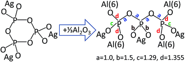
We present a detailed investigation of the effects of synthesis conditions on glasses xAgI–(1 − x)AgPO3 with 0 ≤ x ≤ 0.4. Raman and infrared spectroscopy of glasses synthesized in platinum crucibles showed that their metaphosphate structure remains largely unaffected by increasing the amount of AgI. This result contradicts recent findings on similar glasses synthesized by melting in alumina crucibles, which were found to exhibit a strongly changing phosphate structure with addition of AgI. The glass transition temperature and ionic conduction properties of the latter glasses also show strong deviation from the analogous properties of glasses melted in platinum crucibles, as in this work. To reveal the origin of such effects, glasses with the nominal composition 0.4AgI–0.6AgPO3 were synthesized in alumina crucibles and their structures were compared with glasses prepared in platinum crucibles with composition yAl2O3–(1 − y)[0.4AgI–0.6AgPO3] and y = 0.0, 0.04 and 0.07. Vibrational spectroscopy showed that melting in alumina crucibles, especially in the presence of P2O5, leads to doping the AgI–AgPO3 glass with Al2O3 leached from the crucible. The bond valence approach was employed to assist assigning the new Raman bands that develop when Al2O3 modifies the metaphosphate structure, with an exponential relationship describing the dependence of the P–O stretching frequency on bond strength. Combination of this relationship with bond valence principles allowed us to associate the formation of aluminum-triphosphate species Al2/3Ag3P3O10 with the Raman trends observed for AgI–AgPO3 glasses doped with Al2O3 either intentionally or unintentionally, i.e. when melted in alumina crucibles for long times.


1. Introduction

Ion conducting glasses attract much attention for fundamental studies of their ion transport mechanism and for technological applications in solid state electrochemical devices.1–25 Some of the best glassy electrolytes, known as superionic conductive glasses, result from doping silver metaphosphate glass (AgPO3) with silver halide salts; this process leads to room temperature ionic conductivity as high as 10−2 S cm−1.26–32 This conductivity value is about five orders of magnitude higher than the conductivity of AgPO3 base glass and analogous to that of molten salts, e.g. KNO3.3 Over the years, research efforts have been focused on understanding the origins of the remarkably enhanced ionic conductivity in glasses doped with metal halide salts, in order to control the short- and/or medium-range order structures that favor ionic conduction in doped glasses.

Early studies in the field suggested that the doping salt forms its own conduction pathways1,3,6 or microdomains/clusters26,33,34 which percolate through the base oxide glass and facilitate ion transport. Later investigations showed that the incorporated doping salts expand the glass matrix, and this creates additional free volume30,35 or pathway volume36 available for ion transport and, thus, leads to higher ionic conductivity as a result of the increased charge carrier mobility in the expanded glass matrix. In a different approach the emphasis was placed on the local coordination of Ag ions in AgI-based glasses, where the evolution of mixed I/O environment around Ag ions and the establishment of O–Ag–I conduction sites were suggested to be the main factors controlling the mobility of the Ag ion carriers.37 On the other hand, a correlation between ionic conductivity and the Ag–I distance was demonstrated in families of AgI-doped glasses, and glasses having longer Ag–I distance were found to exhibit lower activation energy and higher ionic conductivity.38 In a unified treatment for ion transport in glassy and polymer electrolytes, it was proposed that very similar forces are involved in both ion migration and structural relaxations processes in such highly ion conducting materials, e.g. AgI-doped glasses.18 A recent analysis of conductivity data for glasses AgI–AgPO3 showed that the mobility of Ag ions does not vary significantly with AgI content; it is rather the increase in the effective Ag ion carrier concentration that causes the enhancement in ionic conductivity.39

Despite their differences, all models cited above highlight the crucial role of AgI in enhancing ionic conductivity. In addition to this role of AgI, other studies emphasized the importance of the synthesis method on structure and properties of glasses xAgI–(1 − x)AgPO3. Thus, it was found40,41 that when the AgPO3 glass is prepared under dry ambient synthesis environments it exhibits enhanced direct current ionic conductivity (σdc) by almost two orders of magnitude and remarkably elevated glass transition temperature (Tg) by as much as 95 °C, relative to AgPO3 glasses developed in common laboratory ambient environments.26–32 This impressive increase in σdc and Tg was attributed to the absence of water traces when the handling of starting materials is performed under dry ambient conditions.40,41 Similarly prepared glasses xAgI–(1 − x)AgPO3 were found to display three distinct regimes of conductivity defined by thresholds at xc1 = 0.09 and xc2 = 0.38, which correlate well with the three elastic phases determined from calorimetric measurements.41–43 Thus, it was suggested that the reversibility window 0.09 < x < 0.38 is suppressed or completely diminished in the presence of water traces because of the depolymerization of phosphate chains and the reduction in network connectivity. The Raman spectra of dry AgI–AgPO3 glasses showed a strong dependence on the AgI content; this was interpreted in terms of changes in glass structure from chain-like to ring-like upon increasing AgI content41,43 and found to contradict earlier Raman results for glasses synthesized under common laboratory ambient conditions which showed little or no change in the phosphate structure as AgI is added to AgPO3.31,44,45 It is noted that AgI-induced changes in the topology of the phosphate network, from long chains to large and small rings,41,43 were considered to be at the origin of the excess constraints in AgI–AgPO3 glasses.46

The fundamental question whether the method of preparation affects structure and properties of AgI-containing phosphate glasses continues to attract interest, in view also of new applications of such materials in the field of photonics.47 In this context, we note a recent investigation focusing on glass preparation by high- and low-temperature routes, i.e. melt-quenching versus mechano-synthesis by ball-milling.48 This study revealed differences in thermal properties (e.g., Tg) and Raman spectra of AgI–AgPO3 glasses prepared by the two different synthesis methods, and attributed them to different energy transfer mechanisms involved in the two synthesis techniques.

In a previous brief report,49 we studied the effect of synthesis methods on structure and properties of the AgPO3 base glass by varying the atmosphere of handling the starting materials, i.e. dry versus normal laboratory ambient conditions, starting materials, melting time and temperature, and type of crucible used for melting (i.e., Pt versus alumina). A key finding was that melting in alumina crucibles, which is a practice used widely for the synthesis of superionic glasses, gives AgPO3 glasses being unintentionally contaminated with up to ca. 3 mol% Al2O3. This Al2O3-doping was found to cause enhanced phosphate network connectivity and drastically increased σdc and Tg relative to Al2O3-free AgPO3 glasses prepared in Pt crucibles. In view of these findings for AgPO3, we have studied and report here on the structure and properties of glasses xAgI–(1 − x)AgPO3 with 0 ≤ x ≤ 0.4. The synthesized glasses were investigated by combining Raman and infrared spectroscopic techniques as they provide complementary structural information, while glass transition temperature and ionic conductivity were also measured as a function of AgI content and compared with literature data. To investigate the role of crucible on the AgI–AgPO3 system, we studied glasses 0.4AgI–0.6AgPO3 melted in alumina and Pt crucible, and compared their vibrational spectra with those recorded for glasses yAl2O3–(1 − y)[0.4AgI–0.6AgPO3] prepared in Pt crucibles, with y = 0.0, 0.04 and 0.07. A key finding was that synthesis in alumina crucibles leads to unintentional doping of the 0.4AgI–0.6AgPO3 glass with aluminum oxide, a process causing the increase of the O/P ratio and, consequently, the progressive change of the phosphate structure as its modification exceeds the metaphosphate composition (O/P = 3.0). Application of the bond valence approach allowed to propose the formation of Al-triphosphate, Al2/3Ag3P3O10, as a main structure formed when the metaphosphate glass is doped with Al2O3 either intentionally or unintentionally when using alumina crucibles and long melting times. These results are discussed in relation to previous studies on AgI–AgPO3 glasses, and contribute to answering open questions in the field of ion conducting glasses.

2. Experimental

2.1 Glass synthesis

As noted in the Introduction, our earlier study on the AgPO3 base glass showed that melting in alumina crucibles leads to contamination with Al2O3.49 To avoid this problem, Pt crucibles were employed in this work to synthesize glasses in the series xAgI–(1 − x)AgPO3, 0 ≤ x ≤ 0.4, using as starting materials dry NH4H2PO4 (99.995%), AgNO3 (99.995%) and AgI (99.9%). As for the case of AgPO3 glass, we performed all weighting and mixing manipulations of the starting materials in a glove bag purged with dry N2 gas, i.e. under dry ambient conditions (ca. 3–5% relative humidity). Each batch was then transferred quickly to an electrical furnace held at 150 °C and slowly calcinated up to 300 °C to remove the volatile products which result from the decomposition of the reactants. The temperature of the furnace was then increased to 700–900 °C depending on composition; the melts were repeatedly stirred and kept at the melting temperature for two hours. For convenience, the synthesis parameters of the studied glasses are summarized in Table 1 with their nominal compositions. Glasses in this series were obtained in the form of ca. 0.5–1.0 mm-thick discs with ca. 10–15 mm of diameter by quenching the melts in stainless steel pre-forms. This procedure gives smooth glass surfaces and suitable specimen sizes for infrared reflectance and electrical conductivity measurements when the AgI content is up to about x = 0.4. All glass specimens were stored under vacuum, with no additional heat or surface treatment to avoid exposing the samples to lab ambient for long periods of time.
Table 1 Synthesis parameters for glasses xAgI–(1 − x)AgPO3 and values determined in this work for glass transition temperature, Tg, dc ionic conductivity at 293 K, σdc, activation energy for ionic conductivity, Ea, infrared absorption at 2815 cm−1, αmax, and water content, CH2O. Density data, ρ, are from ref. 39 and are used to estimate the water content in glass in wt%, WH2O. The starting materials for glass synthesis glasses were NH4H2PO4, AgNO3 and AgI, which were handled under dry ambient conditions
Glass x Crucible Synthesis conditions Tg (°C) ± 1 log[thin space (1/6-em)]σdc (S cm−1) ± 0.03 Ea (eV) ± 0.01 αmax (cm−1) ± 0.1 CH2O (mol L−1) ± 0.01 ρ (g cm−3) WH2O (wt%) ± 0.005
A1 0 Pt 700 °C, 2 h 193 −6.23 0.53 31.0 0.09 4.35 0.037
A2 0.1 Pt 800 °C, 2 h 183 −5.50 0.49 19.2 0.06 4.56 0.022
A3 0.2 Pt 900 °C, 2 h 171 −4.77 0.42 17.7 0.05 4.77 0.019
A4 0.3 Pt 900 °C, 2 h 141 −4.11 0.39 13.7 0.04 4.98 0.014
A5 0.4 Pt 900 °C, 2 h 120 −3.45 0.36 12.3 0.04 5.20 0.012


A second series of glasses was prepared with composition yAl2O3–(1 − y)[0.4AgI–0.6AgPO3] as summarized in Table 2; the first glass (B1) being identical to glass A5 of Table 1. Two additional glasses with starting composition also 0.4AgI–0.6AgPO3 were prepared by quenching melts from alumina crucibles using appropriate batches of NH4H2PO4, AgNO3 and AgI (glass B2) and P2O5, AgNO3 and AgI (glass B3). For glasses B4 (y = 0.04) and B5 (y = 0.07) we used Pt crucibles, the same starting materials NH4H2PO4, AgNO3, AgI and Al2O3, as well as the same temperature and melting time (Table 2). Glasses in this series were prepared also under dry ambient conditions for the weighting and mixing manipulations of the starting materials.

Table 2 Synthesis parameters for glasses yAl2O3–(1 − y)[0.4AgI–0.6AgPO3]. Starting materials were handled under dry ambient conditions
Glass y Crucible Starting materials Synthesis conditions
B1 0 Pt NH4H2PO4, AgNO3, AgI 900 °C, 2 h
B2 0 Alumina NH4H2PO4, AgNO3, AgI 900 °C, 1.5 h
B3 0 Alumina P2O5, AgNO3, AgI 700 °C, 0.5 h
B4 0.04 Pt NH4H2PO4, AgNO3, AgI, Al2O3 1100 °C, 0.5 h
B5 0.07 Pt NH4H2PO4, AgNO3, AgI, Al2O3 1100 °C, 0.5 h


To assist Raman and infrared assignments, we prepared silver-pyrophosphate (Ag4P2O7) by melting appropriate amounts of dried NH4H2PO4 and AgNO3 in Pt crucible. It was found that melt quenching results in polycrystalline Ag4P2O7, while glass formation using the same quenching technique required addition of at least 20 mol% AgI (i.e., glass 0.2AgI–0.8Ag4P2O7). Commercially available silver-orthophosphate (Ag3PO4) in polycrystalline form was considered also for Raman and infrared measurements.

2.2 Spectroscopic techniques

Raman spectra were measured in vacuum at the backscattering geometry (Renishaw inVia Raman Microscope), using the 514.5 nm line of an Ar ion laser for excitation. All spectra were recorded at room temperature with 2 cm−1 resolution. Examination of glass specimens under the Raman microscope at the end measurements revealed no signs of laser-induced modifications at the employed irradiation conditions, i.e. 0.1 mW μm−2 at the sample. The Raman spectra reported here are in the form of temperature-reduced intensity, Ired, calculated from the expression Ired = I(ν)/[n(ν) + 1] where I(ν) is the measured Raman intensity and n(ν) is the Bose–Einstein factor, n(ν) = 1/[exp(hcν/kBT) − 1], where ν is the Raman shift in cm−1, c is the speed of light, T is the temperature in K, and kB and h are the Boltzmann and Planck constants, respectively.

Infrared (IR) spectra were measured on a vacuum Fourier transform spectrometer (Bruker, Vertex 80v), in quasi-specular reflectance and in transmittance modes. For each spectrum, 200 scans were collected at room temperature with 2 cm−1 resolution and averaged for evaluation. Reflectance spectra were measured separately in the far- and mid-IR range and then merged to form a continuous spectrum in the range 30–7000 cm−1. Analysis of such reflectance spectra by Kramers–Kronig transformation yielded the absorption coefficient spectra, α(ν), from the expression α(ν) = 4πνk(ν) where ν is the infrared frequency in cm−1 and k(ν) is the imaginary part of the complex refractive index.14

2.3 Electrical and thermal properties

Impedance spectroscopy was employed to determine the ionic conductivity of glasses xAgI–(1 − x)AgPO3. Circular gold electrodes were evaporated on the parallel surfaces of each disc-shaped glass specimen. The sample was then placed in a sample holder and isothermal conductivity curves were obtained for temperatures 20–160 °C in the frequency range 10 MHz to 5 Hz using an HP4192A impedance analyzer. Sufficient time was allowed between measurements to ensure thermal stability and constant readings. The direct current ionic conductivity, σdc, was deduced at each temperature from the low-frequency plateau of the corresponding curve, and the activation energy for ionic conduction, Ea, was obtained from the Arrhenius dependence of ionic conductivity vs. inverse temperature using the expression Ea = −kB[dln[thin space (1/6-em)]σdc/d(1/T)], where kB is the Boltzmann constant and T is the temperature in K.

The glass transition temperature, Tg, was determined by Differential Scanning Calorimetry (DSC, TA Q200 instrument) from the first heating circle at a scan rate of 10 °C min−1. Glasses for DSC measurements were sealed in Al pans and measured under dry N2. At least two different samples were run for each glass composition. Tg was determined from the inflection point of the DSC curve and was found reproducible within ±1 °C.

3. Results and discussion

3.1 Glasses xAgI–(1 − x)AgPO3

All glasses in the series AgI–AgPO3 were synthesized under identical conditions, apart from some variations in melting temperature (Table 1). Therefore, changes in glass structure and properties should be associated with the amount of AgI added to the AgPO3 base glass.
3.1.1 On the structure of glasses xAgI–(1 − x)AgPO3. Reduced Raman scattering and IR absorption coefficient spectra shown in Fig. 1a and b, respectively, manifest clearly their complementary nature due to differences in selection rules. The spectra are presented normalized on the strongest band to facilitate comparison; Raman spectra are normalized on the 1140 cm−1 band and IR spectra on the band at 1235 cm−1.
image file: c6ra00162a-f1.tif
Fig. 1 (a) Temperature reduced Raman, and (b) infrared absorption coefficient spectra of glasses xAgI–(1 − x)AgPO3. For comparison, Raman spectra were normalized on the strongest band at ca. 1140 cm−1 and infrared spectra on the 1235 cm−1 band.

As discussed earlier for glass AgPO3,49 the picture emerging from the consideration of its Raman and infrared spectra is that based mainly on a polymer-like metaphosphate chain structure which is formed by linked phosphate tetrahedra having two bridging (Ø) and two terminal (O) oxygen atoms, (PØ2O2). This structure is consistent with earlier studies of AgPO3 glass and is widely accepted in the literature.31,40,44,45,50–60 Using the phosphate Qi terminology where i is the number of bridging oxygen atoms per phosphate tetrahedron, the (PØ2O2) tetrahedron is termed Q2 and has the PO3 stoichiometry. The presence of Q2 units explains key features of the Raman and infrared spectra in Fig. 1a and b for AgPO3 glass, x = 0. The strongest Raman band at 1144 cm−1 originates from the symmetric stretching of the terminal PO2 groups in Q2 units, νs(PO2), while the band at 676 cm−1 is due to the symmetric stretching of P–O–P bridges within the phosphate backbone, νs(P–O–P). Likewise, the strongest infrared band at 1235 cm−1 is due to the asymmetric stretching of PO2, νas(PO2), while the corresponding mode of P–O–P in chains of Q2, νas(P–O–P), gives the band at 895 cm−1. Besides these bands, the infrared spectrum shows at high-frequencies additional features at ca. 990 and 1058 cm−1. Based on previous works,51,59 the 990 cm−1 band is attributed to νas(P–O–P) for Q2 units in metaphosphate rings and the one at 1058 cm−1 to the asymmetric stretching of PO32− groups, νas(PO32−), which terminate the metaphosphate chains. For convenience, the assignments of infrared and Raman bands are summarized in Table 3.

Table 3 Frequencies and assignments of Raman and infrared bands measured in this work for metaphosphate glasses xAgI–(1 − x)AgPO3, pyrophosphate glass 0.2AgI–0.8Ag4P2O7, and orthophosphate crystal Ag3PO4
Raman (cm−1) Infrared (cm−1) Assignment References
a Band intensity: vs = very strong, s = strong, m = medium, w = weak, vw = very weak, sh = shoulder.b Vibration mode: ν = stretching, δ = bending, νs, ν1 = symmetric stretching, νas, ν3 = asymmetric stretching, ν2 = symmetric bending, ν4 = asymmetric bending.c Qi: a phosphate tetrahedron with i bridging and 4-i terminal oxygen atoms.
Metaphosphate glasses xAgI–(1 − x)AgPO3
1220–1225 (vw)a 1235 (vs) νas(PO2)b, Q2c 21, 31, 43 and 49–60
1144–1138 (vs) 1140 (sh, vw) νs(PO2), Q2 21, 43–45 and 49–60
  1090 (sh, w) νas(PO32−), Q1 21, 59 and 60
  1058 (s) νas(PO32−), end chains 21, 31, 43 and 49–59
1000 (vw) νs(PO32−), Q1 21, 53, 56 and 57
  990 (s) νas(P–O–P), Q2 in rings 51, 59 and 60
  895 (s) νas(P–O–P), Q2 in chains 21, 31, 43 and 49–60
680 (s), 710, 778 (sh) 710 (m), 778 (m) νs(P–O–P), Q2 21, 43 and 49–60
475 (s), 518 (s), 585 (m) δ(PO2) + δ(PO32−) 21 and 61
128–122 ν(Ag–O), ν(Ag–O/I) 14, 21, 51 and 55
120 (m-s) ν(Ag–I) 44 and 45
45 (m-s) Boson mode 44 and 45
[thin space (1/6-em)]
Pyrophosphate glass 0.2AgI–0.8Ag4P2O7
1075 (m), 1130 (sh) 1085 (vs) νas(PO32−), Q1 21, 59, 61 and 66
1002 (vs) 1000 (vw) νs(PO32−), Q1 21, 55, 57–59 and 61
912 (w) ν1(PO43−), Q0 21, 61 and 67
903 (s) νas(P–O–P), Q1 21, 57–59, 61 and 66
710 (m) 705 (m) νs(P–O–P), Q1 21, 55, 57–59 and 61
  527 (s), 555 (s), 590 (vw) δas(PO32−), Q1 species 61 and 66
345 (w) 415 (vw), 475 (w), 510 (s) δs(PO32−), Q1 species 61 and 66
  207 (s) δ(P–O–P), Q1 species 61
  140 (s) ν(Ag–O) 14, 51 and 55
48 (m) Boson mode 44 and 45
[thin space (1/6-em)]
Orthophosphate crystal Ag3PO4
955 (vw), 1015 (vw) 960 (vs) ν3(PO43−), Q0 21, 59, 61 and 67
910 (vs) ν1(PO43−), Q0 21, 57, 58, 61 and 67
550 (vw) 552 (s) ν4(PO43−), Q0 21, 59, 61 and 67
405 (w) ν2(PO43−), Q0 21, 61 and 67
  198 (m) ν(Ag–O) 14, 51 and 55


Addition of AgI to AgPO3 glass affects clearly the low-frequency Raman spectra with bands developing progressively at ca. 45 and 120 cm−1 (Fig. 1a). Similar features were observed in earlier studies and were associated with the boson band and Ag–I stretching, respectively.44,45 However, the higher-frequency range (>250 cm−1) of the Raman spectra remains largely unaffected by AgI addition except for a small shift of νs(PO2) from 1144 cm−1 for x = 0 to 1138 cm−1 for x = 0.4 and the development of very weak Raman activity at ca. 900–1050 cm−1 with a peak discernible at ca. 1000 cm−1 for x = 0.4. The present results are in agreement with earlier Raman studies on xAgI–(1 − x)AgPO3 glasses44,45 but differ substantially from those of recent reports41,43 that show large changes in the Raman spectra; these were manifested mainly by the progressive loss of intensity of the νs(PO2) band at 1140 cm−1 and its nearly disappearance for x ≥ 0.4 as well as by the growth of a new band at 1008 cm−1 which dominates eventually the Raman spectrum for x ≥ 0.4. The origin of such pronounced differences in the Raman spectra of AgI–AgPO3 glasses will be examined in Section 3.2.

Evidence for the incorporation of AgI in the glass structure is provided also by infrared absorption below ca. 250 cm−1 where the vibrations of Ag ions against their sites are active. For the AgPO3 glass a broad band is measured at 128 cm−1 (Fig. 1b, x = 0) and can be attributed to Ag–O vibrations in oxide sites,14,51,55 while doping of AgPO3 with AgI causes the downshift of this band to 122 cm−1 for x = 0.4. We note that crystalline β-AgI (wurtzite) and α-AgI (superionic phase) show their main infrared absorption at ca. 110 cm−1 due to Ag–I stretching in tetrahedral iodide site hosting the silver ion.62,63 Therefore, the downshift of the 128 cm−1 band is consistent with the formation of mixed I/O coordination environments for Ag ions upon increasing AgI content. This is in line with the proposed formation of O–Ag–I conduction sites and their key role in controlling the mobility of Ag ions.37

The higher-frequency infrared spectrum is affected slightly by AgI addition (Fig. 1b). This is manifested in several ways including the decrease of relative intensity at 990 cm−1, the parallel increase in intensity at 895 and 1058 cm−1 and the gradual growth of a shoulder at ca. 1090 cm−1. These spectral changes are systematic and should mirror variations in the metaphosphate structure. They can be explored on the basis of assignments in Table 3 and include mainly a partial destruction of metaphosphate rings (990 cm−1 band), the opening of which would give metaborate chains (bands at 895 and 1058 cm−1). To search for the origin of such changes we consider first the formation of AgPO3 glass, which can be described by the following reaction driven by thermal decomposition of the AgNO3 and NH4H2PO4 starting materials:64,65

 
AgNO3 + NH4H2PO4 → AgPO3 + (3/2)H2O↑ + NH3↑ + NO2↑ + (1/4)O2 (1)

In the presence of AgI, part of the evolved water in reaction (1) may lead to the opening of metaphosphate rings as follows:

 
image file: c6ra00162a-t1.tif(2)

The above reaction is consistent with the observed variations in relative intensity of the infrared bands at 990, 895 and 1058 cm−1. Water may induce also the formation of shorter metaphosphate chains and pyro-phosphate species as described below:

 
(n + 2)Ag+[PO3.5–(PO3)n−2–PO3.5]−(n+2) + 2AgI + H2O → nAg+[PO3.5–(PO3)n−4–PO3.5]n + 4Ag+P2O74− + 2HI↑ (3)

Evidence for the formation of isolated pyro-phosphate species, (P2O7)4− or (PO3–O–PO3)4− = 2Q1, is provided by the shoulder developing in the IR at ca. 1090 cm−1 which can be attributed to the asymmetric stretch of pyro-phosphate terminal P–O bonds,21,59,60 denoted νas(PO32−) in Table 3. Indeed, the IR spectrum of the pyro-phosphate glass 0.2AgI–0.8Ag4P2O7 in Fig. 2 shows its strongest band at ca. 1085 cm−1. Formation of a minor amount of (P2O7)4− by reaction (3) is also consistent with the observed weak Raman feature at ca. 1000 cm−1 (Fig. 1a, x = 0.4), with the strongest Raman signal of (P2O7)4− being measured at 1002 cm−1, νs(PO32−) (Fig. 2 and Table 3).


image file: c6ra00162a-f2.tif
Fig. 2 (a) Temperature reduced Raman, and (b) infrared absorption coefficient spectra of pyrophosphates in glassy, 0.2AgI–0.8Ag4P2O7, and polycrystalline, Ag4P2O7, forms.

In summary, incorporation of AgI in glasses AgI–AgPO3 leaves the metaphosphate structure practically unaffected. Nevertheless, some small but systematic spectral changes were observed to develop in the Raman and infrared spectra with AgI addition and these could be associated with the opening of metaphosphate rings and the creation of shorter metaphosphate chains and minor amount of isolated pyro-phosphate units. It was proposed that such structural changes are induced by water which is evolved in situ during thermal decomposition of NH4H2PO4 used for AgPO3 glass synthesis (reactions (1–3)). In any case, the minor changes in the Raman and infrared spectra detected in this study cannot explain the strong influence of AgI on the phosphate structure reported recently for AgI–AgPO3 glasses.41,43

3.1.2 Properties of glasses xAgI–(1 − x)AgPO3. Addition of AgI to AgPO3 decreases drastically the glass transition temperature (Tg) and enhances remarkably the direct current ionic conductivity (σdc) as seen in Table 1. It is clear that the increase of ionic conductivity is due to the corresponding reduction in activation energy (Ea) with AgI content. The evolution of properties observed here as a function of AgI content is compared in Fig. 3 with literature data for Tg,26,29,30,43,68 σdc,26,29–31,43,69 and Ea.26,29–31,43,68,69 Starting with Tg (Fig. 3a) we note that values reported in ref. 41 and 43 are considerably larger than those found in earlier studies and in the present work. A large difference in Tg is noted already for the AgPO3 base glass (x = 0) and it spans the range from ΔTg = 94 °C in comparison to ref. 29 to ΔTg = 61 °C in comparison to this work. Novita et al.40–43 attributed such large differences in Tg, as well as in ionic conduction properties (Fig. 3b and c), to large differences in residual water content. It was argued that their method for glass preparation gives dry glasses in contrast to other methods used in literature which produce glasses with, presumably, higher water content.
image file: c6ra00162a-f3.tif
Fig. 3 Comparison of results obtained in this work with literature data for (a) glass transition temperature, Tg, (b) room temperature dc ionic conductivity, σdc, and (c) activation energy, Ea, measured as a function of AgI content for glasses xAgI–(1 − x)AgPO3.

Because residual water would certainly influence glass properties, it is of interest to investigate the presence of water in glasses synthesized in the present work. For this purpose we used IR spectroscopy which is a very sensitive technique to determine water in glass. Following the approach employed for AgPO3 glass,49 we have measured IR transmittance spectra, T(ν), and present in Fig. 4 the corresponding absorption coefficient spectra, α(ν), as a function of AgI content. The α(ν) spectra were calculated using the expression:

 
α(ν) = −ln[T(ν)/T(5000)]/d (4)
where d is the sample thickness and the transmittance T(ν) is normalized to the transmittance at 5000 cm−1 to account for reflection losses.70 The spectra in Fig. 4 show that it is nearly impossible to prepare absolutely dry AgI–AgPO3 glasses with the glass preparation conditions employed in this study, as manifested by the presence of the ca. 2815 cm−1 band which is due to O–H stretching of water molecules.70–72 Nevertheless, the values of the maximum absorption at 2815 cm−1, αmax, in Table 1 show that addition of AgI to AgPO3 reduces the tendency of glass to retain water.


image file: c6ra00162a-f4.tif
Fig. 4 Infrared absorption spectra in the region of O–H stretching for glasses prepared in this work in the system xAgI–(1 − x)AgPO3.

The experimental αmax values can be used to evaluate the water content in glass, CH2O = αmax/(2.303εH2O), but this requires knowledge of the molar extinction coefficient (εH2O) for the band at 2815 cm−1. For the AgPO3 glass we employed previously49 the value εH2O = 180 L mol−1 cm−1 derived from the conversion factor of about 100 ppm OH/cm−1 reported for phosphate glasses,70 while in an earlier study73 of calcium-metaphosphate glasses the value εH2O = 120 L mol−1 cm−1 was used for the absorption band at about 3000 cm−1. This suggests that an average value of εH2O = 150 L mol−1 cm−1 could be used in the present study. We note also that εH2O data have been collected, for boro-silicate glasses mainly, to evaluate water content in glass by IR spectroscopy.74 The derived εH2O vs. frequency curve indicates the value εH2O = 155 L mol−1 cm−1 at 2815 cm−1 in good agreement with the average value noted above. Therefore, we use the value εH2O = 150 L mol−1 cm−1 to estimate the water content in AgI–AgPO3 glasses assuming that the molar extinction coefficient is independent of AgI content. The estimated water content is given in Table 1 and shows a decrease from 0.09 to 0.04 moles H2O per liter of glass from x = 0 to x = 0.4.

The density data reported in ref. 39 allow for the estimation of water content also in wt% (Table 1), to facilitate comparison with NMR results of Mustarelli et al.75 on the effect of water on the thermal properties of AgPO3 glass. It was found that the water content of glasses prepared in Pt crucibles using NH4H2PO4 and AgNO3 (as in the present study) decreases by increasing temperature of melt and melting time, leading at the same time to an increase of Tg. The driest AgPO3 glass was melted at 700 °C and it was found to be NMR-water-free with Tg = 187 °C, while a glass with water content of 0.41 wt% had Tg = 172 °C.75 While there are differences in sensitivity and way of calibration when NMR and IR data are used to evaluate water content in glass, we note that our AgPO3 base glass with IR-evaluated water content of 0.04 wt% has glass transition higher by ΔTg = 6 °C than the NMR-water-free glass of ref. 75. On these grounds we conclude that the synthesis method employed here gives glasses with very low water content even for the base glass, and the water content decreases further when AgI is added to AgPO3. This trend should be due mainly to the use of less amount of NH4H2PO4 when synthesizing glasses with increased AgI content, resulting in less water being evolved in situ during thermal decomposition of the NH4H2PO4 starting material (reaction (1)). Therefore, the decrease of Tg by 73 °C upon increasing the amount of AgI from x = 0 to x = 0.4 (Table 1) cannot be related to traces of water but it should reflect a strong plasticizing effect of AgI which, upon incorporation in the glass structure, loosens the overall connectivity of the metaphosphate backbone.3,7

To understand the origin of differences in Tg and conduction properties between earlier reports and the Novita et al.40–43 data for AgPO3 base glass (see Fig. 3), we synthesized in our previous study49 AgPO3 glasses using P2O5 and Ag3PO4 as starting materials like in ref. 40–43. When Pt crucible was used for melting at 900 °C for 2 h, the resulted glass had practically identical Raman and IR spectra, as well as Tg and conductivity values, with the glass prepared from NH4H2PO4 and AgNO3 in Pt crucible under milder conditions (melting at 700 °C for 2 h). However, melting P2O5 and Ag3PO4 in alumina crucible at 700 °C for 22 h, similarly to Novita et al.,40–43 resulted in AgPO3 glass with Tg = 255 °C and improved electrical conductivity. This was attributed to the unintentional contamination of AgPO3 with up to about 3 mol% Al2O3, as was verified by comparison with spectra and properties of glasses xAl2O3–(1 − x)AgPO3 (x = 0.01, 0.03, 0.05) prepared from NH4H2PO4, AgNO3 and Al2O3 in Pt crucibles.49

Based on these findings, we return now to AgI–AgPO3 glasses and summarize in Table 4 the synthesis protocols employed by authors whose Tg and conductivity data are compared in Fig. 3. While a variety of crucibles was used for melting, what really differentiates the synthesis method of ref. 43 from the others is the combination of alumina crucible with corrosive P2O5 and very long melting time (i.e., overnight). This synthesis protocol and the previous discussion for the AgPO3 base glass, suggest that the use of alumina crucible with P2O5 and long melting times may cause unintentional contamination of AgI–AgPO3 glasses with Al2O3 extracted from the crucible during melting. This possibility is examined in the following section.

Table 4 Summary of synthesis protocols for glasses xAgI–(1 − x)AgPO3 with properties shown in Fig. 3 as a function of AgI content
Crucible Starting materials Melting temperature Melting time References
Alumina AgPO3 + AgI 530–600 °C 2 h 26
Not specified NH4H2PO4 + AgNO3 + AgI 527 °C Several h 29
Pyrex NH4H2PO4 + AgNO3 + AgI 500 °C Not specified 30
Pt NH4H2PO4 + AgNO3 + AgI 700 °C 30 min 31
Not specified Ag3PO4 + P2O5 + AgI 900 °C Not specified 41
Alumina Ag3PO4 + P2O5 + AgI 700 °C Overnight 43
Porcelain NH4H2PO4 + AgNO3 + AgI Not specified 15 min 68
Not specified AgPO3 + AgI 600–700 °C 1–2 h 69
Pt NH4H2PO4 + AgNO3 + AgI 700–900 °C 2 h This work


3.2 Glasses yAl2O3–(1 − y)[0.4AgI–0.6AgPO3]

Glasses with composition yAl2O3–(1 − y)[0.4AgI–0.6AgPO3] were prepared following the synthesis protocols outlined in Table 2 and their Raman and infrared spectra are presented and discussed below.
3.2.1 Vibrational spectra of glasses yAl2O3–(1 − y)[0.4AgI–0.6AgPO3]. The Raman and IR spectra of glasses yAl2O3–(1 − y)[0.4AgI–0.6AgPO3] are reported in Fig. 5a and b, respectively. Starting with the Raman spectra of the binary glasses B1 and B2, because they have the same nominal composition 0.4AgI–0.6AgPO3 and were prepared from the same starting materials, we find that the use of alumina crucible results in well-defined spectral changes for glass B2. These are manifested at high frequencies by the broadening and downshift of the νs(PO2) band to 1135 cm−1 and the enhancement of features at 958, 1002, 1023, and 1082 cm−1, while the 713 cm−1 shoulder gains relative intensity in the region of νs(P–O–P). All these changes become more pronounced for glass B3, for the preparation of which alumina crucible was combined with the use of P2O5. The similarities of the Raman features of the ‘binary’ B3 glass with those of the ternary B4 glass with 4 mol% Al2O3 are striking and suggest similar structural characteristics, which are definitely different from those of the binary metaphosphate glass B1. Based on the Raman profiles, we suggest that glass B3 is unintentionally contaminated with Al2O3 leached from the crucible and that it contains more than 4 mol% Al2O3, while glass B2 should be contaminated with a lower amount of Al2O3. This difference between B3 and B2 should be attributed to the fact that P2O5 being the anhydride of the strong phosphoric acid is corrosive and reacts effectively with the basic Al2O3. On the other hand, NH4H2PO4 is a salt of the dihydrogen phosphate ion, H2PO4, which is the conjugate base of phosphoric acid and, as a weaker acid, is relatively less reactive to Al2O3. For the ternary glass B5, with 7 mol% Al2O3, the features at 958, 1023, 1082 and 1120 cm−1 have almost disappeared while the 1002 cm−1 band dominates the Raman spectrum and weaker features appear at 713 and 914 cm−1. Comparison with the Raman spectrum of pyrophosphate species in Fig. 2a suggests that glass B5 should contain mainly isolated pyrophosphate species (P2O7)4− with characteristic bands at 1002 cm−1, νs(PO32−), and 713 cm−1, νs(P–O–P), a smaller amount of other phosphate species giving the 914 cm−1 Raman band, and no metaphosphate-type units.
image file: c6ra00162a-f5.tif
Fig. 5 (a) Temperature reduced Raman, and (b) infrared absorption coefficient spectra of glasses yAl2O3–(1 − y)[0.4AgI–0.6AgPO3]. For glass preparation conditions see Table 2.

A continuous evolution of glass structure is suggested also by the corresponding infrared spectra in Fig. 5b. Starting with the metaphosphate band of B1 at 1235 cm−1, νas(PO2), we note its progressive downshift and reduction in intensity from B1 to B4 and its disappearance for B5. In agreement with Raman spectrum, the IR spectrum of B5 shows the dominant presence of isolated pyrophosphate species with νas(PO32−) at 1085 cm−1, νs(PO32−) at ca. 1000 cm−1, νs(P–O–P) at 712 cm−1 and νas(P–O–P) at 900 cm−1. The IR shoulder at ca. 958 cm−1 is indicative of a minor amount of a different type of phosphate species, which should exist also in glasses B2 to B4.

We note at this point that the evolution of the Raman and IR spectra in Fig. 5 is very similar to that exhibited by the Raman41,43 and IR43 spectra of glasses xAgI–(1 − x)AgPO3 which were prepared in alumina crucibles using P2O5 and long melting times. Based on our findings in Fig. 5, we suggest that the structure and properties of glasses prepared and studied in ref. 41 and 43 deviate from those of the pristine xAgI–(1 − x)AgPO3 metaphosphate series and should rather correspond to alumino-phosphate glasses with composition changing progressively with increasing x from metaphosphate toward pyrophosphate; this being probably the result of Al2O3 leaching from the alumina crucible at prolong melting. Thus, deviations from the metaphosphate composition and structure should be the main reason for the observed differences in Fig. 3 between physical properties of ref. 41 and 43 and those of earlier studies and this work.

3.2.2 Modifying role of Al2O3 in glasses yAl2O3–(1 − y)[0.4AgI–0.6AgPO3]. To explain the effect of Al2O3 on the phosphate structure of glasses yAl2O3–(1 − y)[0.4AgI–0.6AgPO3] which were prepared here in Pt crucibles with y = 0 (B1), y = 0.04 (B4) and y = 0.07 (B5), we consider their nominal O/P ratio assuming that Al2O3 acts as typical glass modifier. Then the O/P ratio increases from 3.0 (B1) to 3.21 (B4) and to 3.38 (B5). Recalling that the O/P value is 3.0 and 3.5 for metaphosphate and pyrophosphate compositions respectively, it is clear that the nominal composition of B4 and B5 is between those of meta- and pyrophosphate with glass B5 approaching the pyrophosphate composition. Thus, the phosphate network of B4 and B5 is over-modified with respect to B1; the same should hold for glasses B2 and B3 judging from their Raman and IR spectra. The effect of Al2O3 incorporation in the structure of Ag-metaphosphate glasses appears similar to that of In2O3 in Zn-phosphate glasses of composition (50 − x)ZnO–xIn2O3–50P2O5.76 As shown by Koudelka et al.76 replacement of ZnO by In2O3 leads to the progressive depolymerization of metaphosphate chains due to the increase in the O/P ratio. Besides the same effect of Al2O3, one could not exclude here the possibility of some additional phosphate modification by reactions similar to reaction (3) which may lead to some losses of iodine by conversion of AgI to Ag2O and HI.

Having considered the modifying role of Al2O3, which is introduced either unintentionally (B2, B3) or intentionally (B4, B5) in the phosphate structure, we explore next the origin of the Raman bands at 914, 958, 1023, 1082 and 1120 cm−1 in Fig. 5a to understand the nature of phosphate species formed by Al2O3-induced modification of the phosphate structure of B1. In this context, we note that the evolution of the Raman spectra in Fig. 5a is very similar to that found for Ag-phosphate glasses Ag2O–nP2O5 with 0.66 ≤ n ≤ 1.77 Besides some differences in peak frequencies, there is good resemblance of the Raman spectra of glasses B4 and B3 with those of Ag2O–nP2O5 having n = 0.75 (O/P = 3.17) and n = 0.66 (O/P = 3.26) respectively.

As the Ag2O content increases above the metaphosphate composition in glasses Ag2O–nP2O5 (i.e., n < 1) new Raman bands develop at 964, 1008, 1090 and 1120 cm−1.77 The bands at 1008 and 1090 cm−1 were attributed to P–O stretch in end groups of metaphosphate chains, the band at 1120 cm−1 to νs(PO2) in metaphosphate chains, and the 964 cm−1 band to the totally symmetric stretch of orthophosphate PO43− units (Q0).77 In regards to this 964 cm−1 band, the closest band in Fig. 5a is the one at 958 cm−1. Since the latter band is not present in the Raman spectrum of B5 which has a more modified phosphate network, the 958 cm−1 band should arise from phosphate species other than Q0. Interestingly, a weak band also at 958 cm−1 develops in the IR spectra of Fig. 5b and exists as a shoulder for glass B5. This observation shows clearly that the Raman and IR bands at 958 cm−1 could not originate from the same phosphate species.

Considering the previous attribution of the 964 cm−1 band to PO43− units,77 we present in Fig. 6 the Raman and IR spectra of polycrystalline silver-orthophosphate (Ag3PO4). The totally symmetric stretch of PO43− units, ν1(PO43−), gives rise to the strong Raman band at 910 cm−1 while the asymmetric stretch, ν3(PO43−), has very strong IR activity at 960 cm−1 (see Table 3). Therefore, the presence of orthophosphate PO43− units in glasses developed in this study would be signaled by the 914 cm−1 Raman band and the IR feature at 958 cm−1.


image file: c6ra00162a-f6.tif
Fig. 6 (a) Raman, and (b) infrared absorption coefficient spectra of polycrystalline silver-orthophosphate, Ag3PO4.
3.2.3 P–O bonding in glasses yAl2O3–(1 − y)[0.4AgI–0.6AgPO3]: a bond valence approach. The origin of the remaining Raman bands at 958, 1023, 1082 and 1120 cm−1 can be sought in terms of the bond valence (BV) approach introduced by Brow and coworkers to explain the decrease in the Raman stretching frequency of terminal P–O bonds, ν(P–O), upon increasing ZnO content in glasses xZnO–(1 − x)P2O5 with 0.50 ≤ x ≤ 0.71.57 In their approach, ν(P–O) was correlated with the P–O bond strength expressed in valence units (vu). To obtain the P–O bond strength in a phosphate tetrahedron the valence of phosphorus (+5) is distributed along the four P–O bonds, with the restriction that the sum of the vu values assigned to the bonds connected to an oxygen atom must equal two in order to satisfy the valence requirements of oxygen. Along these lines, the strength of the P–O terminal bond decreases from 1.50 vu in metaphosphate (Q2) to 1.33 vu in pyrophosphate (Q1) and to 1.25 vu in orthophosphate (Q0) structures, and ν(P–O) correlates linearly with the strength of the terminal P–O bond, s(P–O).57 As discussed by Brow and co-workers, the resulting valence unit values for P–O bonds are not expected to be quantitatively accurate. However, they give useful relative bond strengths and thus relative Raman stretching frequencies of the corresponding bonds, and this allows probing the evolution of structure and bonding in phosphates systems.

It is of interest to examine whether the vibrational response of neutral Q3 tetrahedra fits also in the above BV approach, considering that Q3 constitutes the structure building unit of v-P2O5 and remains a basic structural unit in ultraphosphate glasses with metal oxide content below 50 mol%.58 For the Q3 tetrahedron, it is straightforward to realize that each of the three bridging P–O(P) bonds has strength of 1.0 vu while the terminal P[double bond, length as m-dash]O bond has strength of 2.0 vu. The corresponding Raman stretching frequencies of these bonds in v-P2O5 are ν(P–O–P) = 635 cm−1 and ν(P[double bond, length as m-dash]O) = 1380 cm−1 according to ref. 78. The ν(P–O) versus s(P–O) data for Q2, Q1, and Q0 units in Ag-phosphate glasses studied in this work are shown in Fig. 7 together with data for v-P2O5. Data are included also for Zn-phosphate57 and Li-phosphate79 glasses for which characteristic Raman bands have been identified for Q2, Q1, and Q0 units.


image file: c6ra00162a-f7.tif
Fig. 7 Correlation between the Raman frequency of phosphorus–oxygen stretch, ν(P–O) in cm−1, and the bond strength, s(P–O) in valence units, for Li-, Zn- and Ag-phosphate glasses. The lines are exponential fits to the data according to eqn (5).

As shown in Fig. 7 the symmetric Raman stretching frequency ν(P–O) is found to correlate well with the strength s(P–O) of the bond, with the v-P2O5 data forming nicely the lower and upper limits in the correlations of the three phosphate glass systems. The correlation between ν(P–O) (in cm−1) and s(P–O) (in vu) can be described by the exponential equation:

 
ν(P–O) = ab[thin space (1/6-em)]exp[−ks(P–O)] (5)

The derived fitting parameters of eqn (5) in the three M-phosphate systems are in the order M = Ag, Li, Zn: a = 1627.8, 1531.4, 1496.1 cm−1, b = 4049.3, 5450.6, 6741.0 cm−1, and k = 1.40, 1.80, 2.05 vu−1 (R2 = 0.998, 0.998, 0.996). It is noted that an exponential expression was found to describe also the correlation between B–N stretching frequency and bond length in boron–nitrogen materials including boron–oxynitride amorphous thin films.80

Having established eqn (5) for metal-phosphate glasses, we return now to bands at 958, 1023, 1082 and 1120 cm−1 measured in the Raman spectra of glasses yAl2O3–(1 − y)[0.4AgI–0.6AgPO3] (Fig. 5). Since eqn (5) was derived for symmetric stretching vibrations, we examine first the polarization characteristics of the bands in question before applying eqn (5). Polarized Raman spectra in the VV and VH backscattering geometries for glasses with y = 0.04 and 0.07 are shown in Fig. 8. It is clear that the bands at 958, 1023, 1082 and 1120 cm−1 are polarized and thus they correspond to totally symmetric modes, as is the case with all other bands already assigned to symmetric stretching vibrations, i.e. νs(PO2) at ca. 1140 cm−1, νs(PO32−) at ca. 1000 cm−1, and νs(P–O–P) in the region 630–780 cm−1.


image file: c6ra00162a-f8.tif
Fig. 8 Polarized Raman spectra of glasses yAl2O3–(1 − y)[0.4AgI–0.6AgPO3] for y = 0.04 (a), and y = 0.07 (b). The Raman spectra were measured in the backscattering geometry with polarizations VV and VH, where the first letter indicates the polarization of the exciting laser beam and the second the polarization of the collected scattered light.

Application of eqn (5) for Ag-phosphate glasses, with s = ln[b/(aν)]/k, gives for the Raman bands at 1120 and 1082 cm−1 terminal P–O bond strengths of 1.48 and 1.43 vu, which are just below the nominal 1.50 vu strength in the PO2 unit of metaphosphate tetrahedra, Q2. This suggests that the 1120 and 1082 cm−1 Raman bands can be attributed to νs(PO2) in shorter (fragmented) metaphosphate chains caused by Al2O3-induced modification. The large bandwidth of the 1120–1082 cm−1 envelop is indicative of a distribution of P–O bond strengths and chain lengths, as was the case for modified zinc-phosphate glasses.57 Therefore, the broadening and downshift of the νs(PO2) mode from 1138 to 1120 cm−1 and to 1082 cm−1 in Fig. 5a suggests a progressive weakening of the terminal P–O bond in PO2 units as Al2O3 is introduced in the phosphate structure. This point of view is consistent with earlier NMR studies on sodium-phosphate glasses, where the shift of the Q231P peak to less negative values was correlated with a decreasing bonds order of terminal P–O bonds upon increasing Na2O content.81

The Raman features at 1023 cm−1 and 958 cm−1, which show similar dependence on Al2O3 content, correspond to terminal P–O bond strengths of 1.36 and 1.29 vu respectively. These values are above and below the nominal P–O strength of 1.33 vu in PO32− units of isolated pyrophosphate species (P2O7)4−, for which eqn (5) gives a frequency value of 1002 cm−1 for the bond strength of 1.33 vu. Therefore, while the Raman band at 1002 cm−1 is the clear signature of isolated pyrophosphate species, the bands at 1023 and 958 cm−1 could signal the presence of PO32− end groups in short phosphate fragments, just before they convert to isolated (P2O7)4− species at higher Al2O3 contents.

To examine this scenario, we explore possible structures in glasses yAl2O3–(1 − y)[0.4AgI–0.6AgPO3] which would be compatible with the Raman features at 1023 and 958 cm−1. For simplicity, we consider the composition y = 0.0625 which is between y = 0.04 and 0.07 and, when ignoring AgI, gives the stoichiometry Al2O3–9AgPO3. A structural unit corresponding to this stoichiometry is shown in Fig. 9; this unit resulting as the product of Al2O3-induced opening of three-member metaphosphate rings. A similar scheme has been proposed by Brow et al.82 for Al2O3-modified NaPO3 glass with 10 mol% Al2O3. Evidence for the gradual destruction of three-member metaphosphate rings, (P3O9)3−, is provided by the intensity reduction from B1 to B5 of the ca. 778 cm−1 IR band (Fig. 5b). According to Rulmont et al.59 an IR band in the region 765–780 cm−1 is characteristic of νs(P–O–P) in cyclotriphosphates, (P3O9)3−. For reasons of stoichiometry, aluminum should be six-fold coordinated to oxygen, Al(6), in the proposed aluminum-triphosphate Al2/3Ag3P3O10 structure (Fig. 9). This is in line with NMR spectroscopy on Al2O3–NaPO3 glasses which showed that octahedral Al sites are the dominant aluminate sites in glasses with Al2O3 content lower than 12.5 mol%.82


image file: c6ra00162a-f9.tif
Fig. 9 Schematic presentation of a three-member metaphosphate ring, 3Ag+(P3O9)3−, which opens by interaction with Al2O3 to form aluminum-triphosphate, Al2/3Ag3P3O10, where aluminium is six-fold coordinated to oxygen, Al(6). The assignment of P–O bond strengths ad (in vu) in the Al2/3Ag3P3O10 structure was based on the combination of the bond valence approach with experimental Raman frequencies in Fig. 5 and eqn (5).

We apply now the BV approach to the Al2/3Ag3P3O10 structure and assign P–O bond strengths in vu values as shown schematically in Fig. 9. Starting with the four P–O(P) bridging bonds, each bond should have strength of a = 1.0 vu to satisfy the oxygen valence. This leaves strength of b = 1.5 vu for each P–O terminal bond in the PO2 unit in order for the phosphorus atom in Q2 to have a sum of 5.0 vu. To satisfy the valence of the two terminal oxygen atoms in PO2, the charge-balancing Ag+ ion contributes 1/2 = 0.5 vu to each Ag–O bond. We consider next the two PO32− end groups, where each Al(6) contributes 3/6 = 0.5 vu to the Al(6)–O(P) bond. The question now is how to distribute the remaining phosphorus valence of 4.0 vu in the PO32− end groups among three bonds, i.e. two P–O(Al) and one P–O(Ag) bond. To tackle this question we use the experimental Raman frequencies of 1023 and 958 cm−1 and make the assumption that the 958 cm−1 band corresponds to the symmetric stretching of the two terminal P–O(Ag) bonds. Then eqn (5) gives strength of c = 1.29 vu for the P–O(Ag) bond, and this requires a contribution of 0.71 vu from Ag to the Ag–O(P) bond to satisfy the oxygen valence. For charge neutrality, the remaining valence on Ag (i.e., 0.29 vu) should be equally distributed to the oxygen atoms in the two Al(6)–O(P) bonds. Therefore, the strength of each P–O(Al) bond should be equal to d = 2–0.5–(0.29/2) = 1.355 vu. This gives for phosphorus in the PO32− end groups a sum of valences equal to 2 × 1.355 + 1.29 + 1.0 = 5.0 vu as expected. To check the compatibility of the estimated P–O(Al) bond strength of d = 1.355 vu with experiment, we use eqn (5) to calculate the Raman frequency for the corresponding P–O(Al) symmetric stretch. The result is 1020 cm−1, in very good agreement with the experimental Raman value of 1023 cm−1. For convenience, the final assignments of Raman and infrared bands for glasses yAl2O3–(1 − y)[0.4AgI–0.6AgPO3] are summarized in Table 5.

Table 5 Frequencies and assignments of Raman and infrared bands for the Al2O3-containing glasses yAl2O3–(1 − y)[0.4AgI–0.6AgPO3] studied in this work
Raman (cm−1) Infrared (cm−1) Assignment
a Vibration mode: ν = stretching, δ = bending, νs, ν1 = symmetric stretching, νas, ν3 = asymmetric stretching, ν2 = symmetric bending, ν4 = asymmetric bending.b Qi: a phosphate tetrahedron with i bridging and 4-i terminal oxygen atoms.
1180–1260 1235 νas(PO2)a, Q2b in meta-phosphate chains/rings
  1225–1202 νas(PO2), Q2, in shorter metaphosphate chains
1138   νs(PO2), Q2, in meta-phosphate chains/rings
1120, 1082   νs(PO2), Q2, in shorter metaphosphate chains
1023   νs(P–O(Al)), in Al2/3Ag3P3O10
1002 1000 νs(PO32−), Q1
958   νs(P–O(Ag)), in Al2/3Ag3P3O10
  958 ν3(PO43−), Q0
914   ν1(PO43−), Q0
  900 νas(P–O–P), Q1
713 712 νs(P–O–P), Q1
  530, 555, 590 δas(PO32−), Q1
350 410, 470, 505 δs(PO32−), Q1
  170 ν(Ag–O) + δ(P–O–P), Q1
120   ν(Ag–I)
45   Boson mode


We note that assignment of the 1023 cm−1 band to the symmetric stretch of the two P–O(Ag) bonds in the PO32− end groups, and application of the approach described above would give strength of 1.32 vu for the P–O(Al) bonds. According to eqn (5) the corresponding Raman band would be at 990 cm−1, but this does not agree with the experimental spectra of Fig. 5a.

In conclusion, the Al2/3Ag3P3O10 structure of Fig. 9, with its assigned bond valences, is consistent with the measured Raman spectra. Participation of Al in P–O–Al(6) bonding causes the splitting of the P–O stretching mode in PO32− groups into two modes; one at 958 cm−1 due to P–O(Ag) stretching and the other at 1023 cm−1 for the P–O(Al) stretching. Therefore, combination of Raman spectra with the bond valence approach provides solid evidence for the incorporation of Al in the structure of glasses B2–B5 synthesized according to the protocols in Table 2.

4. Conclusions

Glasses xAgI–(1 − x)AgPO3, 0 ≤ x ≤ 0.4, were synthesized using AgI, AgNO3 and NH4H2PO4 starting materials, handled under dry ambient conditions and melted in Pt crucibles. Glass transition temperature, ionic conductivity, Raman, IR reflectance and transmission measurements were performed to evaluate properties, structure and residual water as a function of AgI content.

Raman and IR reflectance spectroscopy showed that AgI additions to the AgPO3 glass leave the metaphosphate structure largely unaffected, in line with earlier Raman studies on similar glasses prepared under ambient laboratory conditions.31,44,45 Some small changes in the Raman and IR spectra were observed in this work to develop with AgI content and were attributed to the opening of metaphosphate rings and the creation of shorter metaphospate chains, due to interaction with water evolving in situ from the thermal decomposition of NH4H2PO4 used in glass synthesis. Quantification of the water content by IR transmission spectroscopy showed that the method employed here gives glasses with very low water content which decreases further as AgI is added to AgPO3, i.e. from 0.09 to 0.04 moles H2O per liter of glass from x = 0 to x = 0.4. Such minor changes detected in the Raman and IR spectra of this study cannot explain the strong influence of AgI on the phosphate structure observed earlier41,43 for AgI–AgPO3 glasses prepared by combining alumina crucibles with corrosive P2O5 and long melting times.

To explore the origin of such pronounced differences, we synthesized glasses in the system yAl2O3–(1 − y)[0.4AgI–0.6AgPO3] with y = 0.0, 0.04 and 0.07 using Pt and alumina crucibles, different starting materials (including P2O5) and different melting temperatures and times. Raman and IR spectroscopy showed that glasses prepared in alumina crucibles with y = 0.0 exhibit spectral trends which are very close to those of glasses prepared in Pt crucibles with y = 0.04 and 0.07. These results demonstrate clearly that prolong melting in alumina crucibles, especially in the presence of P2O5, leads to doping the 0.4AgI–0.6AgPO3 glass with Al2O3 leached from the crucible, a process causing the increase of the O/P ratio and, thus, the progressive change of composition, structure and properties from metaphosphate toward pyrophosphate. These findings explain the observed deviations in glass transition temperature and ionic conduction properties of glasses AgI–AgPO3 prepared in alumina crucibles under corrosive conditions41,43 from those reported in this work and in previous studies on glasses prepared under milder conditions.

To understand the nature of alumino-phosphate species formed by Al2O3-induced modification of the metaphosphate structure and to assist in assigning key Raman features, we employed the bond valence approach introduced in Raman studies of phosphate glasses.57 By extending this approach to Raman data from v-P2O5, it was found that an exponential relationship describes the dependence of the P–O symmetric stretching frequency on P–O bond strength in Li-, Zn- and Ag-phosphate glasses. Application of this relationship, in combination with bond valence principles, allowed the proposition of the Al-triphosphate species Al2/3Ag3P3O10 as the main species formed when the glass 0.4AgI–0.6AgPO3 is doped intentionally (y = 0.04 and 0.07) or unintentionally (alumina crucible) with Al2O3.

Acknowledgements

Partial support by the Greek General Secretariat for Research and Technology and the European Commission, through the European Social Fund for Regional Development NSRF 2007–2013 action ‘Development of Research Centres – KPHΠIΣ’, project 447963 ‘New Multifunctional Nanostructured Materials and Devices – POLYNANO’, is gratefully acknowledged. I. K. acknowledges also support from the NHRF/EC grant HPMD-CT-2000-00033.

References

  1. T. Minami, J. Non-Cryst. Solids, 1985, 73, 273–284 CrossRef CAS .
  2. H. L. Tuller and M. W. Basroum, J. Non-Cryst. Solids, 1985, 73, 331–350 CrossRef CAS .
  3. M. D. Ingram, Phys. Chem. Glasses, 1987, 28, 215–234 CAS .
  4. J. L. Souquet, Solid State Ionics, 1988, 28–30, 693–703 CrossRef .
  5. W.-K. Lee, J. F. Liu and A. S. Nowick, Phys. Rev. Lett., 1991, 67, 1559–1561 CrossRef CAS PubMed .
  6. M. Tatsumisago, Y. Shinkuma and T. Minami, Nature, 1991, 354, 217–218 CrossRef CAS .
  7. C. A. Angell, Annu. Rev. Phys. Chem., 1992, 43, 693–717 CrossRef CAS .
  8. K. Funke, Prog. Solid State Chem., 1993, 22, 111–195 CrossRef CAS .
  9. E. I. Kamitsos, J. A. Kapoutsis, G. D. Chryssikos, G. Taillades, A. Pradel and M. Ribes, J. Solid State Chem., 1994, 112, 255–261 CrossRef CAS .
  10. M. Tatsumisago, N. Torata, T. Saito and T. Minami, J. Non-Cryst. Solids, 1996, 196, 193–198 CrossRef CAS ; M. Tatsumisago, T. Saito and T. Minami, J. Non-Cryst. Solids, 2001, 293, 10–18 CrossRef .
  11. J. W. Zwanziger, K. K. Olsen and S. L. Tagg, Phys. Rev. B: Condens. Matter Mater. Phys., 1993, 47, 14618–14621 CrossRef CAS ; K. K. Olsen, J. W. Zwanziger, P. Hartmann and C. Jager, J. Non-Cryst. Solids, 1997, 222, 199–205 CrossRef .
  12. J. Kincs and S. W. Martin, Phys. Rev. Lett., 1996, 76, 70–73 CrossRef CAS PubMed .
  13. R. Lewandowska, K. Krasowski, R. Bacewicz and J. E. Garbarczyk, Solid State Ionics, 1999, 119, 229–234 CrossRef CAS ; J. E. Garbarczyk, T. K. Pietrzak, M. Wasiucionek, A. Kaleta, A. Dorau and J. L. Nowinski, Solid State Ionics, 2015, 272, 53–59 CrossRef .
  14. C. P. Varsamis, E. I. Kamitsos, N. Machida and T. Minami, J. Phys. Chem. B, 1997, 101, 3734–3741 CrossRef CAS ; C. P. Varsamis, E. I. Kamitsos and G. D. Chryssikos, Phys. Rev. B: Condens. Matter Mater. Phys., 1999, 60, 3885–3898 CrossRef .
  15. H. Jain, S. Krishnaswami and O. Kanert, J. Non-Cryst. Solids, 2002, 307, 1017–1023 CrossRef .
  16. K. Funke, R. D. Banhatti and C. Cramer, Phys. Chem. Chem. Phys., 2005, 7, 157–165 RSC .
  17. P. Vinatier and Y. Hamon, Applications of Ion Transport in Disordered Solids, in Charge Transport in Disordered Solids, ed. S. Baranovski, Wiley and Sons, 2006, pp. 403–433 Search PubMed .
  18. M. D. Ingram, C. T. Imrie, J. Ledru and J. M. Hutchinson, J. Phys. Chem. B, 2008, 112, 859–866 CrossRef CAS PubMed ; M. D. Ingram and C. T. Imrie, Solid State Ionics, 2011, 196, 9–17 CrossRef .
  19. S. Bhattachaya and A. Ghosh, J. Phys. Chem. C, 2010, 114, 5745–5750 CAS ; B. Deb and A. Ghosh, J. Phys. Chem. C, 2011, 115, 14141–14147 Search PubMed .
  20. F. Behrends and H. Eckert, J. Phys. Chem. C, 2011, 115, 17175–17183 CAS ; J. Tsuchida, J. Schneider, M. T. Rinke and H. Eckert, J. Phys. Chem. C, 2011, 115, 21927–21941 Search PubMed .
  21. C. P. E. Varsamis, E. I. Kamitsos, T. Minami and N. Machida, J. Phys. Chem. C, 2012, 116, 11671–11681 CAS .
  22. A. Sanson, A. C. Armellini, R. Grissenti and P. Fornasini, J. Phys. Chem. C, 2013, 117, 6081–6087 CAS .
  23. R. Christensen, G. Olson and S. W. Martin, J. Phys. Chem. B, 2013, 117, 2169–2179 CrossRef CAS PubMed .
  24. M. Tatsumisago and A. Hayashi, Int. J. Appl. Glass Sci., 2014, 5, 226–235 CrossRef CAS ; T. Hakari, M. Nagao, A. Hayashi and M. Tatsumisago, Solid State Ionics, 2014, 262, 147–150 CrossRef .
  25. K. Funke, R. D. Banhatti, P. Grabowski, J. Nowinski, W. Wrobel, R. Dinnebier and O. Magdysyuk, Solid State Ionics, 2015, 271, 2–9 CrossRef CAS .
  26. J. P. Malugani, A. Wasniewski, M. Doreau, G. Robert and A. A. Rikabi, Mater. Res. Bull., 1978, 13, 427–433 CrossRef CAS .
  27. S. W. Martin and A. Schiraldi, J. Phys. Chem., 1985, 89, 2070–2076 CrossRef CAS ; S. W. Martin, J. Am. Ceram. Soc., 1991, 74, 1767–1783 CrossRef ; S. W. Martin, Eur. J. Solid State Inorg. Chem., 1991, 28, 163–205 Search PubMed .
  28. B. Roling, M. D. Ingram, M. Lange and K. Funke, Phys. Rev. B: Condens. Matter Mater. Phys., 1997, 56, 13619–13622 CrossRef CAS .
  29. M. B. M. Mangion and G. P. Johari, Phys. Chem. Glasses, 1988, 29, 225–234 CAS .
  30. D. L. Sidebottom, Phys. Rev. B: Condens. Matter Mater. Phys., 2000, 61, 14507–14516 CrossRef CAS .
  31. S. Bhattacharya and A. Ghosh, Phys. Rev. B: Condens. Matter Mater. Phys., 2004, 70, 177703 Search PubMed ; S. Bhattacharya, D. Dutta and A. Ghosh, Phys. Rev. B: Condens. Matter Mater. Phys., 2006, 73, 104201 CrossRef .
  32. H. Takahashi, H. Nakanii, T. Sakuma and Y. Onoda, Solid State Ionics, 2008, 179, 2137–2141 CrossRef CAS ; H. Takahashi, H. Nakanii and T. Sakuma, Solid State Ionics, 2005, 176, 1067–1072 CrossRef .
  33. G. Carini, M. Cutroni, A. Fontana, G. Mariotto and F. Rocca, Phys. Rev. B: Condens. Matter Mater. Phys., 1984, 29, 3567–3572 CrossRef CAS .
  34. M. Tachez, R. Mercier, J. P. Malugani and A. J. Dianoux, Solid State Ionics, 1986, 20, 93–98 CrossRef CAS ; C. Rousselot, M. Tachez, J. P. Malugani, R. Mercier and P. Chieux, Solid State Ionics, 1991, 44, 151–158 CrossRef .
  35. J. D. Wicks, L. Borjesson, G. Bushnell-Wye, W. S. Howells and R. L. McGreevy, Phys. Rev. Lett., 1995, 74, 726–729 CrossRef CAS PubMed ; J. Swenson and L. Borjesson, Phys. Rev. Lett., 1996, 77, 3569–3572 CrossRef PubMed .
  36. S. Adams and J. Swenson, Phys. Rev. Lett., 2000, 84, 4144–4147 CrossRef CAS PubMed ; A. Hall, J. Swenson, S. Adams and C. Meneghini, Phys. Rev. Lett., 2008, 101, 195901 CrossRef PubMed .
  37. P. Mustarelli, C. Tomasi and A. Magistris, J. Phys. Chem. B, 2005, 109, 17417–17421 CrossRef CAS PubMed .
  38. A. Sanson, F. Rocca, C. Armellini, G. Dalba, P. Fornasini and R. Grisenti, Phys. Rev. Lett., 2008, 101, 155901 CrossRef CAS PubMed .
  39. A. C. M. Rodrigues, M. L. F. Nascimento, C. B. Bragatto and J. L. Souquet, J. Chem. Phys., 2011, 135, 234504 CrossRef PubMed .
  40. D. I. Novita and P. Boolchand, Phys. Rev. B: Condens. Matter Mater. Phys., 2007, 76, 184205 CrossRef .
  41. D. I. Novita, P. Boolchand, M. Malki and M. Micoulaut, Phys. Rev. Lett., 2007, 98, 195501 CrossRef PubMed .
  42. M. Micoulaut, M. Malki, D. I. Novita and P. Boolchand, Phys. Rev. B: Condens. Matter Mater. Phys., 2009, 80, 184205 CrossRef .
  43. D. I. Novita, P. Boolchand, M. Malki and M. Micoulaut, J. Phys.: Condens. Matter, 2009, 21, 205106 CrossRef PubMed .
  44. J. P. Malugani and R. Mercier, Solid State Ionics, 1984, 13, 293–299 CrossRef CAS .
  45. A. Fontana, F. Rossi, C. Armellini, G. D'Angelo, G. Tripodo and A. Bartolotta, Philos. Mag. B, 1999, 79, 2073–2080 CrossRef CAS .
  46. B. P. Rodrigues and L. Wondraczek, J. Chem. Phys., 2013, 138, 244507 CrossRef CAS PubMed .
  47. I. Konidakis, G. Zito and S. Pissadakis, Opt. Lett., 2012, 37, 2499–2501 CrossRef CAS PubMed ; I. Konidakis, G. Zito and S. Pissadakis, Opt. Lett., 2014, 39, 3374–3377 CrossRef PubMed ; I. Konidakis and S. Pissadakis, Materials, 2014, 7, 5735–5745 CrossRef .
  48. P. Grabowski, J. L. Nowinski and K. Kwatek, J. Non-Cryst. Solids, 2015, 415, 51–56 CrossRef CAS .
  49. I. Konidakis, C. P. E. Varsamis and E. I. Kamitsos, J. Non-Cryst. Solids, 2011, 357, 2684–2689 CrossRef CAS .
  50. R. F. Bartholomew, J. Non-Cryst. Solids, 1972, 7, 221–235 CrossRef CAS .
  51. E. I. Kamitsos, J. A. Kapoutsis, G. D. Chryssikos, J. M. Hutchinson, A. J. Pappin, M. D. Ingram and J. A. Duffy, Phys. Chem. Glasses, 1995, 36, 141–149 CAS .
  52. G. B. Rouse, P. J. Miller and W. M. Risen, J. Non-Cryst. Solids, 1978, 28, 193–207 CrossRef CAS .
  53. G. J. Exarhos, P. J. Miller and W. M. Risen, J. Chem. Phys., 1974, 60, 4145–4155 CrossRef CAS .
  54. B. N. Nelson and G. J. Exarhos, J. Chem. Phys., 1979, 71, 2739–2747 CrossRef CAS .
  55. G. J. Exarhos, Vibrational Studies of Glass Structure and Localized Interactions, in Structure and Bonding in Non-Crystalline Solids, ed. G. E. Walrafen and A. G. Revesz, Plenum, New York, 1986, pp. 203–217 Search PubMed .
  56. A. Matic and L. Borjesson, Philos. Mag. B, 1998, 77, 357–362 CrossRef CAS .
  57. R. K. Brow, D. R. Tallant, S. T. Myers and C. C. Phifer, J. Non-Cryst. Solids, 1995, 191, 45–55 CrossRef CAS .
  58. R. K. Brow, J. Non-Cryst. Solids, 2000, 263 & 264, 1–28 Search PubMed .
  59. A. Rulmont, R. Cahay, M. Liegeois-Duyckaerts and P. Tarte, Eur. J. Solid State Inorg. Chem., 1991, 28, 207–219 CAS .
  60. L. L. Velli, C. P. E. Varsamis, E. I. Kamitsos, D. Moncke and D. Ehrt, Phys. Chem. Glasses, 2005, 46, 178–181 CAS .
  61. S. D. Ross, Inorganic Infrared and Raman Spectra, Mc Graw Hill, London, 1972, ch. 7 and 10 Search PubMed .
  62. G. L. Bottger and A. L. Geddes, J. Chem. Phys., 1967, 46, 3000–3004 CrossRef CAS .
  63. G. Burns, F. H. Dacol and M. W. Shafer, Phys. Rev. B: Solid State, 1977, 16, 1416–1425 CrossRef CAS .
  64. C. Weiping and Z. Lide, J. Phys.: Condens. Matter, 1997, 9, 7257–7267 CrossRef CAS ; R.-C. Wang, Y.-S. Gao and S.-J. Chen, Nanotechnology, 2009, 20, 375605 CrossRef PubMed .
  65. K. Wakasa and M. Yamaki, J. Mater. Sci.: Mater. Med., 1992, 3, 141–144 CrossRef CAS .
  66. E. Steger and B. Käβner, Spectrochim. Acta, Part A, 1968, 24, 447–456 CrossRef CAS .
  67. M. Mroczkowska, J. L. Nowinski, G. Z. Zukowska, A. Mroczkowska, J. E. Garbarczyk, M. Wasiucionek and S. Gierlotka, J. Power Sources, 2007, 173, 729–733 CrossRef CAS .
  68. A. Schiraldi, E. Pezzati and P. Baldini, Phys. Chem. Glasses, 1986, 27, 190–193 CAS .
  69. V. Clement, D. Ravaine and C. Deportes, Solid State Ionics, 1988, 28–30, 1572–1578 CrossRef .
  70. P. R. Ehrmann, K. Carlson, J. H. Campbell, C. A. Click and R. K. Brow, J. Non-Cryst. Solids, 2004, 349, 105–114 CrossRef CAS .
  71. H. Ebendorff-Heidepriem, W. Seeber and D. Ehrt, J. Non-Cryst. Solids, 1993, 163, 74–80 CrossRef CAS .
  72. A. M. Efimov and V. G. Pogareva, J. Non-Cryst. Solids, 2000, 275, 189–198 CrossRef CAS .
  73. J. C. Arriagada, W. Burckhardt and A. Feltz, J. Non-Cryst. Solids, 1987, 91, 375–385 CrossRef CAS .
  74. Y. Abe and D. E. Clark, J. Mater. Sci. Lett., 1990, 9, 244–245 CrossRef CAS .
  75. P. Mustarelli, C. Tomasi, A. Magistris and S. Scotti, J. Non-Cryst. Solids, 1999, 163, 97–103 CrossRef .
  76. L. Koudelka, A. Racicky, P. Mosner, I. Rösslerova, L. Montagne and B. Revel, J. Mater. Sci., 2014, 49, 6967–6974 CrossRef CAS .
  77. M. Scagliotti, M. Villa and G. Chiodelli, J. Non-Cryst. Solids, 1987, 93, 350–360 CrossRef CAS .
  78. R. K. Brow, D. R. Tallant, J. J. Hudgens, S. W. Martin and A. D. Irwin, J. Non-Cryst. Solids, 1994, 177, 221–228 CrossRef CAS .
  79. M. Tatsumisago, Y. Kowada and T. Minami, Phys. Chem. Glasses, 1988, 29, 63–66 CAS .
  80. M. Dussauze, E. I. Kamitsos, P. Johansson, A. Matic, C. P. E. Varsamis, D. Cavagnat, P. Vinatier and Y. Hamon, J. Phys. Chem. C, 2013, 117, 7202–7213 CAS .
  81. R. K. Brow, R. J. Kirkpatrick and G. L. Turner, J. Non-Cryst. Solids, 1990, 116, 39–45 CrossRef CAS .
  82. R. K. Brow, R. J. Kirkpatrick and G. L. Turner, J. Am. Ceram. Soc., 1990, 73, 2293–2300 CrossRef CAS .

Footnotes

Present address: Foundation for Research and Technology-Hellas (FORTH), Institute of Electronic Structure and Laser (IESL), P.O. Box 1385, 71110, Heraklion, Crete, Greece.
Present address: Piraeus University of Applied Sciences (TEI of Piraeus), Department of Electrical Engineering, 250 Thivon & P. Ralli Str. 12244 Egaleo-Athens, Greece.

This journal is © The Royal Society of Chemistry 2016