Preparation and electromagnetic wave absorption properties of FeNi3 nanoalloys generated on graphene–polyaniline nanosheets

Xiao Dinga, Ying Huang*a, Suping Lia and Jianguo Wangb
aDepartment of Applied Chemistry and the Key Laboratory of Space Applied Physics and Chemistry, Ministry of Education, School of Science, Northwestern Polytechnical University, Xi'an 710072, PR China. E-mail: yingh@nwpu.edu.cn; Tel: +86 29 88431636
bSchool of Materials Science and Engineering, Northwestern Polytechnical University, Xi'an 710072, PR China

Received 28th December 2015 , Accepted 9th March 2016

First published on 11th March 2016


Abstract

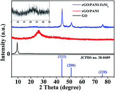
The ternary nanocomposites of rGO–PANI–FeNi3 were successfully synthesized by combining polymerization with hydrothermal reduction reaction. Graphene and polyaniline unite with FeNi3 to form ternary composite for the first time and the paraffin composite containing 20 wt% rGO–PANI–FeNi3 exhibit excellent electromagnetic wave absorption properties. The maximum reflection loss of −43.17 dB is obtained at 6.2 GHz and the absorption bandwidth (<−10 dB) of the reflection loss value can be obtained in the whole frequency range (2.96–18 GHz) with the sample thickness varying from 2.0 to 6.0 mm. The results demonstrate that rGO–PANI–FeNi3 nanocomposites containing magnetic loss and dielectric loss materials have potential applications in the electromagnetic wave absorbing area and can be widely used in developing electromagnetic wave absorption materials.


1. Introduction

In recent years, with modern technology, such as digital systems, fast processors, and radar and microwave telecommunication systems developing rapidly, electromagnetic pollution has become globally harmful to public society and is not to be ignored.1–4 To solve expanded electromagnetic interference problems, electromagnetic (EM) wave absorbing materials have attracted considerable attention.5 EM wave absorption materials are those that can absorb electromagnetic waves effectively and convert EM energy into thermal energy or energy dissipated by interference.6 An ideal absorber is required to have a wide absorption frequency range, strong absorption properties, low density, good thermal stability, and antioxidant capability.7 To date, EM absorption properties of various nanostructures, such as oxides of iron series elements and composites,8–10 traditional magnetic fillers (carbonyl-iron, Fe, Co, Ni),11–14 carbon materials,15,16 conductive polymers,17 and composite absorbents, have been investigated. With the drastic increase in the utilization of EM waves in the gigahertz range, metallic magnetic materials have been investigated in recent years.18 Nanoscale magnetic materials, such as Fe–Ni alloys, have received widespread attention due to the unique characteristics of their high saturation magnetization, high Curie temperature, low magnetostriction and small coercive forces.19,20 However, they have a few disadvantages. Their high density and easy oxidation in air limit their potential applications in the fields of both scientific studies and industrial applications. Wang and co-workers prepared surface modified Fe50Ni50 nanoparticles–reduced graphene oxide epoxy composites; the minimum RL was up to −23.9 dB with a layer thicknesses of 3.0 mm.21 Yang et al. fabricated γ-FeNi-decorated carbon nanotube composites and the epoxy-based nanocomposites exhibited a maximum reflection loss of −15.4 dB at 16.5 GHz with 1.6 mm thickness at low particle loading (2 wt%) of specimen.22 Feng et al. prepared flaky Fe-50 wt% Ni alloy particles by mechanical milling; the minimum RL and peak frequency of the 3 mm absorber were −8.0 dB and 1.3 GHz, respectively.23

Carbon-based materials are good dielectric loss absorbers and it is noteworthy that carbon-based materials are lightweight materials, especially the graphene materials. Graphene, which is a one atom thick two-dimensional planar sheet of carbon, has a number of unique physical properties due to the high surface area and it is one of the most promising candidates for achieving the lightweight and high efficiency EM wave absorbers.24–26 Polyaniline (PANI) is one of the most promising conducting polymers with good dielectric properties, high environmental stability and relatively low density.27–29 Protonic acid plays a role of doping agent in the process of aniline polymerization. Sulfuric acid,30 hydrochloric acid31 and perchloric acid32 can be used as doping agents. Relative permittivity of the composite can be adjusted by adding polyaniline. Therefore, PANI composites can be used as superior microwave absorbers and over the past decade much attention has been paid to their EM absorption properties.33–36

In this study, we prepared the rGO–PANI–FeNi3 ternary composites as excellent absorption materials due to the unique properties of metallic magnetic materials, PANI and graphene with multi-interfacial polarization and impedance matching. In addition, most researches on magnetic alloy absorbers are binary composites. For rGO–PANI–FeNi3 ternary nanocomposites, FeNi3 nanoalloy is used as the magnetic loss material and graphene and PANI as dielectric loss materials. Moreover, graphene nanosheets can serve as an ideal substrate for deposition of the nanoalloy. Because of their synergistic effect, interfacial polarization induced by multiple interfaces in the composites,37 rGO–PANI–FeNi3 ternary nanocomposite exhibits excellent electromagnetic wave absorption performance.

2. Experimental

All chemicals and reagents are of analytical grade and used without further purification. High purity water (resistivity of >18 MΩ cm) was freshly prepared and used for all experiments.

2.1. Synthesis of rGO–PANI–FeNi3 composites

Graphene oxide (GO) was synthesized by Hummers' method according to the literature.38 GO–PANI nanocomposites were prepared as follows. 100 mg of graphene oxide was dispersed in 1 M HClO4 solution under ultrasonic treatment for 0.5 h. The mixture was cooled to 0–3 °C under stirring and then 0.05 mL of aniline monomer was added into the solution and stirred for 30 min. (NH4)2S2O8 (the molar ratio of APS to aniline was 1[thin space (1/6-em)]:[thin space (1/6-em)]1.2) was dissolved in 10 mL 1 M HClO4 solution and slowly added dropwise to the abovementioned mixture with vigorous stirring. The mixture was stirred for 24 h at 0–3 °C and the rGO–PANI nanocomposites products were centrifuged and washed with water and ethanol several times. The products were dispersed in 60 mL water for subsequent experiments. Preparation of rGO–PANI–FeNi3 nanocomposites was carried out by the hydrothermal reduction reaction of Fe2+ and Ni2+ in the presence of GO–PANI. In brief, 0.34 g FeSO4·7H2O, 0.89 g NiCl2·6H2O, 2.0 g PEG-2000 and 1.0 mL C6H6 were dissolved in 100 mL of water under a nitrogen atmosphere. The GO–PANI solution was dispersed in the abovementioned solution under stirring and sodium hydroxide (2.0 g NaOH) and hydrazine (20 mL N2H4·H2O) were added. The mixture was transferred into a 200 mL Teflon-lined stainless steel autoclave and kept in an oven at 180 °C for 18 h. After cooling to room temperature, the final products were washed with water several times until the pH became neutral and freeze dried overnight. For comparison, rGO–PANI nanocomposite was prepared via a similar hydrothermal process.

2.2. Characterization

The phase structures and morphology of the as-prepared products were determined by powder X-ray diffraction (XRD, D/max-2500, 40 kV, 100 mA of Cu-Kα, Rigaku) and transmission electron microscopy (TEM; Tecnai F30 G2, FEI, USA). The thermal stabilities of the composites were determined on a Q50 thermogravimetric analyzer in air and nitrogen atmosphere with a heating rate of 20 °C min−1. The magnetic properties were measured using a vibrating sample magnetometer (VSM, Riken Denshi, BHV-525) with a maximum applied field of 13[thin space (1/6-em)]500 Oe at room temperature. The samples were prepared by uniformly mixing 20 wt% of the sample with a paraffin matrix and pressed into toroidal samples with φout 7.00 mm and φin 3.04 mm. Electromagnetic parameter determinations (complex relative dielectric permittivity and magnetic permeability) of paraffin composites were carried out by a vector network analyzer (NA, HP8720ES) in the range from 2 to 18 GHz.

3. Results and discussion

The typical XRD patterns of prepared samples are shown in Fig. 1. The diffraction peak of GO is sharp and centered at 2θ = 9.38° corresponding to an interlayer spacing of 0.94 nm due to the layers containing oxygen functional groups. For rGO/PANI, the disappearance of the peak at 2θ = 9.38° indicates that oxygen groups have been removed and the peak at 25.68° belongs to the (002) plane of a graphitic structure. It illustrates the successful reduction of GO through the hydrothermal route. The peaks of the PANI have not been obviously observed due to their low content and non-crystalline nature.39 For rGO–PANI–FeNi3, nanoscale FeNi3 alloys matched the cubic-phase FeNi3 with peaks corresponding to its (111), (200) and (220) reflections (JCPDS, no. 38-0409). The expanded curve shows a weak and broad band at 23.2° belonging to the (002) plane of rGO. Compared with rGO–PANI composites, the interlayer spacing of the composite graphitic structures is larger due to the presence of FeNi3 crystallites.
image file: c5ra27905d-f1.tif
Fig. 1 XRD patterns of GO, rGO–PANI, and rGO–PANI–FeNi3 nanocomposites.

The morphology and structure of the synthesized rGO–PANI and rGO–PANI–FeNi3 nanocomposites were examined by transmission electron microscopy (TEM) and the results are presented in Fig. 2. In Fig. 2a, it can be clearly observed that the GO are transparent silky nanosheets with some wrinkles on the surface. In Fig. 2b, it can be observed that the surface of GO becomes rough, which indicates that a thin layer of PANI covers the GO nanosheets. As shown in Fig. 2c, GO nanosheets are decorated with large scale FeNi3 nanoparticles via a hydrothermal method. Most of the FeNi3 nanoparticles range from 80–100 nm, apart from a few conglomerated nanoparticles with sizes around 160 nm (Fig. 2d–f). It is evident that the surface of rGO–PANI is decorated by a large quantity of FeNi3 nanoparticles and both the outline of rGO–PANI and FeNi3 nanoparticles can be clearly observed. In addition, these FeNi3 nanoparticles are firmly attached to the rGO–PANI, even after sonication applied during the preparation of TEM specimens, indicating that an excellent adhesion between rGO–PANI and FeNi3 nanoparticles is obtained. The lattice planes of the nanoparticles are measured to be 0.20 and 0.17 nm (Fig. 2h), corresponding to the spacing of (111) and (220) planes of FeNi3. Furthermore, the selected-area electron diffraction (SAED) pattern (Fig. 2i) clearly shows a ring pattern arising from cubic FeNi3, further confirming the crystalline nature of the FeNi3 nanoalloys.


image file: c5ra27905d-f2.tif
Fig. 2 TEM images of (a) GO and (b) rGO–PANI; (c–f) TEM images, (g and h) HRTEM images and (i) SAED pattern of rGO–PANI–FeNi3 nanocomposites.

To study the component contents of the compound, TG analyses in air and nitrogen atmosphere are carried out and the curves are presented in Fig. 3. The minor weight loss can be assigned to the evaporation of the absorbed water and solvent residues in the samples when the temperature is below 100 °C. For GO, the weight loss in the range of 200–300 °C is due to the decrease of oxygenated functional group content. A significant weight loss occurs at 500–550 °C and the weight loss becomes stable at about 550 °C, which can be ascribed to the oxidation and decomposition of rGO.40 For rGO–PANI, the major weight loss, from 350–450 °C, is accounted for the by degradation of the polymeric backbone and residual oxygenated groups of rGO. For rGO–PANI–FeNi3, the two peaks of weight loss from 200–350 °C and 400–450 °C are supposedly due to the decomposition of oxygenated functional groups and the polymer, respectively. However, when the temperature is increasing, the mass of the compound increases due to alloy oxidation reactions in the air. In addition, the residual weight for rGO–PANI–FeNi3 nanocomposites is more than 100%. TG curves in nitrogen were investigated to calculate the content of FeNi3 in the nanocomposites. As shown in Fig. 3c, when the temperature increases to 800 °C, the weight loss of rGO–PANI and rGO–PANI–FeNi3 are about 43.75% and 17.14%, respectively. The calculated FeNi3 content is 61.93% based on the weight loss of rGO–PANI and rGO–PANI–FeNi3.


image file: c5ra27905d-f3.tif
Fig. 3 TG curves of (a) GO and rGO–PANI, (b) rGO–PANI–FeNi3 nanocomposites in air, (c) TG curves of rGO–PANI and rGO–PANI–FeNi3 in nitrogen atmosphere.

The field-dependent magnetizations for FeNi3 and rGO–PANI–FeNi3 nanocomposites were measured by vibrating sample magnetometer at room temperature. As shown in Fig. 4, the MH curve indicates the ferromagnetic behavior of the FeNi3 nanoparticles and the saturation magnetization of FeNi3 is 112.2 emu g−1. The saturation magnetization, coercivity, and remnant magnetization of the ternary nanocomposite are 80.88 emu g−1, 80.0 Oe and 4.5 emu g−1, respectively.


image file: c5ra27905d-f4.tif
Fig. 4 Magnetization curve of FeNi3 and rGO–PANI–FeNi3 nanocomposite at room temperature.

The electromagnetic parameters (relative complex permittivity, εr = ε′ − jε′′, and relative complex permeability, μr = μ′ − jμ′′) were measured over 2–18 GHz and are shown in Fig. 5a and b. The electromagnetic wave absorption property of an absorber is generally determined by the electromagnetic parameters. Fig. 5a and b present the real parts (ε′, μ′) and the imaginary parts (ε′′, μ′′) of the rGO–PANI and rGO–PANI–FeNi3 nanocomposites, respectively. For rGO–PANI, the ε′ values are in the range of 61.87–21.81 with some fluctuations. Values of ε′′ at first decrease sharply and then slightly as the frequency is increased ranging from 174.47 to 16.37. In addition, the μ′ and μ′′ values float in the range from 1.42 to 0.65 and 0.52 to −0.09, respectively. For rGO–PANI–FeNi3, it can be observed that the value of ε′ decreases from 8.34 to ∼6.0 in the testing frequency range and the trends of ε′′ with frequency are similar to ε′ and vary between 2.78 and 1.57. As can be observed in Fig. 5b, μ′ decreases from 1.29 to 0.88 and the μ′′ values also decrease gradually from 0.86 to −0.096 in the frequency range of 2–14 GHz. Interestingly, the μ′′ values are very negative from about 15 to 18 GHz with a minimum value of −0.096.


image file: c5ra27905d-f5.tif
Fig. 5 Frequency dependence of the complex permittivity and complex permeability of (a) rGO–PANI and (b) rGO–PANI–FeNi3; the loss tangent of (c) rGO–PANI and (d) rGO–PANI–FeNi3 nanocomposites.

In general, the relative permittivity and the permeability represent the dynamic dielectric and magnetic properties of materials and the real part and imaginary parts stand for the storage capability and dissipation of energy, respectively. According to the free electric theory, ε is related to the electrical conductivity by the relation as follows:

 
image file: c5ra27905d-t1.tif(1)
where σ is the electrical conductivity of the absorber, ε0 is the permittivity of free space and f is the frequency of the electromagnetic wave. For rGO–PANI, electronic transmission on the graphite-like structure is easy, indicating that it possesses high conductivity. When the FeNi3 nanoparticles are introduced to the rGO–PANI nanosheets, the resistivity is enhanced due to the graphite structure being destroyed by FeNi3 nanoparticles. The electrical conductivities of the samples were measured by four-probe conductivity meter.41,42 The conductivity of rGO–PANI and rGO–PANI–FeNi3 are 230.7 S m−1 and 30.72 S m−1, respectively. For rGO–PANI–FeNi3, the lower conductivity may be due to the addition of FeNi3 nanoparticles, which destroys graphite structure. Thus, the higher ε′′ values may be determined by its high conductivity. From Fig. 5a and b, it can be observed that both ε′ and ε′′ values of rGO–PANI are higher than those of rGO–PANI–FeNi3 because of the introduction of FeNi3 nanoalloys has lowered εr of rGO–PANI to improve the level of impedance matching.43 It is noteworthy that partial values of μ′′ are negative in the range from 16 to 18 GHz, and the minimum value reached is −0.096. Typically, this phenomenon can be considered as the radiation magnetic energy caused by the induced magnetic field.44,45 The dissipation factors containing dielectric loss (tan[thin space (1/6-em)]δε = ε′′/ε′) and magnetic loss (tan[thin space (1/6-em)]δμ = μ′′/μ′) of rGO–PANI and rGO–PANI–FeNi3 nanocomposites are shown in Fig. 5c and d, respectively. Both rGO and PANI are dielectric loss absorbers and therefore dielectric loss makes a major contribution to electromagnetic loss of rGO–PANI. In addition, the dielectric loss of the composite comes from polarization, interfacial polarization and leakage conductance in the microwave frequency range. For rGO–PANI nanocomposites, the high surface area and the existence of PANI are conductive to the interfacial polarization. As for rGO–PANI–FeNi3, it can be observed that the values of tan[thin space (1/6-em)]δμ decreased in the testing frequency range. There are resonance peaks over 2–5 GHz in the curves of tan[thin space (1/6-em)]δμ, which can be assigned to the natural resonance. In general, the frequency of natural resonance should be around several tens of megahertz.18 However, the natural resonance frequency shifts to higher frequency in this study probably because of the nanosize effect.46 According to the natural resonance equation, 2πfr = rHa, Ha = 4|K1|/3μ0Ms, where r is the gyromagnetic ratio, Ha is the anisotropy energy and |K1| is the anisotropy coefficient. The anisotropy energy of rGO–PANI–FeNi3 is remarkably increased due to the surface anisotropic field by the small size effect.21 The higher anisotropy energy is useful for the improvement of microwave absorption properties.

The tan[thin space (1/6-em)]δμ has negative values due to the negative μ′′ values of μ′′/μ′ over the frequency range from 15 to 18 GHz. The negative values are assigned to the induced magnetic field caused by the eddy current. To verify the existence of eddy the current, we take a further analysis of the experimental results. For eddy current loss, the contribution to imaginary part μ′′ of the complex permeability can be expressed by the equation as follows:

 
μ′′ = 2πμ0(μ′)2σ × d2f/3 (2)
where μ0 is the permeability of vacuum, σ is the electric conductivity and d is the thickness of the composites. Thus, C0 can be described by the equation as follows:
 
C0 = μ′′(μ′)−2f−1 = 2πμ0σd2/3 (3)

If the magnetic loss only results from the eddy current loss, the values of C0 should be constant when the frequency varies.38 As shown in Fig. 6, the values of C0 first decrease sharply as the frequency is increased and then maintain nearly constant as the frequency increases. Thus, we can conclude that the eddy current loss is the main magnetic loss at high frequency. In addition, the induced magnetic energy caused by the eddy current may contribute the complementary magnetic loss with a conversion of the incident wave to other forms of energy.46 Therefore, magnetic loss of the rGO–PANI–FeNi3 nanocomposites was mainly caused by the natural resonance in the frequency range of 3–5 GHz and the eddy current effect in the higher frequency range from 10.0 to 18.0 GHz.


image file: c5ra27905d-f6.tif
Fig. 6 The values of C0 as a function of frequency for rGO–PANI–FeNi3 nanocomposites.

To characterize the electromagnetic wave absorption properties of the rGO–PANI and rGO–PANI–FeNi3 composites, the reflection loss (RL) curves at a given frequency range were calculated according to the transmit-line theory given below as follows:47

 
image file: c5ra27905d-t2.tif(4)
 
image file: c5ra27905d-t3.tif(5)
where Zin is the input impedance of the absorber, μr and εr are the relative complex permeability and the relative complex permittivity, respectively, f is the frequency, d is the thickness and c is the velocity of electromagnetic waves in free space. The dependence of RL on varying thickness in the microwave frequency in the range of 2–18 GHz is presented in Fig. 7. As can be observed from Fig. 7a and b, the electromagnetic wave absorption performances of binary composite rGO–PANI and FeNi3 nanoalloy are poor. For rGO–PANI, the maximum value of RL is about −2.7 dB and the maximum RL value of FeNi3 is −4.2 dB. However, the rGO–PANI–FeNi3 nanocomposites exhibit excellent electromagnetic wave absorption properties in the testing frequency range. The rGO–PANI–FeNi3 absorber with a thickness of 4.8 mm exhibits a maximum RL value, which is −43.17 dB at 6.2 GHz and an RL value below −10 dB can be obtained in the whole frequency range (2.96–18 GHz) with the sample thickness varying from 2.0 to 6.0 mm. It is noteworthy that the maximum RL value of rGO–PANI–FeNi3 nanocomposites shifts to lower frequency with increasing thickness of the layer. Comparing with rGO–PANI, the enhanced absorption properties of the as-prepared rGO–PANI–FeNi3 nanocomposites can be ascribed to the matching dependence43 between the dielectric absorber and the magnetic material. rGO and PANI are dielectric loss absorbents and the FeNi3 nanocrystals have outstanding magnetic characteristics at the same time. The ternary nanocomposites have good impedance matching and are of benefit to electromagnetic wave absorption performance. The abovementioned results demonstrate that the rGO–PANI–FeNi3 nanocomposites could be used as a high-efficiency electromagnetic wave absorbing material.


image file: c5ra27905d-f7.tif
Fig. 7 Reflection loss curves of (a) rGO–PANI, (b) FeNi3 and (c) rGO–PANI–FeNi3 nanocomposites.

4. Conclusions

In conclusion, the rGO–PANI–FeNi3 ternary nanocomposites were successfully synthesized by combining polymerization with a hydrothermal reduction reaction. As EM wave absorbers, the paraffin composite containing 20 wt% ternary nanocomposites exhibit enhanced microwave absorption properties. The maximum reflection loss of rGO–PANI–FeNi3 is −43.17 dB at 6.2 GHz with a thickness of 4.8 mm. The absorption bandwidth (<–10 dB) of the reflection loss value can be obtained over the whole frequency range (2.96–18 GHz) with the sample thickness varying from 2.0 to 6.0 mm. It is believed that such composites containing magnetic loss materials and dielectric loss materials such as rGO–PANI–FeNi3 composites will find widespread application in the microwave absorbing area.

Acknowledgements

This study was supported by the Spaceflight Innovation Fund of the People's Republic of China and the Spaceflight Support Technology Fund of China (No. 2014-HT-XGD).

References

  1. L. Kong, X. Yin, M. Han, L. Zhang and L. Cheng, Ceram. Int., 2015, 41, 4906 CrossRef CAS.
  2. M. Jazirehpour and S. A. Seyyed Ebrahimi, J. Alloys Compd., 2015, 639, 280 CrossRef CAS.
  3. Y. Ren, L. Yang, L. Wang, T. Xu, G. Wu and H. Wu, Powder Technol., 2015, 281, 20 CrossRef CAS.
  4. G. Li, L. Wang, W. Li and Y. Xu, Microporous Mesoporous Mater., 2015, 211, 97 CrossRef CAS.
  5. X. Qi, J. Xu, Q. Hu, W. Zhong and Y. Du, J. Mater. Sci. Eng. B, 2015, 198, 108 CrossRef CAS.
  6. B. P. Li, C. G. Wang, W. Wang, M. J. Yu, R. Gao, Y. Chen and Y. Xiang Wang, J. Magn. Magn. Mater., 2014, 365, 40 CrossRef CAS.
  7. L. Wang, Y. Huang, X. Sun, H. Huang, P. Liu, M. Zong and Y. Wang, Nanoscale, 2014, 6, 3157 RSC.
  8. M. Zong, Y. Huang, H. Wu, Y. Zhao, Q. Wang and X. Sun, Mater. Lett., 2014, 114, 52 CrossRef CAS.
  9. Z. Wang, L. Wu and J. Zhou, Nanoscale, 2014, 12298 RSC.
  10. S. Hazra, B. K. Ghosh, M. K. Patra, R. K. Jani, S. R. Vadera and N. N. Ghosh, Powder Technol., 2015, 279, 10 CrossRef CAS.
  11. Y. Yang, Z. W. Li, C. P. Neo and J. Ding, J. Phys. Chem. Solids, 2014, 75, 230 CrossRef CAS.
  12. Z. Zhu, X. Sun, H. Xue, H. Guo, X. Fan, X. Pan and J. He, J. Mater. Chem. C, 2014, 2, 6582 RSC.
  13. J. T. McKeown, Y. Wu, J. D. Fowlkes, P. D. Rack and G. H. Campbell, Adv. Mater., 2015, 27, 1060 CrossRef CAS PubMed.
  14. G. Chen, Y. Su, P. Kuang, Z. Liu, D. Chen, X. Wu, N. Li and S. Qiao, Chem.–Eur. J., 2015, 21, 4614 CrossRef CAS PubMed.
  15. G. Liu, L. Wang, G. Chen, S. Hua, C. Ge, H. Zhang and R. Wu, J. Alloys Compd., 2012, 514, 183 CrossRef CAS.
  16. T. Wang, Z. Liu, M. Lu, B. Wen, Q. Ouyang, Y. Chen, C. Zhu, P. Gao, C. Li, M. Cao and L. Qi, J. Appl. Phys., 2013, 113, 24314 CrossRef.
  17. H. Yu, T. Wang, B. Wen, M. Lu, Z. Xu, C. Zhu, Y. Chen, X. Xue, C. Sun and M. Cao, J. Mater. Chem., 2012, 22, 21679 RSC.
  18. Y. Yang, C. Xu, Y. Xia, T. Wang and F. Li, J. Alloys Compd., 2010, 493, 549 CrossRef CAS.
  19. A. G. Kolhatkar, I. Nekrashevich, D. Litvinov, R. C. Willson and T. R. Lee, Chem. Mater., 2012, 25, 1092 CrossRef PubMed.
  20. X. Wei, G. Zhu, J. Zhou and H. Sun, Mater. Chem. Phys., 2006, 100, 481 CrossRef CAS.
  21. J. Wang, J. Wang, R. Xu, Y. Sun, B. Zhang, W. Chen, T. Wang and S. Yang, J. Alloys Compd., 2015, 653, 14 CrossRef CAS.
  22. Q. Yang, L. Liu, D. Hui and M. Chipara, Composites, Part B, 2016, 87, 256 CrossRef CAS.
  23. Y. Feng and T. Qiu, J. Magn. Magn. Mater., 2012, 324, 2528 CrossRef CAS.
  24. X. Ding, Y. Huang and J. Wang, RSC Adv., 2015, 80, 64878 RSC.
  25. Y. Huang, L. Wang and X. Sun, Mater. Lett., 2015, 144, 26 CrossRef CAS.
  26. M. Zong, Y. Huang and N. Zhang, Mater. Lett., 2015, 145, 115 CrossRef CAS.
  27. H. Gu, J. Guo, X. Yan, H. Wei, X. Zhang, J. Liu, Y. Huang, S. Wei and Z. Guo, Polymer, 2014, 55, 4405 CrossRef CAS.
  28. B. Qiu, J. Guo, Y. Wang, X. Wei, Q. Wang, D. Sun, A. Khan, P. Young, R. O'Connor, X. Huang, X. Zhang, L. Weeks, S. Wei and Z. Guo, J. Mater. Chem. C, 2015, 3, 3989 RSC.
  29. Y. Zhao, H. Wei, M. Arowo, X. Yan, W. Wu, J. Chen, Y. Wang and Z. Guo, Phys. Chem. Chem. Phys., 2015, 17, 1498 RSC.
  30. P. Liu, Y. Huang and X. Zhang, Compos. Sci. Technol., 2015, 107, 54 CrossRef CAS.
  31. X. Zhang, S. Wei, N. Haldolaarachchige, A. Colorado, Z. Luo and Z. Guo, J. Phys. Chem. C, 2012, 116, 15731 CAS.
  32. L. Wang, Y. Huang, C. Lia, J. Chen and X. Sun, Synth. Met., 2014, 198, 300 CrossRef CAS.
  33. J. Luo, Y. Xu, W. Yao, C. Jiang and J. Xu, Compos. Sci. Technol., 2015, 117, 315 CrossRef CAS.
  34. L. Wang, Y. Huang, C. Li, J. Chen and X. Sun, Compos. Sci. Technol., 2015, 108, 1 CrossRef CAS.
  35. F. Guo, W. Zi, G. Ji, L. Zou and S. Gan, J. Polym. Res., 2015, 22, 1 CrossRef CAS.
  36. Y. Xu, J. Luo, W. Yao, J. Xu and T. Li, J. Alloys Compd., 2015, 636, 310 CrossRef CAS.
  37. Y. Ren, H. Wu, M. Lu, Y. Chen, C. Zhu, P. Gao, M. Cao, C. Li and Q. Ouyang, ACS Appl. Mater. Interfaces, 2012, 4, 6436 CAS.
  38. J. A. R. E. William and S. Hummers, J. Am. Chem. Soc., 1958, 80, 1339 CrossRef.
  39. H. Huang, Y. Huang, M. Wang, X. Chen, Y. Zhao, K. Wang and H. Wu, Electrochim. Acta, 2014, 147, 201 CrossRef CAS.
  40. M. Zong, Y. Huang, Y. Zhao, X. Sun, C. Qu, D. Luo and J. Zheng, RSC Adv., 2013, 3, 23638 RSC.
  41. Q. He, T. Yuan, X. Zhang, X. Yan, J. Guo, D. Ding, A. Khan, P. Young, A. Khasanov, Z. Luo, J. Liu, T. Shen, X. Liu, S. Wei and Z. Guo, J. Phys. Chem. C, 2014, 118, 24784 CAS.
  42. J. Zhu, S. Wei, N. Haldolaarachchige, P. David and Z. Guo, J. Phys. Chem. C, 2011, 115, 15304 CAS.
  43. X. Sun, J. He, G. Li, J. Tang, T. Wang, Y. Guo and H. Xue, J. Mater. Chem. C, 2013, 1, 765 RSC.
  44. J. Zhou, J. He, T. Wang, G. Li, Y. Guo, J. Zhao and Y. Ma, J. Alloys Compd., 2011, 509, 8211 CrossRef CAS.
  45. S. Guo, L. Wang and H. Wu, Adv. Powder Technol., 2015, 26, 1250 CrossRef CAS.
  46. H. Wu, L. Wang, Y. Wang, S. Guo and Z. Shen, J. Alloys Compd., 2012, 525, 82 CrossRef CAS.
  47. S. S. Kim, S. B. Jo, K. I. Gueon, K. K. Choi, J. M. Kim and K. S. Chum, IEEE Trans. Magn., 1991, 27, 5462 CrossRef CAS.

This journal is © The Royal Society of Chemistry 2016
Click here to see how this site uses Cookies. View our privacy policy here.