DOI:
10.1039/C5RA26052C
(Paper)
RSC Adv., 2016,
6, 13032-13039
Facile fabrication of tea tree oil-loaded antibacterial microcapsules by complex coacervation of sodium alginate/quaternary ammonium salt of chitosan†
Received
7th December 2015
, Accepted 21st January 2016
First published on 25th January 2016
Abstract
In this study, flavour tea tree oil (TTO)-loaded antibacterial microcapsules were developed based on the complex coacervation of sodium alginate (SA) and a quaternary ammonium salt of chitosan (HACC). The optimum preparation conditions of the TTO-loaded microcapsules determined using the response surface methodology (RSM) were as follows: a weight ratio of the core to wall material (core-wall ratio) of 1![[thin space (1/6-em)]](https://www.rsc.org/images/entities/char_2009.gif) :1, a pH value of 6.0 and a mass concentration of CaCl2 solution of 0.6 w/v%, at which the resultant microcapsules showed the greatest actual encapsulation efficiency (EE) of 66.06% ± 2.53%. Thereafter, the resultant microcapsules were characterized in terms of morphology, size, components and thermal stability using scanning electron microscopy (SEM), a laser particle diameter analyzer (LPDA), Fourier transform infrared spectroscopy (FTIR), and thermal gravity-differential thermal analysis (TG-DTA) and differential scanning calorimetry (DSC), respectively. Furthermore, both the in vitro drug release and antimicrobial properties of the microcapsules were also assessed. The results displayed that the TTO-loaded microcapsules had a spherical shape with the particle sizes in the range from 1.91 μm to 13.18 μm. The microcapsules possessed outstanding performances with respect to the thermal stability, sustained release activity, antimicrobial effect and long-term inhibition activity. The release profiles of TTO from the microcapsules could be well described by the Ritger–Peppas model. Based on the above research results, the simple and environmentally-friendly microencapsulation formulation could effectively improve the stability and prolong the antimicrobial efficacy of TTO. The microcapsules may find applications in food, cosmetics and medicine.
:1, a pH value of 6.0 and a mass concentration of CaCl2 solution of 0.6 w/v%, at which the resultant microcapsules showed the greatest actual encapsulation efficiency (EE) of 66.06% ± 2.53%. Thereafter, the resultant microcapsules were characterized in terms of morphology, size, components and thermal stability using scanning electron microscopy (SEM), a laser particle diameter analyzer (LPDA), Fourier transform infrared spectroscopy (FTIR), and thermal gravity-differential thermal analysis (TG-DTA) and differential scanning calorimetry (DSC), respectively. Furthermore, both the in vitro drug release and antimicrobial properties of the microcapsules were also assessed. The results displayed that the TTO-loaded microcapsules had a spherical shape with the particle sizes in the range from 1.91 μm to 13.18 μm. The microcapsules possessed outstanding performances with respect to the thermal stability, sustained release activity, antimicrobial effect and long-term inhibition activity. The release profiles of TTO from the microcapsules could be well described by the Ritger–Peppas model. Based on the above research results, the simple and environmentally-friendly microencapsulation formulation could effectively improve the stability and prolong the antimicrobial efficacy of TTO. The microcapsules may find applications in food, cosmetics and medicine.
1. Introduction
In recent years, volatile essential oils have gradually gained wide attention for the development of natural health products because of their special performances, which are beneficial for the enhancement of people’s living standards. Among various volatile essential oil-based natural health products, environmentally-friendly antimicrobial products have been favored by more and more people, because every person inevitably comes into contact with all sorts of harmful bacteria, fungi, viruses and other microbes. Tea tree oil (TTO), from the leaves of Melaleuca alternifolia (family Myrtaceae), is widely used not only in traditional Australian medicine, but also as an important raw material in pharmaceutical, perfume and cosmetics industries,1 due to its broad-spectrum antibacterial, antifungal, anti-inflammatory, anti-allergy, antioxidant and aromatic properties.2–6 TTO is mainly composed of terpenoids, especially monoterpenes, sesquiterpenes, and their associated oxygenated analogues.7 However, TTO is volatile, has poor water-solubility and is chemically unstable in the presence of air, light and high temperature, which can obviously limit its practical application.
Microcapsules have attracted considerable interests in both academic and industrial areas, because they are very effective micrometer-sized containers to deliver active ingredients. Microcapsules can not only protect active ingredients from hostile environments, but can also prolong the release of encapsulated ingredients.8,9 In recent years, several approaches have been proposed to prepare microcapsules, including solvent evaporation,10–12 complex coacervation,13,14 solvent extraction,15 spray drying,16,17 layer-by-layer assembly18 and phase separation.19,20 Especially, complex coacervation, one of the most versatile preparation methods of microcapsules, is based on the simultaneous desolvation of oppositely charged polyelectrolytes by media modifications.21 This method can afford microcapsules with a good control over their particle size, loading, wall permeability and release properties, resulting in broadening the application of the complex coacervation method to prepare drug-loaded microcapsules. Hence, it seems to be a good choice to solve the above-mentioned problem of TTO using a complex coacervation method to load TTO in microcapsules, which can decrease the release rate, enhance the stability, prolong the functional efficiency and expand the potential applications of TTO.
Chitosan is a cationic polysaccharide derived from chitin, which is abundant in the shells of crustaceans and the exoskeletons of insects. In addition to its antimicrobial activity, chitosan also has some important advantages such as nontoxicity, biocompatibility, biodegradability, cost-effectiveness, and film-forming ability. Recently, the employment of chitosan for the wall material of drug-loaded microcapsules and antimicrobial agents have been well investigated.22–24 However, the poor water solubility of chitosan has restricted its application fields. As a chitosan derivative, the quaternary ammonium salt of chitosan (HACC) shows the improved water solubility and higher antibacterial activity than chitosan over an entire range of pH values,25 which can increase the potential of chitosan in drug delivery and antimicrobial agent applications.26 Furthermore, sodium alginate (SA), an anionic linear polysaccharide, is composed of (1→4)-β-D-mannuronic acid (M) and (1→4)-α-L-guluronic acid (G) obtained by brown algae and some bacteria, which has abundant carboxyl groups and has also been widely used as wall material of drug-loaded microcapsules.27 Thus, the combination of HACC and SA, oppositely charged polyelectrolytes, can be used as wall materials to prepare TTO-loaded microcapsules on account of the strong electrostatic interaction between the quaternary ammonium groups of HACC and the carboxyl groups of SA.
In this work, TTO-loaded microcapsules were prepared by the complex coacervation of SA and HACC. The optimum preparation condition for encapsulating TTO to obtain the highest encapsulation efficiency was discussed using the response surface methodology (RSM) with Box–Benhnken design (BBD). Thereafter, the morphology, particle size, components and thermal stability of the microcapsules were studied in detail. Furthermore, the antibacterial properties and the sustained release behaviors of the TTO-loaded microcapsules were investigated. The main objective of this study was to develop an effective formulation for the microencapsulation of various volatile oil compounds, which could have extensive applications in food, cosmetics and medicine.
2. Materials and methods
2.1 Materials
HACC (degree of substitution 0.93) was purchased from AK Biotech-Ltd., China. Sodium alginate (SA) was obtained from Guoyao Chemical Reagent Co., China. Tea tree oil (TTO) was provided by Kangshen Medicinal Oil Refinery, China. Acetic acid, calcium chloride, sodium hydroxide, hexane, span-80 and tween-80 were supplied by Tianjin Damao Chemical Reagent Company, China. All chemicals were used as received. The water used in all experiments was distilled water.
2.2 Preparation of TTO-loaded microcapsules
The TTO-loaded microcapsules were fabricated based on the complex coacervation of SA and HACC. The detailed preparation processes of the TTO-loaded microcapsules were performed as follows. Firstly, two solutions were separately prepared. Solution A: 1.125 g of HACC powder was dissolved in 150 mL of 1.0 v/v% acetic acid. Solution B: 0.150 g of SA was dissolved in 50 mL of distilled water at 45 °C and then 0.25 g of tween-80 was added to form a homogeneous solution. Afterwards, the different weights of TTO (1:2, 1:1 or 3:2, with respect to the weight of the wall material) and 0.25 g of span-80 were mixed equally and then added into solution B, followed by homogenization at 2500 rpm for 5 min at room temperature to obtain an oil-in-water (O/W) emulsion. Furthermore, the obtained emulsion was dropwise added into a 500 mL three necked flask containing solution A using a syringe under the speed of 600 rpm, and then 5 wt% sodium hydroxide solution was added to adjust the pH value (pH = 5.0, 6.0 or 7.0) with stirring for another 30 min. In addition, 30 mL of CaCl2 solution (0.3, 0.6 or 0.9 w/v%) was dropwise added within 0.5 h, and the system was stirred for another 2 h in a 45 °C water bath to obtain the microcapsule suspension. Finally, the microcapsule powder was obtained by spray drying the microcapsule suspension at the following conditions: an inlet drying temperature of 120 °C, a peristaltic pump rate of 10%, an aspirator rate of 100% and a spraying air flow rate of 40 L h−1.
2.3 Optimization of the microcapsule preparation conditions by response surface methodology (RSM)
The response surface methodology (RSM), a collection of statistical and mathematical techniques, is useful for developing, improving, and optimizing processes, in which a response of interest is influenced by several variables and the objective is to optimize this response.28 Moreover, it indicates the interaction effects between multiple single factors through three-dimensional response surface graphs and two-dimensional contour graphs relating the measured responses. The former is very advantageous for learning about the main effects and interaction effects of the independent factors, while the latter provides a visual representation of the values of the response.29,30 Meanwhile, the minimum number of experimental trials that are required by means of statistical predictions is decreased through using RSM.31
RSM, a 3-factor-3-level BBD with a 17 factorial, star design and 5 central points, was employed for constructing the optimum conditions for preparing TTO-loaded microcapsules using Design Expert (version 8.0.6.1, Stat-Ease). The weight ratio of the core to the wall material (X1, 1:2 to 3:2), the pH value (X2, 5 to 7) and the mass concentration of CaCl2 (X3, 0.3 to 0.9 w/v%) were selected as the independent variables (factors) varied at three different levels: low (−1), medium (0) and high (+1) levels, while the dependent variable (response) was the encapsulation efficiency (EE, %). The second-order polynomial equation was estimated for predicting the Y variable. The model devised for the response of Y fitted the following equation:
| |
 | (1) |
where
Y (%) is the response for the EE% of TTO-loaded microcapsules.
ρ0,
ρi,
ρii and
ρij are the constant coefficients of the intercept, linear, quadratic and interaction terms, respectively.
Xi and
Xj are uncoded independent variables.
2.4 Characterization of TTO-loaded microcapsules
The morphology of the TTO-loaded microcapsules was determined using a Zeiss EVO 18 scanning electron microscope (SEM). Samples were sputtered with gold and observed at an accelerated voltage of 10 kV. The particle size distribution of the microcapsules dispersed in water was assessed using an LA-920 laser diffraction particle size analyzer (Horiba Co., Japan). Fourier transform infrared spectroscopy (FTIR) spectra of the samples were recorded using a Nicolet AVATAR360 IR instrument. Thermal gravity-differential thermal analysis (TG-DTA) studies were performed using a DTG-60 thermo-analyzer (Shimadzu, Japan) at a heating rate of 10 °C min−1 under a nitrogen atmosphere from 50 to 600 °C. The thermal stabilities of the samples were also investigated using differential scanning calorimetry (DSC) 8000 (Perkin Elmer, USA) in the range of 50 to 400 °C at a heating rate of 10 °C min−1 under a nitrogen atmosphere. The loading capacity and encapsulation efficiency of the prepared microcapsules were detected using a UV-2550 ultraviolet visible spectrophotometer (Shimadzu, Japan).
2.5 Loading capacity and encapsulation efficiency of TTO-loaded microcapsules
The TTO-loading capacity (LC) and actual encapsulation efficiency (EE) of the prepared microcapsules were estimated using the solvent extraction method as follows. 50 mg of the newly prepared microcapsules was added into a sealed conical flask containing 50 mL of hexane, followed by being placed in a vapour-bathing constant temperature vibrator at 37 °C for 24 h to extract TTO from the microcapsules completely. After centrifugation at 3000 rpm for 5 min, the supernatant was collected. This procedure was repeated three times. Afterwards, the absorbance of the supernatant was obtained using an ultraviolet-visible spectrophotometer at 264 nm detection wavelength. Every experiment was carried out in triplicate, and the mean value was calculated in order to obtain the quantitative capacity for precise encapsulation. The mean values of the triplicates of all experiments were calculated in view of the standard curve. The loading capacity (LC) and encapsulation efficiency (EE) were calculated according to the following equations, respectively:where Mt, Mm and Mo are the weight of TTO in the microcapsules, the weight of the microcapsules initially and the weight of TTO initially, respectively.
2.6 In vitro TTO release study
In the in vitro TTO release studies, the required portions of TTO-loaded microcapsules were placed in a constant temperature humidity chamber with a specific temperature and humidity. At predetermined time intervals, the samples were taken out, and then mixed with a certain volume of hexane to extract TTO from the microcapsules completely. After centrifugation at 3000 rpm for 5 min, the supernatant was collected, followed by measuring the absorbance at 264 nm using an ultraviolet-visible spectrophotometer. Each determination was carried out in triplicate, and the mean value was calculated. The cumulative release (CR) of TTO from the microcapsules was obtained using the following formula:where Mx and Mo are the weight of TTO released from the microcapsules and the weight of TTO in the microcapsules initially, respectively.
2.7 Antibacterial properties of the TTO-loaded microcapsules
In order to investigate the antibacterial properties, tests of the TTO-loaded microcapsules against both Staphylococcus aureus (S. aureus) and Escherichia coli (E. coli) were implemented via the plate counting method. Briefly, the inoculated bacterial suspensions containing about 106 to 107 colony forming units (CFU) mL−1 of the bacterial strains were cultured by suspending the bacteria in sterilized broth culture. Afterwards, 0.05 g of TTO-loaded microcapsules and 0.1 mL of the inoculation bacterial suspension were added into 10 mL of sterilized PBS. The suspension was placed in a shaker and then incubated for 24 h at 37 °C. Furthermore, 0.1 mL of the diluted suspension prepared using a gradient descent algorithm using the 10-fold dilution method was extracted and plated on an agar plate. After 24 h culturing at 37 °C, the number of bacterial colonies in the inoculated plate was counted. In addition, the control suspension without the microcapsules served as the blank control. The experimental determinations were performed in triplicate, and the average values were obtained. The degree of antibacterial properties was expressed as the inhibition rate (IR) of bacterial growth according to the following equation:| | |
IR (%) = [(A − B)/A] × 100
| (5) |
where IR represents the inhibition rate, A is the amount of bacterial colonies of the control group, and B is the amount of bacterial colonies of the sample group.
3. Results and discussion
3.1 Process optimum for the preparation of TTO-loaded microcapsules
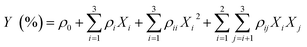
RSM is a favorable way to determine the optimum conditions and level of importance of different factors for the preparation of drug-loaded microcapsules. Herein, we employed RSM to optimize the preparation conditions of TTO-loaded microcapsules based on EE as an optimisation index. The level of the three variables with regard to the actual EE obtained from the experiments and the predicted EE produced by the model would be the most advantageous level of that variable in the preparation of TTO-loaded microcapsules and the information is presented in Table S1.† The regression equation describing the mathematical relationships between the independent (core-wall ratio X1, pH value X2 and the mass concentration of CaCl2 X3) and response variables (EE) is expressed as follows:| | |
Y = 66.06 − 2.89X1 − 2.01X2 + 2.41X3 + 0.77X1X2 + 0.44X1X3 − 0.36X2X3 − 10.02X12 − 8.06X22 − 6.64X32
| (6) |
In order to evaluate the coefficients of the quadratic model statistically, it was necessary to perform an analysis of variance (ANOVA). The P-value (less than 0.05) was considered to be indicative of the significance between the independent variable and the dependent one; however on the contrary, the interaction term indexed as non-significant (P > 0.05).32,33 From the ANOVA of the RSM experiment (Table S2†), the model F-value of 39.53 and the lack of fit F-value of 0.70 implied only a 0.01% chance on the model F-value and a 60.08% chance on the lack of fit F-value could occur due to noise, respectively. The highly significant F-value (P < 0.0001 < 0.001) and the non-significant lack of fit (P = 0.6008 > 0.05) showed that the model was excellent. The favourable value of the correlation coefficient (R2 = 0.9807) further confirmed the splendid fitness of the model, indicating that 98.07% of the variation in the RSM could be explained. The high significance of the model was assured by a satisfactory value of the adjusted coefficient of determination (Adj R2), which was calculated to be 0.9559 (in reasonable agreement with the Pred R2 of 0.8742). Furthermore, the signal to noise ratio (Adeq precision = 16.356 > 4) indicated an adequate signal. The linear term X2 was a prominent factor (P < 0.05), the influence of X1 and X3 was highly prominent (P < 0.01), and X12, X22 as well as X32 signified a highly remarkable influence on EE (P < 0.001). Obviously, the core-wall ratio (X1) and the pH value (X2) were significant parameters in mixed biopolymer systems, because the core-wall ratio affected the balance of macromolecule charges and the intensity of the electrostatic interactions driving the formation of the complexes between the two biopolymers.33 Furthermore, the moderate pH value assisted in the entirely electrostatic interactions between the positively charged HACC and negatively charged SA, which caused the total electrostatic charge to tend to zero.34
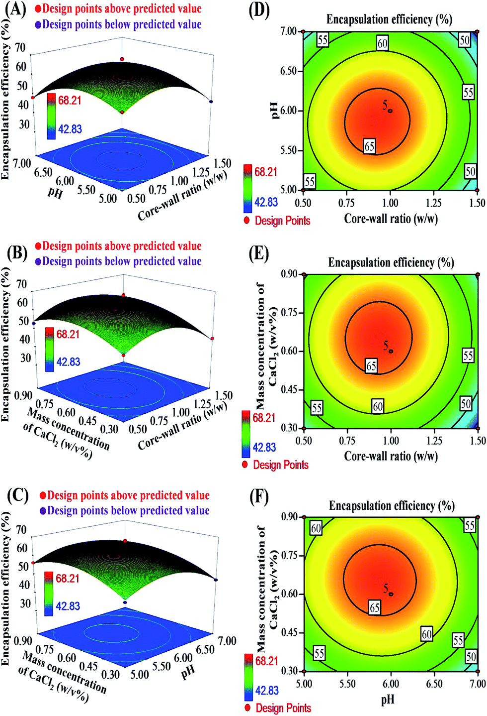
The response surfaces and corresponding contour plots are presented in Fig. 1 to further verify the results of the models. Fig. 1A and D illustrate that the EE of the TTO-loaded microcapsules increased with a decrease in both the core-wall ratio (X1, 1:2 to 1:1) and the pH value (X2, 5 to 6). Increasing the core-wall ratio (X1, 1:2 to 1:1) and the mass concentration of CaCl2 (X3, 0.3 to 0.6 wt%) resulted in an increase of EE (Fig. 1B and E). Similarly, the enhancement of the pH value (X2, 5 to 6) and the mass concentration of CaCl2 (X3, 0.3 to 0.6 wt%) led to the increase of EE (Fig. 1C and F). However, the decrease in EE occurred when the three variables increased together, which showed that the EE would reach the maximum value to a certain extent. The result obtained from the RSM analysis showed that the optimum preparation conditions of the TTO-loaded microcapsules were a core-wall ratio of 1:1, a pH value of 6.0 and a mass concentration of CaCl2 solution of 0.6 wt%, at which the resultant microcapsules showed 66.06% ± 2.53% of the actual embedding rate, 66.62% of the theoretical embedding rate and 33.18% of the LC. The EE of 66.06% ± 2.53% was in accordance with the EE of most of the drug-loaded microcapsules synthesized by complex coacervation,34,35 and the difference in the drug encapsulation efficiency could be caused by the emulsifying style and rate, the emulsifier and the oil type.36 To sum up, it was observed from the RSM analysis that the TTO-loaded microcapsules prepared at the optimal preparation conditions possessed the highest EE.
 |
| | Fig. 1 The contour plots of the response surface methodology: the core-wall ratio and the pH value (A and D); the core-wall ratio and mass concentration of CaCl2 (B and E); the pH value and mass concentration of CaCl2 (C and F). | |
3.2 Morphology and particle size distribution of TTO-loaded microcapsules
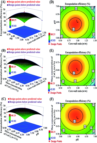
SEM images and particle size distribution curves of the TTO-loaded microcapsules prepared at the optimum conditions are shown in Fig. 2. It was seen that the microcapsules were in nearly spherical shape (Fig. 2A and B), and their particle sizes were in the range from 1.91 to 13.18 μm. Furthermore, it was also observed that the microcapsules possessed a rough wall without observable holes. As we all know, the controlled release of the microcapsules was mainly determined by the integrity of the microcapsule wall. Hence, it was speculated that the TTO-loaded microcapsules should have good controlled drug release properties. In addition, some of the ruptured microcapsules cut by a razor blade were observed using SEM (Fig. 2C). The result confirmed directly that the microcapsules possessed a core–shell structure, and TTO was successfully encased in the microcapsule wall, which could protect TTO from air, light and high temperature, in turn assisting in the enhancement of the TTO stability. Additionally, the particle sizes of the microcapsules estimated by measuring 100 microcapsules from SEM images were comparable to that measured by the laser particle size analyzer (Fig. 2D), which indicated that the microcapsules could easily redisperse into the aqueous medium.
 |
| | Fig. 2 SEM images of the TTO-loaded microcapsules (A): 2000× and (B): 5000×, the ruptured microcapsules (C) and the particle size distributions of the TTO-loaded microcapsule (D). | |
3.3 FTIR determination of TTO-loaded microcapsules
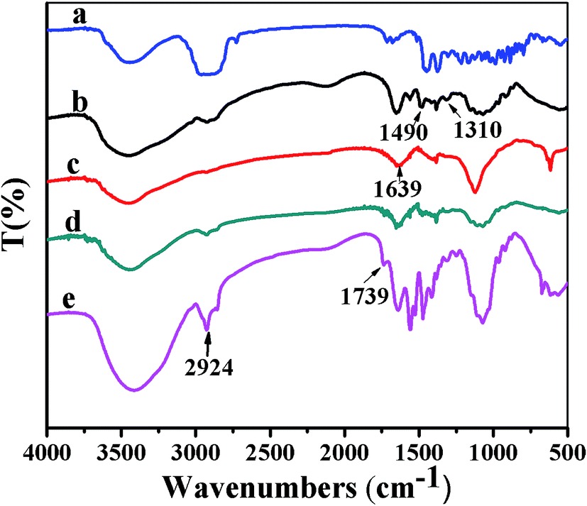
FTIR studies were performed to further verify the successful fabrication of the TTO-loaded microcapsules. Fig. 3 shows the FTIR spectra of TTO (a), HACC (b), SA (c), the microcapsule shell (d) and the TTO-loaded microcapsule (e). In the FTIR spectrum of HACC (Fig. 3b), the peaks at 1490 cm−1 and 1310 cm−1 were attributed to the bending vibration of –CH3 and the stretching vibration of C–N+ for the quaternary ammonium salt group, respectively. In the FTIR spectrum of SA (Fig. 3c), the peak at 1639 cm−1 was assigned to the stretching vibration of the carboxyl group. Compared with the FTIR spectra of HACC and SA, in the FTIR spectrum of the microcapsule shell (Fig. 3d), the peaks at 1490 cm−1 and 1310 cm−1 corresponding to HACC and the peak at 1639 cm−1 derived from SA were shifted to 1476 cm−1, 1303 cm−1 and 1567 cm−1, respectively, which demonstrated that the microcapsule shell was formed via the strong electrostatic interactions between quaternary ammonium groups of HACC and the carboxyl groups of SA. Compared with the FTIR spectrum of the microcapsule shell, the FTIR spectrum of the microcapsule (Fig. 3e) shows that the peak at 2924 cm−1 became stronger and a new sharp peak at 1739 cm−1 appeared, indicating that TTO existed in the microcapsules because the peaks at 2924 cm−1 and 1739 cm−1 were the characteristic peaks of TTO (Fig. 3a). According to the results of the FTIR analysis, the TTO-loaded microcapsules were fabricated successfully via the complex coacervation of HACC and SA.
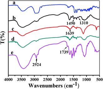 |
| | Fig. 3 FTIR spectra of (a) TTO, (b) HACC, (c) SA, (d) microcapsule shell and (e) TTO-loaded microcapsule. | |
3.4 Thermal stability analysis of TTO-loaded microcapsules
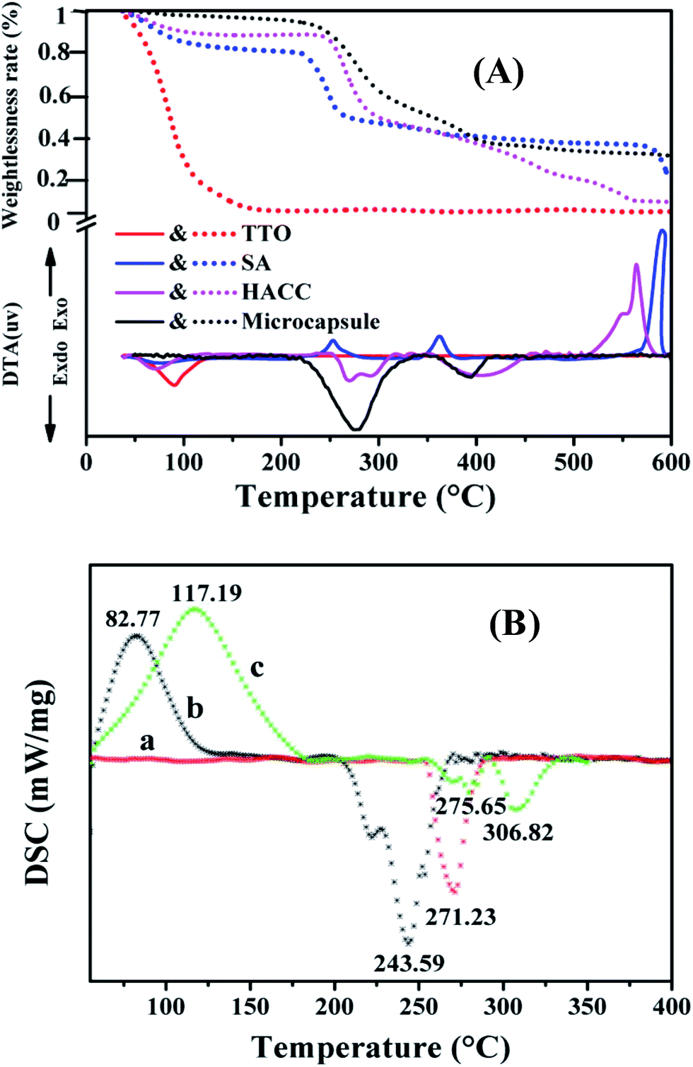
TG-DTA and DSC measurements were conducted to evaluate the thermal stabilities of TTO, SA, HACC and TTO-loaded microcapsules. The results are shown in Fig. 4. From the TG-DTA curves (Fig. 4A), the TTO shows one step of weight loss at about 90 °C, while SA, HACC and the microcapsule presented four steps. The weight of SA, HACC and the microcapsules decreased quickly from 217 °C to 267 °C, 239 °C to 311 °C, and 225 °C to 338 °C, respectively. Therefore, the primary decomposition temperature of the microcapsules was much higher than that of TTO, which suggested that the thermal stability of TTO was obviously improved by the microencapsulation process. Furthermore, it can be seen from Fig. 4B that the DSC thermograms of SA and HACC show peaks at 82 °C and 117 °C owing to the evaporation of structural water, which was identified with the corresponding DTA curves. In addition, the endothermic peaks of SA, HACC and the microcapsules presented at around 243 °C, 306 °C and 271 °C, respectively, were attributed to the decomposition of polymer chains.34 Based on the TG-DTA and DSC analysis results, it was concluded that the TTO-loaded microcapsule revealed excellent thermal stability.
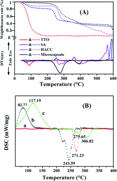 |
| | Fig. 4 (A) TG-DTA curves of TTO, SA, HACC and microcapsules. (B) DSC profiles of (a) microcapsules, (b) SA and (c) HACC. | |
3.5 In vitro release study of TTO-loaded microcapsules
It is well known that the in vitro release of drug-loaded microcapsules can be influenced by many factors, such as the environmental temperature, the environmental humidity and the particle size of the microcapsules. Herein, the release behavior of TTO from the microcapsules prepared at the optimum conditions was investigated at different temperatures and humidities in an incubator. Fig. 5A shows the release profiles of the TTO-loaded microcapsules investigated at different temperatures, whereas Fig. 5B presents the related release profiles studied in different humidities. It was observed that all of the microcapsule samples exhibited a similar TTO sustained release behavior during the whole study period, which exhibited two different phases: a relatively fast release rate in the first stage and an appreciably slow release rate in the second stage. To be specific, the release rate of TTO from the TTO-loaded microcapsules was very significant in the first release phase (within the first 48 h), which was primarily attributed to the fast release of TTO absorbed on the microcapsule surface and/or near the interior of the microcapsule surface.37,38 Afterwards, the cumulative release rate of TTO became sustained slowly and then steady gradually in the second phase, which should be ascribed to the fact that prolonging the release time of the microcapsules increased the diffusion path length of TTO and decreased the concentration difference of TTO between the inside and outside of the microcapsules. Furthermore, it was also seen that after 216 h, the cumulative TTO release rates of the microcapsules were 42.05% at 4 °C, 56.09% at 25 °C and 67.16% at 37 °C, respectively. Similarly, after 216 h, the cumulative TTO release rates of the microcapsules in a relative humidity of 24%, 35% and 50% were 67.16%, 76.32% and 86.43%, respectively. The above results showed that the in vitro release study of the microcapsules was preformed at a higher temperature and relative humidity, TTO possessed a higher release amount and release rate. This phenomenon should be ascribed to the fact that the volatility of TTO would increase with the enhancement of the temperature. Thus, the higher temperature led to easier burst release of TTO from the microcapsules. The microcapsules were placed in high relative humidity conditions, which might result in the partial dissolution of the microcapsule walls composed of hydrophilic polymers, and in turn shorten the diffusion distance of TTO from the microcapsules. Hence, the higher relative humidity resulted in the faster release of TTO from the microcapsules. Based on the above discussion, the TTO-loaded microcapsules should be stored in low temperature and humidity conditions.
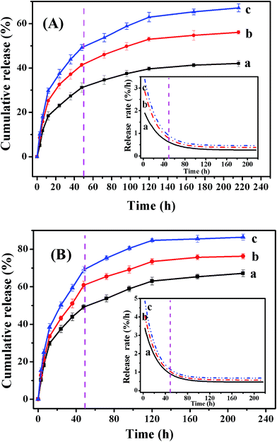 |
| | Fig. 5 In vitro release profiles of TTO from the microcapsules with different environmental (A) temperature: (a) 4 °C, (b) 25 °C, and (c) 37 °C and (B) humidity: (a) 24%, (b) 35%, and (c) 50%. | |
Furthermore, the effect of particle size of the TTO-loaded microcapsules on the in vitro release of the encapsulated TTO was also discussed. The results are shown in Fig. S1.† It was observed that the release rate of the encapsulated TTO decreased with the enhancement of the microcapsule size. The phenomenon was consistent with previous reports.39,40 This should be ascribed to the fact that increasing the particle size of a microcapsule could increase the diffusion path length and decrease the specific surface area, which reduced the TTO release rate, and in turn enhanced the TTO storage stability for long term antisepsis.
In order to assess the TTO release kinetics and discern the TTO release mechanisms, four different kinetic models (the zero-order kinetic model, first-order kinetic model, Higuchi kinetic model and Ritger–Peppas model) were used to fit the release profiles of TTO from the microcapsules prepared at the optimum conditions (Fig. 5). The kinetic model with higher correlation coefficients was regarded as the appropriate kinetic model for the drug release profiles.41 From Table S3,† it can be observed that the values of the correlation coefficient derived from the linear regressions of the Ritger–Peppas model are larger than those from other models. Hence, the Ritger–Peppas model could better fit the TTO release profiles. In addition, the drug release mechanism was related with the specific np values of Ritger–Peppas equation. If the value of np is not more than 0.45, the release follows Fickian diffusion. In this study, the value of np for all the release profiles fitted by the Ritger–Peppas equation were less than 0.45, which indicated that the release of TTO from the microcapsules followed Fickian diffusion.
3.6 The antibacterial activities of TTO-loaded microcapsules
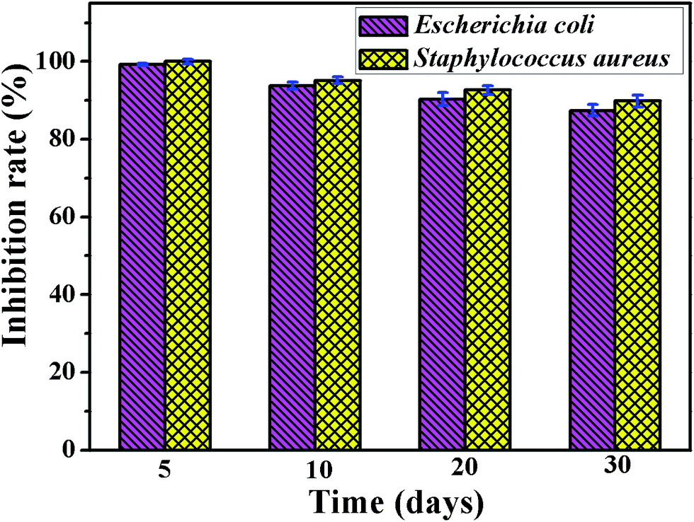
The TTO-loaded microcapsules were stored in an unsealed bottle at room temperature, and the antibacterial activities of the TTO-loaded microcapsules against E. coli (typical Gram negative bacteria) and S. aureus (typical Gram positive bacteria) were evaluated. Fig. 6 shows the inhibition rates of TTO-loaded microcapsules stored at different times. It was observed that the TTO-loaded microcapsules showed a strong antibacterial effect against E. coli (IR = 98.66% at 5 day storage) and S. aureus (IR = 100% at 5 day storage). This result was mainly ascribed to the joint contributions of the antibacterial effect of the wall materials and TTO.42 In particular, TTO could act as a prominent antibacterial agent because it possessed the capacity to denature proteins and alter the structure and function of the cell membrane and wall.43–45 Furthermore, it was also seen that the inhibition rates against both two bacteria gradually declined with the increase in the storage time. This should be ascribed to the release of TTO from the microcapsules resulting in a decrease in the amount of antimicrobial compounds in the microcapsules. Despite all this, after storage for 30 days, the inhibition rate of the microcapsules against E. coli and S. aureus was still maintained up to 87% and 89%, respectively, which proved that the TTO-loaded microcapsules possessed a long-term antimicrobial effect. In addition, the antibacterial effect against S. aureus was slightly superior to that against E. coli. The result should be ascribed to the fact that compared to S. aureus, E. coli possessed two layers of lipid bilayer (cell membrane and specific outer membrane containing lipopolysaccharides and porins), which was beneficial to protect the bacterial cell from being destroyed with the extraneous compounds to a certain degree.46,47
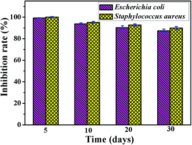 |
| | Fig. 6 The antimicrobial activities of the TTO-loaded microcapsules stored for different times against S. aureus and E. coli. | |
4. Conclusion
In this study, we successfully developed TTO-loaded antibacterial microcapsules by complex coacervation of SA and HACC. The optimum preparation conditions for the microencapsulation of TTO determined using RSM with BBD was attained as follows: a core-wall ratio of 1:1, a pH value of 6.0 and a mass concentration of CaCl2 solution of 0.6 w/v%. The actual EE of 66.06% was in an agreement with the predicted EE value. The microcapsules were nearly spherical in shape with particle sizes in the range from 1.91 to 13.18 μm. TG-DTA and DSC studies showed that the microcapsules possessed excellent thermal stability. Moreover, the in vitro release tests exhibited that the TTO-loaded microcapsules showed a controlled release behavior of TTO, which enhanced with a higher temperature and humidity of the release conditions. The release profiles of TTO from the microcapsules could be well fitted by the Ritger–Peppas model, and the TTO release kinetics followed Fickian diffusion. In addition, the TTO-loaded microcapsules possessed excellent long-term antimicrobial activity, and the bacterial inhibition rates against S. aureus and E. coli maintained as high as 87% even after 30 days of storage. Because of the advantages of the TTO-loaded microcapsules, including the improved thermal stability, the controlled release properties and the long-term antimicrobial effect, we expect that the TTO-loaded microcapsules can possess promising applications as antibacterial agents in food, cosmetics and medicine industries.
Acknowledgements
This work was supported by the Fund of Ministry of Science and Technology of China (13C26214404557), Science and Technology Project Funds of Guangdong Province (2015A010105023) and Guangdong Provincial Key Disciplines of Applied Chemistry.
References
- L. Sánchez-González, M. Vargas, C. González-Martínez, A. Chiralt and M. Cháfer, Food Hydrocolloids, 2009, 23, 2102–2109 CrossRef.
- H. J. Kim, F. Chen, C. Wu, X. Wang, H. Y. Chung and Z. Jin, J. Agric. Food Chem., 2004, 52, 2849–2854 CrossRef CAS PubMed.
- M. S. Dryden, S. Dailly and M. Crouch, J. Hosp. Infect., 2004, 56, 283–286 CrossRef CAS PubMed.
- A. Garozzo, R. Timpanaro, B. Bisignano, P. M. Furneri, G. Bisignano and A. Castro, Lett. Appl. Microbiol., 2009, 49, 806–808 CrossRef CAS PubMed.
- Y. Ge and M. Q. Ge, Fibers Polym., 2015, 16, 308–318 CrossRef CAS.
- D. C. Homeyer, C. J. Sanchez, K. Mende, M. L. Beckius, C. K. Murray, J. C. Wenke and K. S. Akers, Med. Mycol., 2015, 53, 285–294 CrossRef PubMed.
- Y. F. Wong, N. W. Davies, S. T. Chin, T. Larkman and P. J. Marriott, Ind. Crops Prod., 2015, 67, 475–483 CrossRef CAS.
- Y. T. Zhang, C. Tan, S. Abbas, K. Eric, S. Q. Xia and X. M. Zhang, Food Hydrocolloids, 2015, 51, 108–117 CrossRef CAS.
- Y. Hu, Y. Yang, Y. Ning, C. Y. Wang and Z. Tong, Colloids Surf., B, 2013, 112, 96–102 CrossRef CAS PubMed.
- C. Gupta, P. Chawla, S. Arora, S. K. Tomar and A. K. Singh, Food Hydrocolloids, 2015, 43, 622–628 CrossRef CAS.
- Y. L. Yu, M. J. Zhang, R. Xie, X. J. Ju, J. Y. Wang, S. W. Pi and L. Y. Chu, J. Colloid Interface Sci., 2012, 376, 97–106 CrossRef CAS PubMed.
- Y. Hu, S. W. Zou, Y. Yang, Z. Tong and C. Y. Wang, Macromol. Chem. Phys., 2015, 216, 714–720 CrossRef CAS.
- J. Y. Liu, C. H. Liu, Y. J. Liu, M. J. Chen, Y. Hu and Z. H. Yang, Colloids Surf., B, 2013, 109, 103–108 CrossRef CAS PubMed.
- Z. H. Ma, D. G. Yu, C. J. Branford-White, H. L. Nie, Z. X. Fan and L. M. Zhu, Colloids Surf., B, 2009, 69, 85–90 CrossRef CAS PubMed.
- N. A. Wagdare, A. T. M. Marcelis, R. M. Boom and C. J. M. Rijn, J. Colloid Interface Sci., 2011, 355, 453–457 CrossRef CAS PubMed.
- X. Y. Shi and T. W. Tan, Biomaterials, 2002, 23, 4469–4473 CrossRef CAS PubMed.
- H. Rajabi, M. Ghorbani, S. M. Jafari, A. S. Mahoonak and G. Rajabzadeh, Food Hydrocolloids, 2015, 51, 327–337 CrossRef CAS.
- F. J. Rossier-Miranda, K. Schroën and R. Boom, Food Hydrocolloids, 2012, 27, 119–125 CrossRef CAS.
- R. Badulescu, V. Vivod, D. Jausovec and B. Voncina, Carbohydr. Polym., 2008, 71, 85–91 CrossRef CAS.
- M. A. Trojer, Y. Li, M. Wallin, K. Holmberg and M. Nydén, J. Colloid Interface Sci., 2013, 409, 8–17 CrossRef PubMed.
- J. Milanović, L. Petrović, V. Sovilj and J. Katona, FoodHydrocolloids, 2014, 37, 196–202 CrossRef.
- S. Argin, P. Kofinas and Y. M. Lo, Food Hydrocolloids, 2014, 40, 138–144 CrossRef CAS.
- J. D. Chen, P. P. Pan, Y. J. Zhang, S. N. Zhong and Q. Q. Zhang, Colloids Surf., B, 2015, 134, 401–407 CrossRef CAS PubMed.
- H. Zhang, J. D. Chen, Y. J. Zhang, P. P. Pan and Q. Q. Zhang, Mater. Lett., 2014, 119, 143–145 CrossRef CAS.
- H. L. Tan, R. Ma, C. C. Lin, Z. W. Liu and T. T. Tang, Int. J. Mol. Sci., 2013, 14, 1854–1869 CrossRef CAS PubMed.
- Z. X. Peng, L. Wang, L. Du, S. R. Guo, X. Q. Wang and T. T. Tang, Carbohydr. Polym., 2010, 81, 275–283 CrossRef CAS.
- H. Liu, X. Y. Gu, M. Hu, Y. Hu and C. Y. Wang, RSC Adv., 2014, 4, 16751–16758 RSC.
- D. Bas and I. H. Boyac, J. Food Eng., 2006, 78, 836–845 CrossRef.
- A. K. Nayak, D. Pal and K. Santra, Carbohydr. Polym., 2014, 107, 41–50 CrossRef CAS PubMed.
- J. Malakar, A. K. Nayak and D. Pal, Int. J. Biol. Macromol., 2012, 50, 138–147 CrossRef CAS PubMed.
- W. C. Ko, C. K. Chang, H. J. Wang, S. J. Wang and C. W. Hsieh, Food Chem., 2015, 172, 497–503 CrossRef CAS PubMed.
- P. Sutaphanit and P. Chitprasert, Food Chem., 2014, 150, 313–320 CrossRef CAS PubMed.
- X. Y. Qv, Z. P. Zeng and J. G. Jiang, Food Hydrocolloids, 2011, 25, 1596–1603 CrossRef CAS.
- X. Yang, N. Gao, L. D. Hu, J. L. Li and Y. B. Sun, J. Food Eng., 2015, 161, 87–93 CrossRef CAS.
- M. G. Santos, F. T. Bozza, M. Thomazini and C. S. Favaro-Trindade, Food Chem., 2015, 171, 32–39 CrossRef CAS PubMed.
- A. S. Prata and C. R. F. Grosso, J. Am. Oil Chem. Soc., 2015, 92, 1063–1072 CrossRef CAS.
- Y. H. Lee, F. Mei, M. Y. Bai, S. L. Zhao and D. R. Chen, J. Controlled Release, 2010, 145, 58–65 CrossRef CAS PubMed.
- A. S. Prata, M. H. A. Zanin, M. I. Ré and C. R. F. Grosso, Colloids Surf., B, 2008, 67, 171–178 CrossRef CAS PubMed.
- G. H. Ma, J. Controlled Release, 2014, 193, 324–340 CrossRef CAS PubMed.
- D. Li, B. X. Liu, F. Yang, X. Wang, H. Shen and D. C. Wu, Carbohydr. Polym., 2016, 136, 341–349 CrossRef CAS PubMed.
- M. J. Chen, J. Y. Liu, Y. J. Liu, C. Guo, Z. H. Yang and H. Wu, RSC Adv., 2015, 5, 14522–14530 RSC.
- K. H. Liu, Y. Q. Xu and X. C. Wang, J. Food Eng., 2012, 110, 390–394 CrossRef CAS.
- C. C. Liolios, O. Gortzi, S. Lalas, J. Tsaknis and I. Chinou, Food Chem., 2009, 112, 77–83 CrossRef CAS.
- J. A. Cuaron, S. Dulal, Y. Song, A. K. Singh, C. E. Montelongo, Y. Q. Yu, V. Nagarajan, R. K. Jayaswal, B. J. Wilkinson and J. E. Gustafson, Phytother. Res., 2013, 27, 390–396 CrossRef CAS PubMed.
- C. F. Carson, K. A. Hammer and T. V. Riley, Clin. Microbiol. Rev., 2006, 19, 50–62 CrossRef CAS PubMed.
- T. Wu, A. G. Xie, S. Z. Tan and X. Cai, Colloids Surf., B, 2011, 86, 232–236 CrossRef CAS PubMed.
- Y. Ouyang, X. Cai, Q. S. Shi, L. L. Liu, D. L. Wan, S. Z. Tan and Y. S. Ouyang, Colloids Surf., B, 2013, 107, 107–114 CrossRef CAS PubMed.
Footnote |
| † Electronic supplementary information (ESI) available. See DOI: 10.1039/c5ra26052c |
|
| This journal is © The Royal Society of Chemistry 2016 |
Click here to see how this site uses Cookies. View our privacy policy here.