Dehydrochlorination mechanism of γ-hexachlorocyclohexane degraded by dehydrochlorinase LinA from Sphingomonas paucimobilis UT26

Xiaowen Tang, Ruiming Zhang, Qingzhu Zhang* and Wenxing Wang
Environment Research Institute, Shandong University, Jinan 250100, P. R. China. E-mail: zqz@sdu.edu.cn; Fax: +86-531-8836-1990

Received 15th October 2015 , Accepted 1st December 2015

First published on 8th December 2015


Abstract

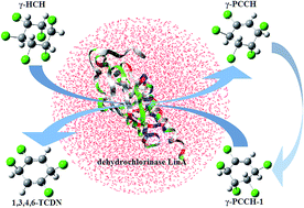
This study investigated the aerobic degradation mechanism of γ-HCH to 1,3,4,6-TCDN catabolized by dehydrochlorinase LinA from Sphingomonas paucimobilis UT26. The enzymatic step was studied by a combined quantum mechanics/molecular mechanics (QM/MM) computation and the nonenzymatic step was investigated by the DFT method. There are three elementary steps involved in the degradation process. Two discontinuous dehydrochlorination reactions with the Boltzmann-weighted average potential barriers of 16.2 and 17.3 kcal mol−1 are connected by a conformational transition with a barrier of 11.1 kcal mol−1. The electrostatic influence analysis of fourteen key residues surrounding the active site has been carried out. The study reveals that Phe68 facilitates the dehydrochlorination of γ-HCH, whereas Leu21 and Cys71 suppress it. Future mutation studies for improving the degradation efficiency of LinA can focus on mutating the amino acids of Leu21 and Cys71.


1. Introduction

Hexachlorocyclohexane (HCH) is an organochlorine compound with several stable isomers. Among all the isomers, only the γ isomer (γ-hexachlorocyclohexane, γ-HCH) has insecticidal properties and has been widely used as a broad-spectrum insecticide to control a wide range of agricultural, horticultural, and public health pests.1–3 Two kinds of γ-HCH products, technical HCH (the content of γ-HCH is 10–15%) and lindane (purified γ-HCH), have been applied and about 600[thin space (1/6-em)]000 tons were released into the environment between the 1940s and 1990s.4,5 Due to its non-target toxicity and environmental persistence, HCH was included in the list of persistent organic pollutants (POPs) according to the Stockholm convention in 2009,6 and has been prohibited in most countries. However, lindane is still used in some developing countries for its high efficiency and low cost.3,7 Therefore, HCH contamination, especially γ-HCH contamination, is a serious continuous threat to the environment.

Microbial degradation of γ-HCH can proceed under either anaerobic or aerobic condition.8–10 Chlorobenzene and benzene will be accumulated when γ-HCH is degraded under the anaerobic condition. The biochemical pathways for anaerobic degradation of γ-HCH are available, but unfortunately the specific genes and enzymes involved in the anaerobic degradation have not been identified yet.4 In contrast, γ-HCH can be degraded completely into nontoxic molecules under the aerobic condition. Researches about the aerobic degradation of γ-HCH are numerous and several HCH-degrading aerobes have been described in details.1,11–15 Most of them belong to the family of Sphingomonadaceae.16 They contain a set of genes called lin genes, which can encode HCH degradation enzymes. The aerobic degradation pathway of γ-HCH is devoted by various enzymes, among which the HCH dehydrochlorinase (LinA) from Sphingobium japonicum UT26 is considered to be significant because it catalyzes the initial step of the γ-HCH aerobic degradation.17,18

LinA catalyzes the dehydrochlorination of γ-HCH to generate an observed intermediate γ-pentachlorocyclohexene (γ-PCCH), which is transformed to a putative product 1,3,4,6-tetrachloro-1,4-cyclohexadiene (1,3,4,6-TCDN) through another dehydrochlorination step.19 During the LinA-catalyzed degradation process, the substrate is released from the enzyme after the first dehydrochlorination reaction and rebinds to the active site of LinA when undergoing the subsequent dehydrochlorination reaction.17 Crystal structure studies revealed that a catalytic dyad formed by His73 and Asp25 is located in the active site of LinA.17,18 During the enzymatic dehydrochlorination reaction, His73 of LinA acts as a general base to grab the proton of substrate, generating a positively charged His73 residue which is stabilized though its interaction with Asp25.20 It is generally considered that the leaving hydrogen and chlorine in the LinA-catalyzed dehydrochlorination reaction should be axial, adjacent and in antiparallel position.21 Hence, LinA exclusively catalyzes the dehydrochlorination reaction of the substrates containing at least one adjacent trans-diaxial H/Cl pair. The biotransformation mechanism from γ-HCH to 1,3,4,6-TCDN is exhibited in Scheme 1A. The hydrogen atom bonded to C1 and chlorine atom bonded to C2, composing an adjacent trans-diaxial H/Cl pair (H1/Cl2), are removed in the dehydrochlorination reaction of γ-HCH by LinA. Enzymatic dehydrochlorination of the newly generated intermediate (γ-PCCH) must proceed through the elimination of H4/Cl5 pair during the transformation from γ-PCCH to 1,3,4,6-TCDN. However, neither of them in γ-PCCH is situated on axial orientation, implying that γ-PCCH is unable to transform to 1,3,4,6-TCDN by enzymatic dehydrochlorination directly. Actually, the formation of 1,3,4,6-TCDN can accomplish through the LinA-catalyzed dehydrochlorination of a PCCH conformer with an adjacent trans-diaxial H4/Cl5 pair, as γ-PCCH-1 presented in Scheme 1. It can be considered as a product of the conformational transition of γ-PCCH. Therefore, the transformation pathways from γ-PCCH to 1,3,4,6-TCDN via γ-PCCH-1 must be at work, in which the LinA-catalyzed dehydrochlorination reaction occurs after the conformational transition of γ-PCCH instead of eliminating the H4/Cl5 pair in γ-PCCH directly.


image file: c5ra21461k-s1.tif
Scheme 1 (A) The degradation pathway from γ-HCH to 1,3,4,6-TCDN catabolized by dehydrochlorinase LinA. The leaving atoms are labeled in bold red. The QM region for LinA-catalyzed dehydrochlorination of γ-HCH (B) and γ-PCCH-1 (C). Several key atoms are numbered and the boundary between the QM and MM regions is indicated by wavy lines.

Although the LinA-catalyzed degradation process of γ-HCH have been established roughly,4,22 the in-depth understanding of its dehydrochlorination reaction still remains indistinct. The transition states and some intermediates as well as some products formed in the catalytic process are impossible to be observed in the general experimental enzyme chemistry, for instance, 1,3,4,6-TCDN, a very short-lived metabolism product, has never been directly detected in experimental characterization.20 Furthermore, the influence of residues Leu21, Ile109, and Thr133 as well as other key residues surrounding the active site of LinA in the γ-HCH dehydrochlorination process is still unknown. Therefore, theoretical calculation can be an alternative. In the present work, the detailed degradation mechanism from γ-HCH to 1,3,4,6-TCDN catalyzed by dehydrochlorinase LinA from Sphingomonas paucimobilis UT26 was investigated by theoretical calculations. The enzymatic step was studied with the aid of a combined quantum mechanics/molecular mechanics (QM/MM) method. QM/MM computations of the enzyme-catalyzed reaction can provide the atomistic details of the enzyme mechanism and is therefore becoming an increasingly important tool to supplement experimental enzyme chemistry.

2. Calculation methods

2.1 System setup and molecular dynamics

The initial enzyme model for the present simulation was built on the basis of the X-ray crystal structure of γ-hexachlorocyclohexane dehydrochlorinase LinA from Sphingomonas paucimobilis UT26 (PDB code: 3A76) obtained from the Protein Data Bank (http://www.rcsb.org).17 It reveals that LinA is a homotrimer with no significant difference in backbone conformation among the three chains and the LinA-catalyzed reaction can be achieved in any chain independently.17 Therefore, chain A of LinA was selected as the initial enzyme model for our present study. The protonation state of ionizable residues was determined on the basis of the pKa values obtained from the PROPKA procedure.23 Missing hydrogen atoms of the crystal structure were supplemented through CHARMM22 force field24 in the HBUILD facility of CHARMM package.25–27 MolProbity software was used to check the flipped Asn/Gln/His residues.28 Substrate models (γ-HCH and γ-PCCH-1) were built by using the Material Studio 4.4 program and then docked with the dehydrochlorinase LinA through a grid-based receptor-flexible docking module (CDOCKER) installed in the Discovery Studio 2.1 program29,30 (Accelrys Software Inc.). The binding site was defined as a sphere with a radius of 5.0 Å (coordinate: −9.806, 22.269, −5.274). Substrates were docked into the binding site with the aid of a CHARMM-based molecular dynamics (MD). Random substrate conformations were generated through high-temperature MD and translated into the binding site. Candidate poses were then created using rigid-body rotations followed by simulated annealing. A final minimization was used to refine the substrate poses. Finally, the substrate poses with interaction energy of 23.7 kcal mol−1 for γ-HCH and 22.8 kcal mol−1 for γ-PCCH-1 were select for our present work. The substrate–LinA binary complex was placed in a water sphere (TIP3P model31) with a diameter of 70.0 Å, which ensures that the complex was completely solvated. Water molecules overlapping within 2.5 Å of the binary complex were deleted. The whole system was neutralized with seven sodium ions at random positions. After that, the system was heated gradually from 0 K to 298.15 K within 50 ps and a trajectory of 500 ps was calculated to reach the thermal equilibration state (1 fs per step). Finally, a 6 ns stochastic boundary molecular dynamic (SBMD) simulation with canonical ensemble (NVT, 298.15 K) was performed to mimic the aqueous environment.32 The leap-frog algorithm and Langevin dynamics attached in the CHARMM package were applied during the simulation.

2.2 QM/MM calculations

The QM/MM calculations were performed with the aid of ChemShell 3.3.01 (ref. 33) integrating Turbomole 6.2 (ref. 34) and DL-POLY35 programs. The hybrid delocalized internal coordinate (HDLC)36 was adopted for the calculation. The MM region was treated with the CHARMM22 force field,24 while the QM region was calculated by the DFT37 method. The boundary was defined by cleaving the covalent bonds between the QM and MM regions. In order to avoid over-polarization of the QM density in the QM region, hydrogen-link atoms were complemented to the QM side with the charge shift model.38 When partitioning the QM region, some essential criteria should be considered, residues participating in bond formation or cleavage and having strong interaction with the reactive center should be classified to the QM region. Therefore, the QM region of the LinA-catalyzed dehydrochlorination reaction system in the present study contains residues Lys20, Asp25, Trp42, His73, Arg129 and the substrate (γ-HCH or γ-PCCH-1). Together with five hydrogen-link atoms, a total of 83 atoms were treated in the QM region. Similarly, 81 atoms were regarded as QM atoms in the γ-PCCH-1 reaction system. For both of the two systems, all the atoms within 18 Å of Nε atom (Scheme 1) from His73 were selected to be the active region (about 3400 movable atoms). Atoms that lie beyond 18 Å of Nε were fixed during the QM/MM calculation. The QM region was optimized by the B3LYP/6-31G(d,p) method with a charge of 1 and a spin multiplicity of 1. The transition state structure was determined by scanning the potential energy profile from the reactant to the product. The corresponding structure with the highest energy along the reaction path was selected and further optimized through microiterative TS optimizer which was supported by partitioned rational function optimizer (P-RFO) algorithm39 and the low-memory Broyden–Fletcher–Goldfarb–Shanno (L-BFGS) algorithm.40 The character of the transition state was validated by analysis of harmonic vibrational frequencies at the B3LYP/6-31G(d,p)//CHARMM22 level. A larger basis set, B3LYP/6-311++G(d,p), was adopted in single point energy calculation. Further details of the QM/MM setup can be found in ESI. In addition, the conformational transition of γ-PCCH was studied by the DFT method with solvation effect which was performed by the polarizable continuum model (PCM)41 of the self-consistent reaction field theory. This method is implemented in the Gaussian 09 package.42 Water was selected as the solvent (ε = 80.0) and the PCM cavity was defined by using the default (UFF) radii. The single point energy was calculated on the basis of the B3LYP/6-31G(d,p) optimized geometries at the B3LYP/6-311++G(d,p) level of theory so that the energetic results of whole degradation process can be obtained on the same scale.

3. Results and discussion

The LinA-substrate binary complex was extracted per picosecond during the 6000 picosecond SBMD simulation. The corresponding root-mean-square deviations (RMSD) of the backbone for the two enzymatic reaction systems were checked and displayed in Fig. S1 of the ESI. Moreover, two distance variations, Oα–Hβ and Nε–H (Nε–H1 for γ-HCH reaction system and Nε–H4 for γ-PCCH-1 reaction system, the superscript can be consulted in Scheme 1), along the 6000 picosecond trajectory were depicted in Fig. S1C and D. The distance of Nε–H1 became stable after 1700 picosecond of the SBMD simulation and the average distance of Nε–H1 and Nε–H4 were 2.75 and 2.70 Å, respectively. It can be concluded that the systems have been stabilized and the substrates meet the condition of dehydrochlorination. The distance between Oα and Hβ is about 1.70 Å for both of two systems, which indicates that a hydrogen bond is formed in the catalytic dyad of LinA.

For more details to identify the reliability of the model used in our present work, three dimensional models for the docked structures, MD snapshots, and QM/MM-optimised structures were exhibited in ESI. For the γ-HCH reaction system (Fig. S2), the substrate keeps its chair conformation with the position staying relatively stationary in the three sections. The relative position with His73 is measured through distance of Nε–H1, which is 2.63 Å in docked structure, an average of 2.75 Å in MD snapshots, and an average of 2.46 Å in QM/MM-optimised structures. Similarly, D1, D2 and D3 are also adopted to estimate the relative position with Trp42, Arg129 and Lys20, which are about 3.50 Å, 5.00 Å and 4.90 Å in the three sections. Analogously, the half-chair conformation γ-PCCH-1 (Fig. S3) is also located in the active site with a relatively stable position. Hence, it might be inferred that the model used in our present work could be credible for the present study.

3.1 Reaction mechanism and energetic results

The rate constant of an enzyme-catalyzed reaction generally exhibits a wide range of fluctuation instead of a constant, according to the room-temperature single molecule experiment.43,44 It is assumed that each snapshot extracted from the dynamics trajectory corresponds to a local rate constant.45 The potential barrier of an enzymatic reaction is supposed to be a statistic value by considering all the fluctuant results. In order to obtain the potential barrier of an enzymatic reaction, the Boltzmann-weighted averaging method is introduced. It can be described by the following equation:46,47
image file: c5ra21461k-t1.tif
where, ΔEea is the Boltzmann-weighted average potential barrier, R is gas constant, T is the temperature, n is the number of snapshots, and ΔEi is the potential barrier of pathway i. In the present study, five different snapshots were extracted every 0.5 ns from 4 to 6 ns from the SBMD simulations. They were labeled as SH-4.0, SH-4.5, SH-5.0, SH-5.5, and SH-6.0 for the γ-HCH dehydrochlorination reaction system and SP-4.0, SP-4.5, SP-5.0, SP-5.5, and SP-6.0 for the γ-PCCH-1 dehydrochlorination reaction system. These structures served as the starting configurations in the following geometry optimization and transition-state search.

The degradation process of γ-HCH covers three elementary steps: dehydrochlorination of γ-HCH, conformational transition of γ-PCCH, and dehydrochlorination of γ-PCCH-1, as indicated in Scheme 1A. Energy profiles of the three steps are calculated and shown in Fig. 1. For the dehydrochlorination of γ-HCH, a substantial potential barrier spread from 12.6 to 21.3 kcal mol−1 is found among different snapshots as listed in Table 1. The large potential barrier fluctuation observed is helpful in understanding the room-temperature single molecule experimental evidence that the reaction rate of a single enzyme molecule exhibits large fluctuation.43,44 The calculated average potential barrier for dehydrochlorination of γ-HCH, 16.2 kcal mol−1, conforms exactly to the experimental result of ∼16 kcal mol−1, which is converted from experimentally determined kcat value (63.5 s−1 (ref. 48)) with the aid of the conventional transition-state theory.49 Similarly, a potential barrier fluctuation spread from 13.4 to 21.5 kcal mol−1 listed in Table 2 is found in the second dehydrochlorination step (dehydrochlorination of γ-PCCH-1) and the calculated average potential barrier is 17.3 kcal mol−1, a slightly higher than that of the dehydrochlorination of γ-HCH. For the conformational transition of γ-PCCH, the calculated potential barrier is 11.1 kcal mol−1. It is worth noticing that all of the three elementary steps are exothermic, the enthalpy of reaction (ΔH, 298.15 K) is −4.7 kcal mol−1 for dehydrochlorination of γ-HCH, −1.0 kcal mol−1 for conformational transition of γ-PCCH and −19.3 kcal mol−1 for dehydrochlorination of γ-PCCH-1. The low potential barrier and strong exothermicity of three elementary steps indicate that they are energetically feasible. Consequently, the assumed metabolic pathway from γ-HCH to 1,3,4,6-TCDN catalyzed by dehydrochlorinase LinA from Sphingomonas paucimobilis UT26 is reasonable.


image file: c5ra21461k-f1.tif
Fig. 1 Energy profiles of three elementary steps along the transformation process from γ-HCH to 1,3,4,6-TCDN. The structures of the reactant (γ-PCCH), transition state (TS-2) and product (γ-PCCH-1) involved in the conformational transition step are exhibited in ball and stick models. The potential barriers of each elementary step are provided in the braces.
Table 1 Potential barriers ΔE (in kcal mol−1) and enthalpy of reaction ΔH (in kcal mol−1) as well as selected internuclear distances (in Å) in the reactant (R), transition state (TS-1) and product (IM-1) involved in the LinA-catalyzed dehydrochlorination of γ-HCH in five pathways. ΔH is calculated at 298.15 K
Pathway Nε–H1 H1–C1 C1–C2 C2–Cl2 ΔE ΔH
R TS-1 IM-1 R TS-1 IM-1 R TS-1 IM-1 R TS-1 IM-1
SH-4.0 2.42 1.23 1.02 1.09 1.57 2.60 1.53 1.48 1.34 1.82 1.95 2.91 12.6 −3.9
SH-4.5 2.41 1.23 1.01 1.09 1.55 2.97 1.53 1.48 1.33 1.83 1.96 2.97 17.4 −5.4
SH-5.0 2.37 1.21 1.01 1.09 1.57 2.60 1.53 1.48 1.33 1.82 1.93 2.92 16.8 −0.2
SH-5.5 2.58 1.24 1.01 1.09 1.53 2.61 1.52 1.48 1.33 1.82 1.93 2.97 12.8 −9.1
SH-6.0 2.53 1.22 1.01 1.09 1.56 2.78 1.53 1.48 1.33 1.82 1.94 3.16 21.3 −4.9


Table 2 Potential barriers ΔE (in kcal mol−1) and enthalpy of reaction ΔH (in kcal mol−1) as well as selected internuclear distances in the reactant (IM-2), transition state (TS-3) and product (P) involved in the LinA-catalyzed dehydrochlorination of γ-PCCH-1 in five pathways. ΔH is calculated at 298.15 K
Pathway Nε–H4 H4–C4 C4–C5 C5–Cl5 ΔE ΔH
IM-2 TS-3 P IM-2 TS-3 P IM-2 TS-3 P IM-2 TS-3 P
SP-4.0 2.40 1.25 1.01 1.10 1.52 2.97 1.53 1.49 1.33 1.82 1.90 4.15 14.9 −19.8
SP-4.5 2.41 1.20 1.01 1.09 1.60 3.15 1.52 1.49 1.34 1.82 1.90 4.01 21.5 −22.8
SP-5.0 2.50 1.25 1.01 1.09 1.52 3.03 1.53 1.50 1.33 1.82 1.91 3.94 19.5 −18.7
SP-5.5 2.48 1.26 1.01 1.09 1.53 2.98 1.53 1.50 1.34 1.82 1.90 4.16 17.3 −16.3
SP-6.0 2.26 1.23 1.01 1.10 1.53 2.81 1.53 1.49 1.34 1.82 1.91 3.78 13.4 −18.9


3.2 Catalytic itinerary and structural details

For convenience of description, several key atoms in the QM region are numbered for the LinA-catalyzed dehydrochlorination of γ-HCH, as presented in Scheme 1B. Some crucial internuclear distances in the reactant, transition state, and product computed at the B3LYP/6-31G(d,p)//CHARMM22 level are provided in Table 1. Since a majority of the catalytic reactions occur through the pathway with the lowest potential barrier, the following investigation towards γ-HCH dehydrochlorination process will mainly focus on the pathway SH-5.5. For a more intuitive observation, the three dimensional structures of R, TS-1, and IM-1 involved in the γ-HCH dehydrochlorination step of the pathway SH-5.5 are displayed in Fig. 2. Obviously, an adjacent trans-diaxial H/Cl pair composed by H1 and Cl2 is situated towards the Nε atom of His73 residue in the reactant, and the distance between H1 and Nε is 2.58 Å. It reveals that the γ-HCH molecule satisfies the condition of dehydrochlorination by LinA. In the process from the reactant to the transition state, the bond length of C1–H1 is stretched from 1.09 Å to 1.53 Å and the distance between Nε and H1 is reduced to 1.24 Å, indicating that H1 is delivered from γ-HCH to His73 residue. The character of the transition state is verified by the vibrational mode and the corresponding imaginary frequency of 770i cm−1. In the product, the length of double bond C1[double bond, length as m-dash]C2 (1.33 Å) and the angles of Cl1–C1–C2 (118.9°), H2–C2–C1 (120.4°) as well as the dihedral angle of H1–C1–C2–Cl2 (0.8°) suggest the formation of γ-PCCH. Meanwhile, the distance of C2–Cl2 (2.97 Å) suggests the formation of a chloride anion. The new-formed chloride anion can be stabilized by a positively charged region constituted by Lys20 and Arg129. It is compelling to note that the hydrogen bond between Oα and Hβ becomes stronger during the process of proton H1 transferring from γ-HCH to His73 residue. Hence, Asp25 can distribute the positive charge in protonated imidazole ring of His73.
image file: c5ra21461k-f2.tif
Fig. 2 The three dimensional structures of the reactant (R), transition state (TS-1), and product (IM-1) involved in the pathway SH-5.5 of the γ-HCH dehydrochlorination step. The QM atoms including link hydrogen atoms are shown in ball and stick representation. The unit of bond distances and imaginary frequency are in Å and cm−1.

For a more detailed description, the internuclear distance and Mulliken population analysis charge variations are introduced. Fig. 3A shows the variations of four crucial internuclear distances along the γ-HCH dehydrochlorination process. It is evident that the dehydrogenation and dechlorination process occur simultaneously, theoretically confirming the fact that LinA catalyzes degradation of γ-HCH via an E2 mechanism. The atomic charge analysis of several key atoms is displayed in Fig. 3B. The negative charge of Nε has been weakened along the process, corresponding to the state of proton transfer. The anion character of Cl2 in the product was further confirmed by its negative charge (−0.48).


image file: c5ra21461k-f3.tif
Fig. 3 Variations of four crucial internuclear distances (A) and atomic charges of several key atoms (B) along pathway SH-5.5 of the γ-HCH dehydrochlorination process.

The intermediate γ-PCCH will diffuse out of the enzyme when the dehydrochlorination of γ-HCH is completed.17 As a consequence, the subsequent conformational transition of γ-PCCH is nonenzymatic. In the present work, the conformational transition step was considered by the DFT method with solvation effect. The structures of reactant, transition state and product optimized at the B3LYP/6-31G(d,p) level are exhibited in Fig. 1. During the conformational transition process, the dihedral angle of C3–C4–C5–C6 varies from −58.8° to 59.9°, indicates that the relative position of C4 and C5 has been inverted. The adjacent diequatorial H4/Cl5 pair is converted to trans-diaxial H4/Cl5 pair. The transformation from one half-chair conformer (γ-PCCH) to another half-chair conformer (γ-PCCH-1) is accomplished. It is worth noting that the dihedral angle of C3–C4–C5–C6 in transition state is approximately 0°, suggests this four carbon atoms are coplanar in the cyclohexene structure. However, all the six carbon atoms of the cyclohexene structure are not situated in the same plane. The dihedral angle of C1–C2–C3–C4 (33.7°) and C2–C1–C6–C5 (−33.2°) reveals that the transition state is a boat form structure. The character of the transition state is also verified by the vibrational mode and the corresponding imaginary frequency of 54i cm−1.

For the dehydrochlorination of γ-PCCH-1, some crucial QM atoms are numbered in Scheme 1C. The degradation process was investigated at the B3LYP/6-31G(d,p)//CHARMM22 level. Four selected internuclear distances in the reactant, transition state and product are provided in Table 2 respectively. Fig. 4 displays the active site structures of IM-2, TS-3, and P in the pathway SP-6.0 as it executes the dehydrochlorination process with the lowest potential barrier. An overall view of the reaction process indicates that the dehydrochlorination of γ-PCCH-1 is accomplished with the same mechanism as that from γ-HCH. The metabolism product 1,3,4,6-TCDN is optimized successfully, theoretically verifying the existence of the putative short-lived product. The distance between the leaving chlorine atom (Cl5) and its interrelated carbon atom (C5) is 3.78 Å. However, the negative charge of the leaving chlorine atom Cl5 (−0.29) is incomprehensibly weaker than that of Cl2 (−0.48). A reasonable explanation is that the chloride anion Cl5 is closer to the positively charged region constituted by Lys20 and Arg129, causing a more sufficient charge dispersion.


image file: c5ra21461k-f4.tif
Fig. 4 The three dimensional structures of the reactant (IM-2), transition state (TS-3), and product (P) involved in the pathway SP-6.0 of the γ-PCCH-1 dehydrochlorination step. The QM atoms including link hydrogen atoms are shown in ball and stick representation. The unit of bond distances and imaginary frequency are in Å and cm−1.

3.3 Individual residue influence

According to previous crystal structure study, the active site of LinA is largely surrounded by fourteen residues.17 They can make an electrostatic influence on the enzyme reaction, though they do not participate in the reaction directly. In order to clarify the electrostatic influence of the residues surrounding the active site, the electrostatic interaction energies of the fourteen residues were estimated towards the two dehydrochlorination processes. The electrostatic influence of an amino acid i can be described as:
ΔEi−0 = ΔEi − ΔE0
where, ΔEi−0 is the changes of the barrier, ΔEi is the potential barrier with charges on residue i set to 0, and ΔE0 is the original values of the potential barrier. During all these energy calculations, the geometry structures of the stationary points were kept unchanged. A positive ΔEi−0 value means that neglecting the influence of the ith residue will increase the potential barrier. In other words, the ith residue can diminish the potential barrier and facilitate the enzyme reaction. Contrarily, a negative ΔEi−0 value denotes that the ith residue can increase the potential barrier and suppress the enzyme reaction.47

The ΔEi−0 values of fourteen residues studied in the current work were schematically described in Fig. 5. For the dehydrochlorination of γ-HCH, the electrostatic influence analysis shows that residue Phe68 facilitates this degradation reaction (ΔEi−0 > 1 kcal mol−1),whereas residues Leu21 and Cys71 suppress it (ΔEi−0 < −1 kcal mol−1). The other eleven residues are found to perform a negligible influence (−1 kcal mol−1 < ΔEi−0 < 1 kcal mol−1) towards the dehydrochlorination of γ-HCH. This electrostatic influence analysis highlights Leu21 and Cys71 as candidate residues for future mutation studies. In addition, all the fourteen residues studied in this analysis are found to have a weaker effect (−1 kcal mol−1 < ΔEi−0 < 1 kcal mol−1) on the dehydrochlorination of γ-PCCH-1.


image file: c5ra21461k-f5.tif
Fig. 5 ΔEi−0 values of fourteen individual residues toward the dehydrochlorination of γ-HCH and γ-PCCH-1.

4. Conclusions

The present work investigated the biotransformation pathway from γ-HCH to 1,3,4,6-TCDN catabolized by dehydrochlorinase LinA from Sphingomonas paucimobilis UT26. The degradation process contains two discontinuous dehydrochlorination reactions. The product of the first dehydrochlorination step undergoes a conformational transition instead of executing the second dehydrochlorination step directly. The electrostatic influence analysis reveals that the residue Phe68 facilitates the degradation reaction most and the residues Leu21 and Cys71 suppress it. It can be a valuable base for rational design of mutants of dehydrochlorinase LinA with a more efficient activity towards the degradation of γ-HCH and further experimental verification would be anticipated.

Acknowledgements

This work was supported by NSFC (National Natural Science Foundation of China, project No. 21337001, 21177077) and the Research Fund for the Doctoral Program of Higher Education of China (project No. 20130131110058).

References

  1. V. Raina, A. Hauser, H. R. Buser, D. Rentsch, P. Sharma, R. Lal, C. Holliger, T. Poiger, M. D. Muller and H. P. E. Kohler, Environ. Sci. Technol., 2007, 41, 4292–4298 CrossRef CAS PubMed.
  2. M. Okai, J. Ohtsuka, L. F. Imai, T. Mase, R. Moriuchi, M. Tsuda, K. Nagata, Y. Nagata and M. Tanokura, J. Bacteriol., 2013, 195, 2642–2651 CrossRef CAS PubMed.
  3. K. L. Willett, E. M. Ulrich and R. A. Hites, Environ. Sci. Technol., 1998, 32, 2197–2207 CrossRef CAS.
  4. R. Lal, G. Pandey, P. Sharma, K. Kumari, S. Malhotra, R. Pandey, V. Raina, H. P. E. Kohler, C. Holliger, C. Jackson and J. G. Oakeshott, Microbiol. Mol. Biol. Rev., 2010, 74, 58–80 CrossRef CAS PubMed.
  5. M. Suar, A. Hauser, T. Poiger, H. R. Buser, M. D. Müller, C. Dogra, V. Raina, C. Holliger, J. R. van der Meer and R. Lal, Appl. Environ. Microbiol., 2005, 71, 8514–8518 CrossRef CAS PubMed.
  6. K. Bala, B. Geueke, M. Miska, D. Rentsch, T. Poiger, M. Dadhwal, R. Lal, C. Holliger and H. P. E. Kohler, Environ. Sci. Technol., 2012, 46, 4051–4058 CrossRef CAS PubMed.
  7. K. Walker, D. A. Vallero and R. G. Lewis, Environ. Sci. Technol., 1999, 33, 4373–4378 CrossRef CAS.
  8. H. R. Buser and M. D. Muller, Environ. Sci. Technol., 1995, 29, 664–672 CrossRef CAS PubMed.
  9. P. J. Middeldorp, W. van Doesburg, G. Schraa and A. J. Stams, Biodegradation, 2005, 16, 283–290 CrossRef CAS PubMed.
  10. T. M. Phillips, A. G. Seech, H. Lee and J. T. Trevors, Biodegradation, 2005, 16, 363–392 CrossRef CAS PubMed.
  11. S. Nagasawa, R. Kikuchi, Y. Nagata, M. Takagi and M. Matsuo, Chemosphere, 1993, 26, 1719–1728 CrossRef CAS.
  12. H. H. M. Rijnaarts, A. Bachmann, J. C. Jumelet and A. J. B. Zehnder, Environ. Sci. Technol., 1990, 24, 1349–1354 CrossRef CAS.
  13. S. K. Sahu, K. K. Patnaik, S. Bhuyan, B. Sreedharan, N. Kurihara, T. K. Adhya and N. Sethunathan, J. Agric. Food Chem., 1995, 43, 833–837 CrossRef CAS.
  14. S. K. Sahu, K. K. Patnaik and N. Sethunathan, Bull. Environ. Contam. Toxicol., 1992, 48, 265–268 CrossRef CAS PubMed.
  15. S. K. Sahu, K. K. Patnaik, M. Sharmila and N. Sethunathan, Appl. Environ. Microbiol., 1990, 56, 3620–3622 CAS.
  16. D. Boltner, S. Moreno-Morillas and J. L. Ramos, Environ. Microbiol., 2005, 7, 1329–1338 CrossRef CAS PubMed.
  17. M. Okai, K. Kubota, M. Fukuda, Y. Nagata, K. Nagata and M. Tanokura, J. Mol. Biol., 2010, 403, 260–269 CrossRef CAS PubMed.
  18. Y. Nagata, K. Miyauchi and M. Takagi, J. Ind. Microbiol. Biotechnol., 1999, 23, 380–390 CrossRef CAS PubMed.
  19. R. Imai, Y. Nagata, M. Fukuda, M. Takagi and K. Yano, J. Bacteriol., 1991, 173, 6811–6819 CAS.
  20. L. Trantírek, K. Hynkova, Y. Nagata, A. Murzin, A. Ansorgova, V. Sklenaŕ and J. Damborsky, J. Biol. Chem., 2001, 276, 7734–7740 CrossRef PubMed.
  21. P. G. Deo, N. G. Karanth and N. G. Karanth, Crit. Rev. Microbiol., 1994, 20, 57–78 CrossRef CAS PubMed.
  22. R. Lal, M. Dadhwal, K. Kumari, P. Sharma, A. Singh, H. Kumari, S. Jit, S. K. Gupta, D. Lal, M. Verma, J. Kaur, K. Bala and S. Jindal, Indian J. Microbiol., 2008, 48, 3–18 CrossRef CAS PubMed.
  23. H. Li, A. D. Robertson and J. H. Jensen, Proteins, 2005, 61, 704–721 CrossRef CAS PubMed.
  24. A. D. MacKerell, D. Bashford, M. Bellott, R. L. Dunbrack, J. D. Evanseck, M. J. Field, J. Gao, H. Guo, S. Ha, D. Joseph-McCarthy, L. Kuchnir, K. Kuczera, F. T. Lau, C. Mattos, S. Michnick, T. Ngo, D. T. Nguyen, B. Prodhom, W. E. Reiher, B. Roux III, M. Schlenkrich, J. C. Smith, R. Stote, J. Straub, M. Watanabe, J. Wiórkiewicz-Kuczera, D. Yin and M. Karplus, J. Phys. Chem. B, 1998, 102, 3586–3616 CrossRef CAS PubMed.
  25. B. R. Brooks, R. E. Bruccoleri, B. D. Olafson, D. J. States, S. Swaminathan and M. Karplus, J. Comput. Chem., 1983, 4, 187–217 CrossRef CAS.
  26. B. R. Brooks, C. L. Brooks III, A. D. Mackerell, L. Nilsson, R. J. Petrella, B. Roux, Y. Won, G. Archontis, C. Bartels, S. Boresch, A. Caflisch, L. Caves, Q. Cui, A. R. Dinner, M. Feig, S. Fischer, J. Gao, M. Hodoscek, W. Im, K. Kuczera, T. Lazaridis, J. Ma, V. Ovchinnikov, E. Paci, R. W. Pastor, C. B. Post, J. Z. Pu, M. Schaefer, B. Tidor, R. M. Venable, H. L. Woodcock, X. Wu, W. Yang, D. M. York and M. Karplus, J. Comput. Chem., 2009, 30, 1545–1615 CrossRef CAS PubMed.
  27. A. D. MacKerell Jr, B. Brooks III, C. L. Brooks, L. Nilsson, B. Roux, Y. Won and M. Karplus, Encyclopedia Comput. Chem., 1998, 1, 271–277 Search PubMed.
  28. V. B. Chen, W. B. Arendall III, J. J. Headd, D. A. Keedy, R. M. Immormino, G. J. Kapral, L. W. Murray, J. S. Richardson and D. C. Richardson, Acta Crystallogr., Sect. D: Biol. Crystallogr., 2010, 66, 12–21 CrossRef CAS PubMed.
  29. G. Wu, D. H. Robertson, C. L. Brooks III and M. Vieth, J. Comput. Chem., 2003, 24, 1549–1562 CrossRef CAS PubMed.
  30. M. Vieth, J. D. Hirst, A. Kolinski and C. L. Brooks III, J. Comput. Chem., 1998, 19, 1612–1622 CrossRef CAS.
  31. W. L. Jorgensen, J. Chandrasekhar, J. D. Madura, R. W. Impey and M. L. Klein, J. Chem. Phys., 1983, 79, 926–935 CrossRef CAS.
  32. C. L. Brooks and M. Karplus, J. Chem. Phys., 1983, 79, 6312–6325 CrossRef CAS.
  33. P. Sherwood, A. H. D. Vries, M. F. Guest, G. Schreckenbach, C. R. A. Catlow, S. A. French, A. A. Sokol, S. T. Bromley, W. Thiel, A. J. Turner, S. Billeter, F. Terstegen, S. Thiel, J. Kendrick, S. C. Rogers, J. Casci, M. Watson, F. King, E. Karlsen, M. Sjovoll, A. Fahmi, A. Schafer and C. Lennartz, J. Mol. Struct.: THEOCHEM, 2003, 632, 1–28 CrossRef CAS.
  34. R. Ahlrichs, M. Bär, M. Häser, H. Horn and C. Kölmel, Chem. Phys. Lett., 1989, 162, 165–169 CrossRef CAS.
  35. W. Smith and T. R. Forester, J. Mol. Graphics Modell., 1996, 14, 136–141 CrossRef CAS.
  36. S. R. Billeter, A. J. Turner and W. Thiel, Phys. Chem. Chem. Phys., 2000, 2, 2177–2186 RSC.
  37. K. Burke, J. Werschnik and E. K. U. Gross, J. Chem. Phys., 2005, 123, 1–9 CrossRef PubMed.
  38. A. H. de Vries, P. Sherwood, S. J. Collins, A. M. Rigby, M. Rigutto and G. J. Kramer, J. Phys. Chem. B, 1999, 103, 6133–6141 CrossRef CAS.
  39. J. Baker, J. Comput. Chem., 1986, 7, 385–395 CrossRef CAS.
  40. D. C. Liu and J. Nocedal, Math. Program., 1989, 45, 503–528 CrossRef.
  41. C. Amovilli, V. Barone, R. Cammi, E. Cances, M. Cossi, B. Mennucci, C. S. Pomelli and J. Tomasi, Adv. Quantum Chem., 1998, 32, 227–261 CrossRef.
  42. M. J. Frisch, G. W. Trucks, H. B. Schlegel, P. W. M. Gill, B. G. Johnson, M. A. Robb, J. R. Cheeseman, T. A. Keith, G. A. Petersson and J. A. Montgomery, GAUSSIAN 03, Pittsburgh, PA, 2003 Search PubMed.
  43. H. P. Lu, L. Y. Xun and X. S. Xie, Science, 1998, 282, 1877–1882 CrossRef CAS PubMed.
  44. W. Min, B. P. English, G. Luo, B. J. Cherayil, S. C. Kou and X. S. Xie, Acc. Chem. Res., 2005, 38, 923–931 CrossRef CAS PubMed.
  45. Y. W. Li, R. M. Zhang, L. K. Du, Q. Z. Zhang and W. X. Wang, RSC Adv., 2015, 5, 13871–13877 RSC.
  46. R. Lonsdale, J. N. Harvey and A. J. Mulholland, J. Phys. Chem. B, 2010, 114, 1156–1162 CrossRef CAS PubMed.
  47. Y. W. Li, X. L. Shi, Q. Z. Zhang, J. T. Hu, J. M. Chen and W. X. Wang, Environ. Sci. Technol., 2014, 48, 5008–5016 CrossRef CAS PubMed.
  48. D. R. B. Brittain, R. Pandey, K. Kumari, P. Sharma, G. Pandey, R. Lal, M. L. Coote, J. G. Oakeshott and C. J. Jackson, Chem. Commun., 2011, 47, 976–978 RSC.
  49. J. R. Alvarez-Idaboy, A. Galano, G. Bravo-Perez and M. E. Ruiz, J. Am. Chem. Soc., 2001, 123, 8387–8395 CrossRef CAS PubMed.

Footnote

Electronic supplementary information (ESI) available: Root-mean-square deviations (RMSD) of the backbone and key distance variations along the molecular dynamic simulations (Fig. S1); the three dimensional structures of the docked structure, the MD snapshot, and the QM/MM-optimised structure in the γ-HCH and γ-PCCH-1 reaction systems (Fig. S2 and S3); additional details on the methods; the coordinates of the docked structures, MD snapshots, QM-optimized structures and QM/MM-optimized structures. See DOI: 10.1039/c5ra21461k

This journal is © The Royal Society of Chemistry 2016
Click here to see how this site uses Cookies. View our privacy policy here.