DOI:
10.1039/C5RA23924A
(Paper)
RSC Adv., 2016,
6, 4193-4199
PVP-derived carbon nanofibers harvesting enhanced anode performance for lithium ion batteries
Received
12th November 2015
, Accepted 8th December 2015
First published on 21st December 2015
Abstract
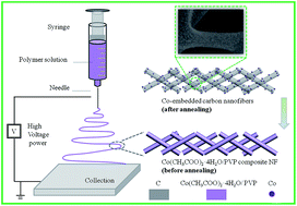
Co-embedded carbon nanofibers were synthesized using electrospinning with polyvinylpyrrolidone (PVP) instead of high cost polyacrylonitrile (PAN). The obtained composite nanofibers as an anode material for lithium ion batteries deliver a reversible capacity of 542.6 mA h g−1 in the 100th cycle at a current density of 100 mA g−1. Moreover, the anode material shows better cycle performance and rate capability in comparison to the resultant product without Co additive. It is believed that the significant improvement is attributed to the nanofiber morphology with high surface-to-volume ratio as well as the existence of Co nanoparticles that enhance the electrical conductivity of the nanocomposites.
1. Introduction
Over the past few decades, rechargeable lithium ion batteries (LIBs) have been widely used in some applications of energy storage devices, such as cell phones, portable electronic devices and electrical vehicles. Carbon materials (i.e. graphite) have been successfully commercialized as anodes in LIBs.1,2 Besides the most pervasive graphite, some other carbon materials have been investigated, including graphene,3 carbon nanotubes,4 porous carbons,5 and carbon nanofibers.6,7 Among these materials, the carbon nanofibers have attracted a lot of attention as one of the promising anode materials, owing to high surface-to-volume ratios, increased lithium ion transmission channel, and more lithium ion insertion sites on the electrode surface.8–13 As a result, carbon nanofibers exhibit improved reversible capacity and cycling stability than non-morphology carbon materials. For instance, carbon nanofibers could show a reversible capacity of about 400 mA h g−1 after 40 cycles at a current of 50 mA g−1.14 In contrast, the specific capacities of non-morphology carbon materials decrease to 100–200 mA h g−1 after 100 cycles.15 However, it is still a significant challenge to synthesize carbon nanofibers with low cost and high LIB performance.
Recently, an electrospinning technique16–20 received extensive attention to produce nanofibers through a simple, inexpensive and highly reproducible process.21 In the earliest stage, the carbon fibers were obtained using viscose fiber for the space shuttle and the high-end field via electrospinning.22 Then, polyacrylonitrile (PAN) as the precursor23–26 was utilized to synthesize the carbon fibers. Subsequently, a series of polymers, such as asphalt, phenolic aldehyde, and polyvinyl alcohol (PVA),23,27 were applied to produce carbon nanofibers. However, higher cost of the aforementioned precursors, as previously reported,28 inevitably increased the price of carbon nanofiber, which highly prevents them from further applications. In contrast, poly(vinylpyrrolidone) (PVP) with its lower cost and environmental friendliness,29,30 has been a potential precursor for fabricating carbon nanofibers. Unfortunately, little research has been reported that focus on PVP-derived carbon nanofibers via electrospinning.
In this study, we synthesize carbon nanofibers using the cheaper precursor PVP via electrospinning. More importantly, some metal Co was successfully introduced into the carbon nanofibers. The Co nanoparticles as catalyst could create some graphitic carbon with high electrical conductivity. It is expected that the nanocomposites increase anode cycling performance with the help of Co introduction into the carbon nanofibers.31
2. Experimental details
2.1. Synthesis of PVP-C and PVP-CNF
Poly(vinylpyrrolidone) (PVP, Aldrich, Mw = 1
300
000 g mol−1) as precursor, and N,N-dimethylformamide (DMF) as the solvent were utilized to produce carbon nanofibers. Cobalt acetate tetrahydrate (Co(CH3COO)2·4H2O) was used as the source of Co. A typical process is as follows. First, 1.5 g of PVP was dissolved into a mixed solution of 5 mL of acetate and 5 mL of DMF for 3 h under magnetic stirring. Second, 0.5 g of cobalt acetate tetrahydrate (Co(CH3COO)2·4H2O) was dissolved in 5 mL of deionized water for 1 h. Then, the two solutions were mixed for 2 h. The mixture was transferred to a plastic injector, and electrospun on a pink film on a clean wiper wrapped around a cylinder collector at a rotation speed of 0.03 mm min−1, at a DC voltage of 20 kV with a flow rate of 0.18 mL h−1. The distance between the collector and the needle was kept at 10 cm. Finally, the as-collected precursor fibers were dried in air at 80 °C overnight. The resultant materials were annealed at 300 °C for 3 h, and then heated to 500 °C for 3 h in Ar atmosphere. The Co-embedded carbon nanofibers were produced, which were marked as PVP-CNF. For comparison, pristine carbon, marked as PVP-C, was obtained by a similar method without the introduction of Co(CH3COO)2·4H2O. In addition, PAN was employed as the carbon source to synthesize carbon nanofibers (PAN-CNF) using similar electrospinning parameters.
2.2. Structural characterizations
Surface morphologies of the materials were observed via a scanning electron microscope (SEM, SU8010, Hitachi Jaoan) equipped with energy dispersive X-ray (EDX, INCAx-sight, Oxford). The phase and crystallinity of the carbon nanofibers were studied by powder X-ray diffraction (XRD), which was recorded on an X-ray diffractometer (DX-2700) with Cu Kα radiation. High resolution images of microstructures and selected area electron diffraction (SAED, JEOL JEM-3000F) patterns were obtained with a transmission electron microscope (TEM, JEOJ JEM-3000F). Thermal properties were tested by a thermal gravimetric analyzer (TGA, Pyris Diamond6000 TG/DTA, PerkinElmer Co, America) in air over a temperature range of room temperature to 800 °C at a heating rate of 10 °C min−1. Raman spectra were recorded using Jobin-Yvon Horiba HR800.
2.3. Electrochemical measurements
The working electrode was prepared by casting a homogeneous slurry consisting of the active materials, conductive carbon black, polyvinylidene difluoride (PVDF) (the weight ratio of 80
:
10
:
10) in N-methyl-2-pyrrolidone (NMP) on copper foil. Then, the electrodes were dried under vacuum at 100 °C overnight. The electrodes were cut in 12 mm diameter disks. The electrolyte was 1 M LiPF6 dissolved in a mixture of ethylene carbonate (EC) and dimethyl carbonate (DMC) in a ratio of 1
:
1 by volume. CR2032-type coin cells were assembled in an argon-filled glovebox. Galvanostatic charge–discharge cycles were performed using a Land battery test system (LANHE CT2001A) in the voltage of 0.01–3 V at room temperature. Electrochemical impedance spectroscopy (EIS) was recorded between 200 kHz and 5 mHz with an amplitude of 5 mV on a VMP3 (Princeton Applied Research).
3. Results and discussion
Scheme 1 shows the formation procedures of the PVP-CNF: (i) to form smooth nanofibers using the precursor solution of Co(CH3COO)2·4H2O and PVP via electrospinning; (ii) to obtain the PVP-CNF by annealing in Ar atmosphere. The carbon content of the PVP-CNF is confirmed by TGA, as shown in Fig. 1a. A sharp weight variation occurs at about 390 °C, indicating the oxidation of carbon with the weight loss. It is noteworthy that the oxidation of Co in air can trigger the weight raise. As shown in Fig. 1a, the content of Co3O4 in the sample is 38.35%. The Co content in the composites is based on the equation 3WCo × XCo3O4/WCo3O4 = XCo, where WCo and WCo3O4 are the relative molecular masses of Co and Co3O4, respectively. XCo3O4 is the residual weight percentage and XCo is the Co content in the composites. The calculated result of XCo is 28.17 wt%; thus, the content of carbon nanofibers in the composites is 71.83 wt%. In addition, the decomposition temperatures of the two materials are different (see Fig. 1a), which illustrates that the existing Co grains result in lower decomposition temperature. Fig. 1b represents the EDX results of the PVP-C and PVP-CNF. The PVP-C consists of C and O elements. In addition to C and O, Co element was observed in the Co-carbon nanofibers. In the Raman spectra of the PVP-CNF (Fig. 1c), two main peaks are situated at 1350 and 1580 cm−1, which are designated as the D band and the G band, respectively. The G band is associated with graphitic carbon, while the D band is attributed to amorphous carbon. In addition, the relative intensity of D band against G band (ID/IG) represents the degree of disorder in the graphite structure. The Raman spectrum of the PVP-CNF shows that the ratio of ID/IG is ∼0.82, which further confirms the existence of graphitic carbon. It is worth noting that the PVP-C suffered from the fluorescence reaction; as a result, the Raman signal for PVP-C is not obvious (see the inset of Fig. 1c). Fig. 1d displays the XRD patterns of the PVP-C and the PVP-CNF. The main peaks of the PVP-CNF can be observed at 2θ of about 44.2°, 51.5°, and 75.9°, corresponding to (111), (200) and (220) lattice planes of Co, respectively. And the broad peak (2θ = 26°) of the PVP-C corresponds to the (002) plane of carbon material.
 |
| Scheme 1 Schematic illustration on the formation mechanism of PVP-CNF. | |
 |
| Fig. 1 (a) The TGA curves and (b) the EDX results of PVP-C and PVP-CNF; (c) Raman spectra of PVP-CNF (the inset is that for PVP-C); (d) XRD patterns of PVP-C and PVP-CNF. | |
Fig. 2 shows the SEM images of the PVP-C and PVP-CNF. Before carbonization, both samples display similar smooth nanowired morphologies in diameters within the range of 400–500 nm (see Fig. 3a and b). After thermal treatment, the nanofibers in Fig. 2a totally disappear, as shown in Fig. 2c. More interestingly, the Co-contained sample still retains the nanowired structure (Fig. 2d) with decreased diameters. It can be also observed that a lot of Co nanograins are uniformly deposited along the nanofibers. The SEM images of PAN-CNF before and after carbonization are shown in Fig. 2e and f. As expected, carbon nanofibers can be obtained when PAN is used as the carbon source via electrospinning, which differs from the product of PVP, as shown in Fig. 2c. Thus, the diameter of PVP-CNF is obviously smaller than that of PAN-CNF. The elemental mappings in Fig. 2g and h prove that the Co nanograins are evenly distributed along the carbon nanofibers. It is worth mentioning that after carbonization, the Co-carbon nanofibers can still maintain the morphology of the nanofibers originating from the existence of the Co nanograins. Moreover, further examination in high resolution TEM revealed that the Co nanograins are evenly distributed in the carbon nanofibers. The size of the Co nanograins is about 10 nm. In addition, as apparent from Fig. 3b, some graphitic carbon is formed around the Co nanograins. This proves that the Co nanograins function as the catalyst, and facilitate the increase of graphitization.
 |
| Fig. 2 SEM images of (a) PVP-C and (b) PVP-CNF before carbonization, (c) PVP-C and (d) PVP-CNF PVP-C as well as (e) and (f) PAN-CNF before and after carbonization; elemental mapping analysis of (g) C element and (h) Co element in PVP-CNF. | |
 |
| Fig. 3 TEM images of PVP-CNF at (a) low and (b) high magnification (the inset in (a) is the selected area electron diffraction). | |
Fig. 4a compares the cycling performance of PVP-CNF, Co3O4 nanofibers and PVP-C at the current density of 100 mA g−1 in the voltage range of 0.01–3 V. Clearly, three samples exhibit different cycling performances. Both Co3O4 nanofibers and PVP-C deliver poor performance. In contrast, the PVP-CNF material shows better cyclical stability and higher reversible capacity. As expected, the introduction of Co in the nanocomposites significantly enhances anode performance. For instance, in the 100th cycle, the reversible capacities of the PVP-C, Co3O4 nanofibers and PVP-CNF are 449.8, 430.5 and 766 mA h g−1, respectively. The PVP-CNF and PVP-C are examined at various current densities of 50, 100, 150, 200, and 250 mA g−1. As shown in Fig. 4b, the PVP-CNF possesses better rate capability and higher capacity retention as compared with PVP-C. It was reported that the suppression of the formation of SEI on the active material by Co nanograins is another pivotal factor to enhance the rate capabilities.32 At each current density, the average discharge capacities of PVP-CNF are 821.8, 707.6, 677.6, 660.2, 650.5 mA h g−1, which are much higher than those of PVP-C (549.7, 476.3, 455.3, 443.7, and 435 mA h g−1), respectively.
 |
| Fig. 4 (a) Cycling performance of the PVP-CNF, Co3O4-CNF and PVP-C; (b) rate capability of PVP-CNF and PVP-C; the charge–discharge profiles of (c) PVP-C and (d) PVP-CNF; (e) electrochemical polarization of the PVP-C and PVP-CNF; and (f) The EIS of PVP-C and PVP-CNF after cycling (the inset is the equivalent circuit). | |
Fig. 4c and d show the charge–discharge behaviors of the PVP-C and PVP-CNF electrodes in the 1st, 2nd, 5th, 10th, 20th and 50th cycles over a voltage ranging from 0.01 to 3.0 V at a current density of 100 mA g−1. The initial discharge and charge capacities of the PVP-C are 1338.7 and 670.2 mA h g−1, respectively. In contrast, the PVP-CNF reveals higher initial discharge and charge capacities of 1770.7 and 1025.7 mA h g−1, respectively. The reason causing the initial irreversible capacity is some side reactions such as the formation of solid electrolyte interface (SEI) film. The PVP-CNF anode exhibits a high reversible capacity of about 932.3 mA h g−1 at the second cycle with a 95.6% coulombic efficiency. After 50 cycles, the coulombic efficiency of PVP-CNF increases to 98.3%, and the capacity decreases to about 639.9 mA h g−1, which corresponds to 68.6% of capacity retention in comparison to that of the second cycle. The average capacity fade rates of PVP-CNF and PVP-C are 0.18% and 0.33% per cycle during 100 cycles. This indicated that the reversible capacity and capacity retention of PVP-CNF are higher than those of the PVP-C, which is attributed to the unique nanostructure of PVP-CNF. The effects of the nanofiber structures and Co nanograins on electrochemical performance are as follows: (i) enhancement of charge transfer and electron conduction and decrease in inner resistance and (ii) more surface area is available for Li ion diffusion, which shortens the Li ion diffusion pathway.
As shown in the inset of Fig. 4c and d, electrochemical polarization can be observed during the rest period of testing cells. The polarization voltage is defined as the difference between 3 V and the voltage onset. The relationship between the polarization voltage and cycle number is shown in the Fig. 4e. As can be seen, the polarization voltage of PVP-C is larger than that of PVP-CNF. This result indicates that the incorporation of Co nanograins within the PVP-CNF can significantly decrease the resistance polarization. Furthermore, to further confirm the improved electrochemical reaction kinetics of the PVP-CNF electrode, EIS studies of PVP-C and PVP-CNF after 5 cycles were carried out within the frequency range of 100 kHz to 10 mHz, and the Nyquist plots are shown in Fig. 4f. To obtain kinetics parameters, we employed the two RC parallel circuits to simulate the impedance spectra on the basis of the potential SEI layer. In the equivalent circuit (inset of Fig. 4f), Rs represents the ohmic resistance of whole reaction system and Rsf is the resistance of the SEI film. Rct is the charge transfer resistance related to the electrochemical reaction. Constant phase element (CPE) was employed in the equivalent circuits instead of pure capacitance owing to the inhomogeneous surface of the working electrode. CPE1 and CPE2 are constant phase elements corresponding to the surface film and double-layer capacitance, respectively. Zw stands for Warburg impedance associated with solid state diffusion.33 Based on the equivalent circuits, the obtained kinetics parameters are listed in Table 1. One can observe that the Rs value of PVP-CNF is 11.3 Ω, which is lower than that of PVP-C (17.1 Ω). This result is consistent with the resistance polarization presented in Fig. 4e. Moreover, it is seen that the Rsf of PVP-CNF is lower, which can be due to the fact that the Co nanograins possibly restrain the formation of the SEI film. PVP-CNF shows lower Rct as compared with the PVP-C, which is attributed to the improved electrical conductivity of PVP-CNF.34,35 These results indicate that the ohmic resistance of the whole reaction system and the charge-transfer resistance of the PVP-CNF anode are smaller than those of the PVP-C, and further illustrate the fact that the electrochemical reaction kinetics of PVP-CNF electrode is improved, which might be one of the key factors contributing to enhanced electrochemical performance of the PVP-CNF.
Table 1 Kinetics parameters of PVP-CNF and PVP-C after 5 cycles
Sample |
Rs (Ω) |
Rsf (Ω) |
Rct (Ω) |
PVP-CNF |
11.3 |
355.0 |
325.6 |
PVP-C |
17.1 |
417.5 |
569.8 |
4. Conclusions
In summary, PVP-CNF was reasonably designed and successfully synthesized using a simple and environmentally benign electrospinning method. Compared with PVP-C, PVP-CNF is proved to be a promising anode, showing higher reversible capacity, superior cycling durability, and increasing rate capability. This can be ascribed to both the existence of Co nanograins, leading to the formation of the carbon nanofibers with high specific surface area, and the Co nanograins embedded in the carbon fibers, which increase the electrical conductivity. The existence of the Co nanograins can result in the formation of carbon nanofibers. The nanofiber composites effectively enhance the electrical conductivity of the electrode and thereby increase cycling performance of the anode materials. Therefore, this study will open an opportunity to create high performance carbon nanofibers as electrodes for lithium-ion batteries.
Acknowledgements
This study was supported by the National Natural Science Foundation of China (51572194), the Key Projects of Tianjin Municipal Natural Science Foundation of China (14JCZDJC32200 and 13JCZDJC33900), the Project 2013A030214 supported by CAEP (China Academy of Engineering Physics), LPMT (Laboratory of Precision Manufacturing Technology), CAEP (KF14006 and ZZ13007), Academic Innovation Funding of Tianjin Normal University (52XC1404), Training Plan of Leader Talent of University in Tianjin and the program of Thousand Youth Talents in Tianjin of China.
References
- W. Zhang and P. N. Pintauro, ChemSusChem, 2011, 4, 1753–1757 CrossRef CAS PubMed.
- J. Li, C. Daniel and D. Wood, J. Power Sources, 2011, 196, 2452–2460 CrossRef CAS.
- G. Wang, X. Shen, J. Yao and J. Park, Carbon, 2009, 47, 2049–2053 CrossRef CAS.
- J. J. Gooding, Electrochim. Acta, 2005, 50, 3049–3060 CrossRef CAS.
- K.-S. Kim and S.-J. Park, J. Power Sources, 2013, 244, 792–798 CrossRef CAS.
- I. Rajzer, R. Kwiatkowski, W. Piekarczyk, W. Biniaś and J. Janicki, Mater. Sci. Eng., C, 2012, 32, 2562–2569 CrossRef CAS.
- X. Su, Q. Wu, X. Zhan, J. Wu, S. Wei and Z. Guo, J. Mater. Sci., 2012, 47, 2519–2534 CrossRef CAS.
- Y. Yu, L. Gu, C. Wang, A. Dhanabalan, P. A. van Aken and J. Maier, Angew. Chem., Int. Ed., 2009, 48, 6485–6489 CrossRef CAS PubMed.
- I. Meschini, F. Nobili, M. Mancini, R. Marassi, R. Tossici, A. Savoini, M. L. Focarete and F. Croce, J. Power Sources, 2013, 226, 241–248 CrossRef CAS.
- Y.-H. Liu, T. Takasaki, K. Nishimura, M. Yanagida and T. Sakai, J. Power Sources, 2015, 290, 153–158 CrossRef CAS.
- R. Elazari, G. Salitra, A. Garsuch, A. Panchenko and D. Aurbach, Adv. Mater., 2011, 23, 5641–5644 CrossRef CAS PubMed.
- Y. Zhang and A. L. Yarin, Langmuir, 2011, 27, 14627–14631 CrossRef CAS PubMed.
- M. H. Kjell, E. Jacques, D. Zenkert, M. Behm and G. Lindbergh, J. Electrochem. Soc., 2011, 158, A1455–A1460 CrossRef CAS.
- J. Yue, X. Zhao and D. Xia, Electrochem. Commun., 2012, 18, 44–47 CrossRef CAS.
- L. Jabbour, M. Destro, D. Chaussy, C. Gerbaldi, S. Bodoardo, N. Penazzi and D. Beneventi, Compos. Sci. Technol., 2013, 87, 232–239 CrossRef CAS.
- J. W. Min, A. K. Kalathil, C. J. Yim and W. B. Im, Mater. Charact., 2014, 92, 118–126 CrossRef CAS.
- F. L. Zhu, J. X. Zhao, Y. L. Cheng, H. B. Li and X. B. Yan, Acta Phys.–Chim. Sin., 2012, 28, 2874–2878 CAS.
- X. Zhang, B. L. Shi and Y. Wang, J. Adhes. Sci. Technol., 2012, 26, 353–360 CAS.
- I. Uslu, T. Tunc, M. K. Ozturk and A. Aytimur, Polym.-Plast. Technol. Eng., 2012, 51, 257–262 CrossRef CAS.
- G. Zhu, L. K. Pan, J. Yang, X. J. Liu, H. C. Sun and Z. Sun, J. Mater. Chem., 2012, 22, 24326–24329 RSC.
- J. Wu, D. Zeng, X. Wang, L. Zeng, Q. Huang, G. Tang and C. Xie, Langmuir, 2014, 30, 11183–11189 CrossRef CAS PubMed.
- E. Jacques, M. H. Kjell, D. Zenkert, G. Lindbergh, M. Behm and M. Willgert, Compos. Sci. Technol., 2012, 72, 792–798 CrossRef CAS.
- C. W. Cho, D. Cho, O. H. Kwon, Y.-G. Ko and I.-K. Kang, Carbon Lett., 2007, 8, 313–320 CrossRef.
- G. Y. Oh, Y. W. Ju, H. R. Jung and W. J. Lee, J. Anal. Appl. Pyrolysis, 2008, 81, 211–217 CrossRef CAS.
- S. A. H. Ravandi and M. Sadrjahani, J. Appl. Polym. Sci., 2012, 124, 3529–3537 CrossRef CAS.
- K. Nasouri, H. Bahrambeygi, A. Rabbi, A. M. Shoushtari and A. Kaflou, J. Appl. Polym. Sci., 2012, 126, 127–135 CrossRef CAS.
- P. Lian, J. Wang, D. Cai, L. Ding, Q. Jia and H. Wang, Electrochim. Acta, 2014, 116, 103–110 CrossRef CAS.
- Y.-H. Liu, T. Takasaki, K. Nishimura, M. Yanagida and T. Sakai, J. Power Sources, 2015, 290, 153–158 CrossRef CAS.
- T. L. Yang, C. T. Pan, Y. C. Chen, L. W. Lin, I. C. Wu, K. H. Hung, Y. R. Lin, H. L. Huang, C. F. Liu, S. W. Mao and S. W. Kuo, Opt. Mater., 2015, 39, 118–124 CrossRef CAS.
- M. Enculescu, A. Evanghelidis and I. Enculescu, J. Phys. Chem. Solids, 2014, 75, 1365–1371 CrossRef CAS.
- X. Yu, Y. Fu, X. Cai, H. Kafafy, H. Wu, M. Peng, S. Hou, Z. Lv, S. Ye and D. Zou, Nano Energy, 2013, 2, 1242–1248 CrossRef CAS.
- J. Liu and A. Manthiram, Chem. Mater., 2009, 21, 1695–1707 CrossRef CAS.
- B. Yan, M. Li, X. Li, Z. Bai, L. Dong and D. Li, Electrochim. Acta, 2015, 164, 55–61 CrossRef CAS.
- R. Song, H. Song, J. Zhou, X. Chen, B. Wu and H. Y. Yang, J. Mater. Chem., 2012, 22, 12369–12374 RSC.
- F. G. Emmerich, Carbon, 1995, 33, 1709–1715 CrossRef CAS.
|
This journal is © The Royal Society of Chemistry 2016 |
Click here to see how this site uses Cookies. View our privacy policy here.