
A novel NIR long phosphorescent phosphor:SrSnO3:Bi2+
Xixi Qinab,
Yang Li*abc,
Dakun Wua,
Yiling Wuab,
Ruchun Chena,
Zhijun Maab,
Shijian Liua and
Jianrong Qiu*ab
aState Key Laboratory of Luminescent Materials and Devices, School of Materials Science and Technology, South China University of Technology, Guangzhou 510640, China. E-mail: qjr@scut.edu.cn; msliyang@scut.edu.cn; Fax: +86-20-87114204; Tel: +86-20-87113646
bGuangdong Provincial Key Laboratory of Fiber Laser Materials and Applied Techniques, Guangzhou 510640, China
cSchool of Chemistry and Chemical Engineering, South China University of Technology, Guangzhou 510640, China
First published on 19th November 2015
Abstract
A novel phosphor with near-infrared (NIR) long persistent luminescence, SrSnO3:Bi2+ was successfully synthesized by traditional solid-state reaction. The phosphor shows deep red-NIR persistent luminescence with a broad emission band from 700 to 900 nm peaking at 808 nm, as well as a persistence time of >30 min. This phosphor has broken the domination of conventional Cr3+, Mn2+ and Mn4+-activated long persistent phosphors and provides a new perspective on the NIR phosphorescence of Bi-doped long phosphorescent phosphors. Meanwhile, the characteristic operational waveband offer the possibility of potentially being applied in vivo bio-imaging of this new NIR phosphorescent material.
1. Introduction
Long persistent phosphorescence, also called long-lasting afterglow, is a phenomenon whereby luminescence can last for a long time after ceasing the excitation. The delay in emission is caused by the presence of defect levels ionized under external field (including irradiation, electron beam, temperature and mechanical strain). Such trap site levels have strong abilities to capture, store and release charge carriers (electrons or holes) in the forbidden gaps.1–3 The works on long phosphorescent phosphors (LPPs) on the early stage were in pursuit of applications in decorations, displays and dials of sulphide phosphors. After discovering a bright green-emitting phosphor SrAl2O4:Eu2+, Dy3+ in 1996 and a remarkable Cr3+ doped Zn3Ga2Ge2O10 near-infrared (NIR) phosphor in 2012,2,3 numbers of optoelectronic applications of LPPs have been burgeoned. Especially for the persistent phosphors in the region of the biologically transparent window (650–1100 nm).4 They are anticipated to open the possibility of advanced optical imaging with high resolution and weak light disturbance for realistic analysis of the structural and functional processes in cells, tissues and other complex systems, owing to the distinctive features of low autofluorescence, high signal-to-noise ratio, and deep tissue penetration.5Over the past few years, substantial achievements have been obtained in the research and development of NIR LPPs. The researches mainly focused on transition metals (Cr3+, Mn2+, Mn4+)-activated NIR LPPs, which have a broad emission band. Some of the most famous were Zn3Ga2Ge2O10:Cr3+, Zn3Ga2SnO8:Cr3+, ZnGa2O4:Cr3+, Zn2SnO4:Cr3+, MAlO3:Mn4+, Ge4+ (M = La, Gd), Ca0.2Zn0.9Mg0.9Si2O6:Eu2+, Dy3+, Mn2+.3,6–10 However, we must deal with the fact that the number of known phosphorescent activators is relatively limited. Besides these main activators, scarcely any other transition metal ion has been found to exhibit NIR persistent luminescence, even though a wide variety of activation ions can be used as NIR luminescent centres.11–16
To address this issue, we expect to develop a new long persistent phosphor with a wondrous activation center. In the context of materials selection, this system successfully avoids the existing ubiquitous reliance on Cr3+, Mn4+, and Mn2+ as activation center. Bi2+ represents the intermediate species of Bi, which typically absorbs in the UV-visible region, and emits from orange to red spectral range. Photoluminescent properties of Bi2+ have been widely investigated in recent years. For example, spectroscopic properties of orange phosphor SrB4O7:Bi2+ have been reported by Peng et al. in 2010.17 Three distinct photoluminescent excitation peaks and featured photoluminescent peaks were observed at 245 nm (2P1/2 → 2S1/2), 478 nm (2P1/2 → 2P3/2(2)), 578 nm (2P1/2 → 2P3/2(2)), and 588 nm (2P3/2(1) → 12P1/2), respectively. Similar results were also observed in Bi2+-doped SrB6O10. Even more remarkable, optical properties of Bi2+ ion strongly depend on ligand field strength of the employed host material. In addition to the yellow luminescence, red luminescence at 627 nm was also investigated in BaSO4:Bi2+ phosphor.18
SrSnO3 is selected as the host candidate to take advantages of the ingenious crystalline structure. Alkaline earth borates, sulfates, phosphates and borophosphates have already considered as the most suitable host materials for Bi2+. Because usual sites for incorporating Bi2+ ions are Ca2+, Sr2+ and Ba2+ ions vacancies, based on the predominance of tetrahedral anion groups. Both compounds are highly efficient for stabilizing the Bi2+-ion over its divalent form.19,20 Therefore, during the last decade, alkaline earth orthostannates (MSnO3, where M = Ca, Sr and Ba) can be considered as the host matrixes for new phosphors because of their stable crystalline structure and high physics-chemical stabilities.
In this paper, we report a novel NIR persistent phosphor SrSnO3:Bi2+ as a promising material for vivo imaging application due to its emission at 808 nm and the wide emission band ranging from 700 to 900 nm. It may largely extend the operation waveband in biological tissue window. Here, we show the capabilities of SrSnO3:5% Bi2+ as an example for investigation.
2. Experimental
Preparation
The phosphor sample of SrSnO3:5% Bi2+ was synthesized by conventional high temperature solid state reaction method. Typically, stoichiometric amounts of starting materials Bi2O3 (99.99%), SnO2 (99.99%) and SrCO3 (analytical reagent) were weighted and mixed thoroughly in an agate mortar. The process comprised a two-step thermal treatment. Initial calcination at 800 °C for 2 h in air, after the prefired powders were thoroughly ground again, secondary calcination at 1200 °C for 4 h in air. After that, the samples were furnace-cooled to room temperature and ground again into powder for subsequent use.Characterization
The crystalline structure of the powder SrSnO3:5% Bi was analyzed by X-ray diffractometer (XRD) (Cu Kα) and the data was collected between 10° and 90°. The data for Rietveld analysis were collected between 10° and 100°, the step scan mode was used with a setup of 40 kV × 40 mA, 0.01° per step and 0.2 s per step. Rietveld refinement of XRD data was performed by using the TOPAS-Academic program. The morphology and particle size of phosphors were observed by Nova Nano SEM 430 Scanning Electron Microscopy (SEM). X-ray photoelectron spectra (XPS, K-Alpha, Thermo Scientific, UK) were measured by the physical electronics 5600 multi-technique system. Achromatic 200W Al Kα X-rays were chosen as the excitation source. The resolution was 0.05 eV. Photoluminescence excitation (PLE), photoluminescence (PL), long-lasting phosphorescence (LLP) spectra and decay curve were measured with a high resolution spectrofluorometer (Edinburgh Instruments FLS920) equipped with a 500 W Xe lamp as excitation source. All PL results were corrected over the lamp intensity with the PMT spectral response. Thermo-luminescence glow curves were measured after irradiation with the UV lamp for 5 min using a FJ-427A TL meter (Beijing Nuclear Instrument Factory) by heating the irradiated samples from room temperature to 650 K with a fixed heating rate of 2 °C s−1. All the measurements of optical properties were carried out at room temperature. Long persistent luminescence imaging, were performed with a modified imaging system including a Germany pco dicam pro camera as the signal collector. All images were taken in a dark room and analysed with home-made software.3. Results and discussion
Phase of the samples
All the main diffraction peaks are indexed to those of the orthorhombic SrSnO3 crystal (Fig. 1, JCPDS no. 70-4389). Obviously, the higher doping amount of bismuth do not change the crystalline phase of SrSnO3, and the pattern is in perfect match with the standard powder peak positions of SrSnO3, indicating the successful incorporation of Bi2+ into SrSnO3 host. Rietveld refinement results of the XRD patterns of SrSnO3:5% Bi are summarized in Tables 2 and 3, and the corresponding refinement profiles are presented in Fig. 2. The lattice of SrSnO3:5% Bi has a cubic symmetry with space group of Pm![[3 with combining macron]](https://www.rsc.org/images/entities/char_0033_0304.gif) m.
m.
 | ||
| Fig. 1 XRD pattern of SrSnO3:5% Bi sample, which is in agreement with JCPDS 70-4389. | ||
| Materials | Excitation peak (nm) | Emission peak (nm) | Ref. | ||
|---|---|---|---|---|---|
| 2P1/2 → 2S1/2 | 2P1/2 → 2P3/2(2) | 2P1/2 → 2P3/2(1) | 2P3/2(1) → 2P1/2 | ||
| SrB4O7:Bi2+ | 245 | 478 | 578 | 660 nm (orange) | 17 |
| SrBPO5:Bi2+ | 234 | 432 | 619 | 688 nm (red) | 25 |
| Sr2P2O7:Bi2+ | 261 | 428 | 628 | 667 nm (red) | 26 |
| SrB6O10:Bi2+ | 286 | 380 | 560 | 660 nm (red) | 27 |
| BaSO4:Bi2+ | 260 | 452 | 592 | 627 nm (red) | 28 |
| Compounds | SrSnO3 |
| Space group | Pmm |
| a (Å) | 4.0339(1) |
| Cell volume (Å3) | 65.641(6) |
| Rwp | 13.70 |
| Compound | SrSnO3 | |||
|---|---|---|---|---|
| Atoms | wyck | x | y | z |
| Sr1/Bi | 1 | 0.5000(0) | 0.5000(0) | 0.5000(0) |
| Sn1 | 1 | 0.0000(0) | 0.0000(0) | 0.0000(0) |
| O1 | 3 | 0.5000(0) | 0.0000(0) | 0.0000(0) |
 | ||
| Fig. 2 The experimental (blue solid line) and calculated (red dotted line) XRD patterns as well as their difference profile (gray line) for the Rietveld structure analysis of SrSnO3:5% Bi sample. | ||
Fig. 3 is the SEM micrograph of SrSnO3:5% Bi powders. It can be clearly seen that the sample has irregular morphology with round edge, and the grain sizes varied from 0.3 to 1 μm. To further investigate the divalent bismuth in SrSnO3 material in the air condition, representative X-ray photoelectron spectroscopy (XPS) at Bi 4f position of the SrSnO3:5% Bi particles is shown in Fig. 4. It is distinctly seen that two peaks around 164 and 159 eV present for the samples sintered in air. The shapes combined with binding energies of the Bi 4f signals agree well with the signals of 4f5/2 and 4f7/2, respectively. It is worth to mention that XPS observed two peaks at 163.66 eV (4f5/2) and 158.25 eV (4f7/2), which were related to the lower-valence bismuth ions, in addition to the peak at 164.56 eV and 159.12 eV for the dominant trivalent Bi3+.21–24 Thus, the XPS spectra reveal the coexistence of Bi2+ and Bi3+ in air condition.
 | ||
| Fig. 3 SEM images of SrSnO3:5% Bi phosphors. | ||
 | ||
| Fig. 4 XPS analysis, showing the presence of Bi2+ in the SrSnO3:5% Bi phosphor. | ||
Optical properties
 | ||
| Fig. 5 (a) Normalized excitation and emission spectra of the SrSnO3:5% Bi phosphor at room temperature. The emission spectrum is acquired under 298 nm light excitation and the excitation spectrum is obtained by monitoring at 808 nm emission. (b) Simplified energy level diagram of SrSnO3:Bi phosphor. | ||
It is known that bismuth exhibits various optoelectronic properties due to the native electron configuration. Bi2+ ion shows the broad absorption band in UV-vis region and emission band from orange, red, to NIR spectral range.25 The normal electronic configuration of Bi2+ is [Xe]4f145d106s26p1. In addition to the ground state 2P1/2, the first excited state 2P3/2 also is derived from 6s26p1 state, which is usually split into two sublevels, 2P3/2(1) and 2P3/2(2). The excited level 2S1/2 derives from 6s27s1. Normally, 2P1/2 → 2P3/2 transition is parity forbidden in contrast to the inter transition 2P1/2 → 2S1/2. In fact, after being incorporated into one crystal lattice, the strong lattice vibrations and electron-phonon coupling will promote the admixture of 2P1/2, 2S1/2, and 2P3/2 wave functions of Bi2+, leading to the permissible transition of 2P1/2 → 2P3/2. Therefore, in a solid matrix, three absorption transitions (2P1/2 → 2P3/2(1), 2P1/2 → 2P3/2(2), and 2P1/2 → 2S1/2, ordered by increasing transition energy) and one emission transition (2P3/2(1) → 2P1/2) should be visible.19 The positions of some optical transitions of Bi2+ in different host lattices are listed in Table 1.
The first one was a clear confirmation of Blasse's postulation of the existence of an excitation peak below 312.5 nm.27 Latter three peaks were consistent with Blasse's observations. Similar excitation bands were observed at 260 nm (2P1/2 → 2S1/2), 452 nm (2P1/2 → 2P3/2(2)) and 592 nm (2P1/2 → 2P3/2(1)) respectively. All the bands were consistent with Hamstra's observations.28
Therefore, three excitation peaks should be assigned to inner transitions of Bi2+ (2P1/2 → 2S1/2), (2P1/2 → 2P3/2(2)) and (2P1/2 → 2P3/2(1)), while the distinct emission band corresponds to the transition (2P3/2(1) → 2P1/2). Fig. 2b shows the simplified energy level diagram of SrSnO3:Bi2+ phosphor.
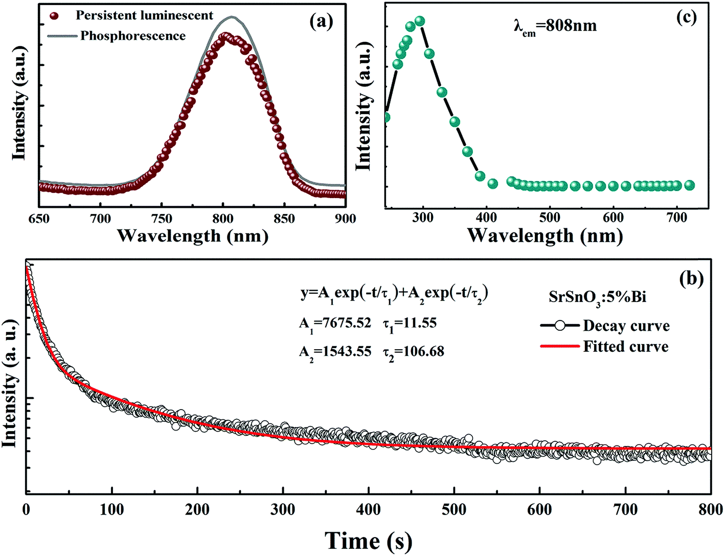 | ||
| Fig. 6 (a) LLP spectrum of SrSnO3:5% Bi after 500 W Xe lamp illumination for 10 min. (b) LLP decay curve of SrSnO3:5% Bi phosphor monitored at 808 nm emission after illumination for 10 min. (c) Afterglow intensity I10 s monitored at 808 nm as a function of the excitation wavelengths over the 250–750 nm spectral range. | ||
The afterglow attenuates exponentially. It contains a rapid decay process and a slow decay process. The decay curve is similar to that of typical long afterglow materials and it can be evaluated by fitting double exponential equation which reflects the trend of the decay. The equation is as follows:29
 | (1) |
We also studied the relationship between afterglow intensity and excitation wavelengths. Fig. 6c shows the afterglow intensity I10 s (afterglow intensity recorded 10 s after the stoppage of irradiation were used as reference points) of SrSnO3:5% Bi2+ powders monitored at 808 nm as a function of excitation wavelengths, indicating that persistent luminescence can be effectively excited by the light from 250 to 430 nm.
The long-lasting NIR afterglow of the SrSnO3:5% Bi phosphor was also visually evaluated using a supersensitive camera in a dark room. Fig. 7a–d shows the changes of NIR emission brightness with a decay time up to 20 min for phosphor after irradiation at ultraviolet lamp for 10 min. The images were taken in a dark room using a Germany pco dicam pro camera for a decay period lasting at least 20 min. It is shown that phosphor can be effectively activated by an ultraviolet lamp and result in persistent NIR emission.
 | ||
| Fig. 7 Long persistent luminescence imagings of sample SrSnO3:5% Bi. (a) 1, (b) 5, (c) 10 and (d) 20 min post-irradiation phosphorescence images. The samples were pre-excited by ultraviolet lamp for 10 min. | ||
Defect investigation
It is well known that trapping centers play an essential and crucial role for photo-energy storage in afterglow phosphors.30,31 Thermoluminescence (TL) spectrum is very useful for evaluating the trap depth, which is an important factor affecting emission of afterglow. In order to investigate trap states of SrSnO3:Bi2+, TL measurement was carried out. Fig. 8 shows normalized TL curve for sample SrSnO3:Bi2+ measured 30 s after irradiation ceased. The TL curve consists of two broad bands with maxima at 335 and 583 K, corresponding to the shallow and deep traps, respectively. At room temperature, shallow traps are easily emptied, whereas deep traps are difficult to be emptied; and parts of captured electrons remain stored there. If a trap is too deep, the captured electrons cannot escape, and no persistent phosphorescence is detected. Engineering a suitable trap depth is essential for achieving room temperature persistent phosphorescence. The depth of traps is estimated using the following equation:32
 | (2) |
 | ||
| Fig. 8 Thermo-luminescence curve of the sample SrSnO3:5% Bi measured at 10 min after the stoppage of irradiation. | ||
4. Conclusions
To the best of our knowledge, such long persistent phosphors with Bi2+ as the phosphorescent center have never been reported. There is also still no any study on the afterglow properties of Bi2+ doped SrSnO3 host. Bi2O3 was just proved to be an effective codopant to enhance red persistent luminescence, for instance, in ZnGa2O4:Cr, Bi spinel or Ca3Ga2Ge3O12:Cr, Bi.33,34But in this paper, we successfully developed a novel NIR persistent phosphor SrSnO3:Bi2+, which exhibits NIR persistent luminescence ranging from 700 nm to 900 nm. The phosphor has broken the domination of the conventional Cr, Mn-activated long persistent phosphors and provides a new perspective on the afterglow of Bi-doped phosphorescent phosphors. This new NIR persistent material has potential applications in vivo bio-imaging. Further research is underway on the mechanism analysis, nanocrystallization and functionalization of LPPs, which are expected to open up new possibilities for visualizing structural and functional processes in cells, tissues and other complex systems.
Acknowledgements
This work was financially supported by National Natural Science Foundation of China (Grant No. 51132004, 51072754, 51472091), Guangdong Natural Science Foundation (Grant No. S2011030001349, 2014A030310444), National Basic Research Program of China (Grant No. 2011CB808100), China Postdoctoral Science Foundation (Grant No. 2015M570707) and Fundamental Research Funds for the Central Universities (Grant No. 2013ZM0001, 2015ZM089). This work was also supported by the Open Fund of the State Key Laboratory of High Field Laser Physics (Shanghai Institute of Optics and Fine Mechanics.).References
- M. Kowatari, D. Koyama, Y. Satoh, K. Linuma and S. Uchida, Nucl. Instrum. Methods Phys. Res., Sect. A, 2002, 480, 431 CrossRef CAS.
- T. Matsuzawa, Y. Aoki, N. Takeuchi and Y. Matsuzawa, J. Electrochem. Soc., 1996, 143, 2670 CrossRef CAS.
- Z. Pan, Y. Lu and F. Liu, Nat. Mater., 2012, 11, 58 CrossRef CAS PubMed.
- J. P. Ryman-Rasmussen, M. F. Cesta, A. R. Brody, J. K. Shipley-Phillips, J. I. Everitt, E. W. Tewksbury, O. R. Moss, B. A. Wong, D. E. Dodd, M. E. Andersen and J. C. Bonner, Nat. Nanotechnol., 2009, 4, 747 CrossRef CAS PubMed.
- A. Smith, M. Mancini and S. Nie, Nat. Nanotechnol., 2009, 4, 710 CrossRef CAS PubMed.
- Y. Li, S. Zhou, Y. Li, K. Sharafudeen, Z. Ma, G. Dong, M. Peng and J. Qiu, J. Mater. Chem. C, 2014, 2, 2657 RSC.
- A. Bessière, Opt. Express, 2011, 19, 10131 CrossRef PubMed.
- Y. Li, Y. Li, R. Chen, K. Sharafudeen, S. Zhou, M. Gecevicius, H. Wang, G. Dong, Y. Wu, X. Qin and J. Qiu, NPG Asia Mater., 2015, 7, e180 CrossRef.
- Y. Li, Y. Li, K. Sharafudeen, G. Dong, S. Zhou, Z. Ma, M. Peng and J. Qiu, J. Mater. Chem. C, 2014, 2, 2019 RSC.
- A. Lecointre, B. Viana, Q. LeMasne, A. Bessière, C. Chanéac and D. Gourier, J. Lumin., 2009, 129, 1527 CrossRef CAS.
- D. Jia, L. A. Lewis and X. Wang, Electrochem. Solid-State Lett., 2010, 13, J32 CrossRef CAS.
- M. Allix, S. Chenu, E. Véron, T. Poumeyrol, E. A. Kouadri- Boudjelthia, S. Alahraché, F. Porcher, D. Massiot and F. Fayon, Chem. Mater., 2013, 25, 1600 CrossRef CAS.
- T. Maldiney, M. Kaikkonen, J. Seguin, Q. le Masne de Chermont, M. Bessodes, K. Airenne, S. Herttuala, D. Scherman and C. Richard, Bioconjugate Chem., 2012, 23, 472 CrossRef CAS PubMed.
- A. Bessiére, A. Lecointre, R. Benhamou, E. Suard, G. Wallez and B. Viana, J. Mater. Chem. C, 2013, 1, 1252 RSC.
- H. Zhou, M. Jiang and Y. Jin, RSC Adv., 2014, 4, 45786 RSC.
- J. Zhang, Z. Hao, J. Li, X. Zhang, Y. Luo and G. Pan, Light: Sci. Appl., 2015, 4, e239 CrossRef CAS.
- M. Peng and L. Wondraczek, J. Am. Ceram. Soc., 2010, 93, 1437 CAS.
- R. Cao, M. Peng and J. Qiu, Opt. Express, 2012, 20, A977 CrossRef PubMed.
- M. Peng and L. Wondraczek, Opt. Lett., 2009, 34, 2885 CrossRef PubMed.
- A. Srivastava, J. Lumin., 1998, 78, 239 CrossRef CAS.
- D. K. Mishra and X. Qi, J. Alloys Compd., 2010, 504, 27 CrossRef CAS.
- M. Qian, C. Yu, J. Cheng, K. Li and L. Hu, J. Lumin., 2012, 132, 2634 CrossRef CAS.
- Y. J. Siao, X. Qi, C. R. Lin and J. C. A. Huang, J. Appl. Phys., 2011, 109, 07A508 CrossRef.
- H. Guan, X. Zhang and Y. Xie, J. Phys. Chem. C, 2014, 118, 27170 CAS.
- M. Peng, N. Da, S. Krolikowski, A. Stiegelschmitt and L. Wondraczek, Opt. Express, 2009, 17, 21169 CrossRef CAS PubMed.
- M. Peng and L. Wondraczek, Opt. Lett., 2010, 35, 2554 Search PubMed.
- G. Blasse, A. Meijerink, M. Nomes and J. Zuidema, J. Phys. Chem. Solids, 1994, 55, 171 CrossRef CAS.
- M. Hamstra, H. Folkerts and G. Blasse, J. Mater. Chem., 1994, 4, 1349 RSC.
- D. Kshatri and A. Khare, J. Alloys Compd., 2014, 585, 488 CrossRef.
- D. Jia, R. Meltzer, W. Yen, W. Jia and X. Wang, Appl. Phys. Lett., 2002, 80, 1535 CrossRef CAS.
- Y. Li, K. Sharafudeen, G. Dong, Z. Ma and J. Qiu, Spectrochim. Acta, Part A, 2014, 123, 7 CrossRef CAS PubMed.
- K. van den Eeckhout, P. Smet and D. Poelman, Materials, 2010, 3, 2536 CrossRef CAS.
- Y. Zhuang, J. Ueda and S. Tanabe, Appl. Phys. Express, 2013, 6, 052602 CrossRef.
- C. Liu, Z. Xia, M. Molokeev and Q. Liu, J. Am. Ceram. Soc., 2015, 98, 1870 CrossRef CAS.
| This journal is © The Royal Society of Chemistry 2015 |