Shengyan Wanga,
Jianing Xua,
Yong Fana,
Jia Jiaa,
Lijuan Gaoa,
Jing Suna,
Liang Shana,
Xiaodong Chena,
Li Wang*a and
Hyo Jin Seo*b
aCollege of Chemistry, Jilin University, Changchun 130012, Jilin, P. R. China. E-mail: lwang99@jlu.edu.cn; Fax: +86-431-85671974; Tel: +86-431-85168439
bDepartment of Physics, Pukyong National University, Busan, Republic of Korea. E-mail: hjseo@pknu.ac.kr; Fax: +82-51-6295549; Tel: +82-51-6295568
First published on 10th December 2015
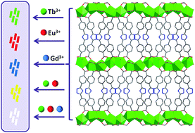
Three isomorphic lanthanide metal–organic frameworks (Ln-MOFs) have been constructed from lanthanide formate skeletons and 3,5-bis(4′-carboxy-phenyl)-1,2,4-triazole (H2bct) connectors. Further, by adjusting the co-doping ratio of different Ln3+ ions into the framework, two doped Ln-MOFs are synthesized and show tunable luminescence emission including white-light emission.
As well known, several factors have great influence on the design and synthesis of Ln-MOFs, such as the choice of organic ligands, the metal/ligand ratio and anions etc. Lanthanide ions have a high affinity and prefer to bond to oxygen atoms, thus, the selection or design of ligands containing appropriate oxygen atom coordination sites is crucial to construct Ln-MOFs.6 So far, many carboxylic acids, such as benzenepolycarboxylates,7 pyridinecarboxylates,8 imidazolecarboxylates9 and pyrazolecarboxylates10 have been widely employed to construct Ln-MOFs. More recently, based on mixed organic or inorganic anion ligands, such as carboxylate–oxalate,11 carboxylate–isonicotinate12 and carboxylate–sulfate,13 carboxylate–acetate,14 various Ln-MOFs have been prepared, in which anions as bridges incorporated in inorganic lattices. Thus, anion not only acts as a contributor to increase complexity but also exerts a synergistic influence in the structural control for self-assembly of Ln-MOFs.15 Especially, formate anion can be in situ generated by the hydrolysis of dimethylformamide (DMF) solvent under solvothermal conditions.16 It provides a new strategy for constructing functional Ln-MOFs.
3,5-Bis(4′-carboxy-phenyl)-1,2,4-triazole (H2bct) endowed with two carboxylate groups and a triazole ring, is a versatile bridging ligand for the construction of multi-dimensional MOFs17 and has conjugated system possess good energy-transfer ability, which can magnify the luminescence of the f–f transitions of Ln3+ ions. In this work, using DMF and H2O as solvents, a new family of isomorphic Ln-MOFs, namely, [Ln2(HCOO)2(bct)2]·H2O (Ln = Tb, Eu, Gd) have been constructed from lanthanide formate skeletons and bct2− connectors, exhibiting a 3D framework with 1D oval channels. Luminescence investigation reveals that 1-Tb and 2-Eu show characteristic green and red emissions of the corresponding Tb3+ and Eu3+ ions, respectively, while 3-Gd exhibits blue emission arising from the bct2− ligand. Then based on their luminescent properties, we have successfully designed and constructed two novel doped Ln-MOFs, Eu3+-doped 1-Tb and Eu3+, Tb3+-doped 3-Gd by adjusting the co-doping ratio of different Ln3+ ions into the same framework as that of 1–3. The emission colors of Eu3+-doped 1-Tb can be tuned from red to green by varying the molar ratios of Eu3+ ion from 10% to 0.02%, while that of Eu3+, Tb3+-doped 3-Gd can be tuned from yellow to white by changing excitation length.
Solvothermal reaction of H2bct with Ln(NO3)3 in a mixed solvent DMF and H2O yields isomorphic Ln-MOFs, [Ln2(HCOO)2(bct)2]·H2O (Ln = Tb (1), Eu (2) and Gd (3)). The corresponding mixed-lanthanide MOF, Eu3+-doped 1-Tb and Eu3+, Tb3+-doped 3-Gd, can be readily synthesized by using mixed lanthanide salts following the same procedure applied to 1–3.
X-ray analyses of 1-Tb, 2-Eu and 3-Gd reveals that they are isomorphous and exhibit a 3D framework with 1D oval channels constructed via dense [Ln2(HCOO)2]∞ 2D layers and bct2− bridging ligands. Hence, only the crystal structure of 1-Tb is described herein as a representative example. 1-Tb crystallizes in the orthorhombic space group Pnma and there are one unique Tb3+ ion, one bct2− ligand, one formate anion, and one lattice water molecule in the asymmetric unit of 1-Tb. As shown in Fig. 1, the central Tb3+ ion is eight-coordinated and described as a slightly distorted dicapped trigonal prism coordination mode: four oxygen atoms from the carboxylate moiety of four bct2− anions and four carboxylate oxygen atoms from two formate anions. The Tb–O bond lengths range from 2.279(7) to 2.589(7) Å which are comparable to those reported in the related Tb3+ compounds.18 The bond angles of O–Tb–O are in the range of 50.5(2) to 157.3(2)°. The formate anions adopt the chelating and bridging coordination modes, while bct2− anions exhibit the bis(monodentate) coordination fashion as depicted in Fig. 1c. All carboxyl groups of these ligands are deprotonated, in agreement with the IR data (Fig. S1†) in which no strong absorption peaks around 1700 cm−1 (–COOH) are observed.19
In the structure of 1-Tb, two Tb3+ centers, Tb1 and Tb1A (A = symmetry code: −x, −y + 2, −z + 1) are directly bridged by two μ2-O(4) atoms from two formate anions to form a [Tb2O2] rhombic SBU with a Tb⋯Tb distance of 3.8948(9) Å as shown in Fig. 2a. These dinuclear SBUs are connected to each other via O(6) atoms from formate anions forming a dense [Tb2(HCOO)2]∞ 2D network in the ac plane (Fig. 2b). As viewed along the a axis, the bct2− ligands further bridge the adjacent layers in a μ2-O coordination mode to result in a 3D framework with 1D oval channels (Fig. 2c).
The luminescent properties of 1-Tb, 2-Eu and 3-Gd and H2bct ligand in the solid state were investigated at room temperature. As shown Fig. 3, upon excitation with 350 nm, the free H2bct ligands show a blue emission band centered around 410 nm, which is assigned to the π–π* transition. Upon excitation at 307 nm, 1-Tb exhibits characteristic emission bands of Tb3+ ions at 488, 546, 582, and 620 nm, associated with the 5D4–7FJ (J = 6–3) transitions. 2-Eu emits red light when excited at 382 nm. The emission peaks at 579, 590, 616, 653, and 700 nm originate from the 5D0–7FJ (J = 0–4) transitions. The characteristic emission from H2bct ligand around 410 nm disappears, indicating that there exists energy transfer from H2bct ligands to Ln3+ ions that “magnifies” the luminescence of the f–f transitions. Under excitation at 380 nm, 3-Gd presents a blue emission with a broad band centered at 430 nm, which is quite similar to that of the free H2bct ligand, except for a slight red shift. As the lowest excited state of the Gd3+ 6P7/2 is too high to accept energy from the ligand, its characteristic 4f–4f transition at 311 nm is not visible. Therefore, it is reasonable to assign this emission to the intra ligand π–π* charge transfer. This phenomenon is similar to that previously reported.20
![]() | ||
Fig. 3 Emission spectra of 1-Tb (a), 2-Eu (b), 3-Gd (c) and H2bct ligand (d) in the solid state at room temperature. |
The characteristics of luminescent emission in 1-Tb and 2-Eu indicate that the antenna effect occurs, that is, energy migration takes place upon H2bct ligand absorption, followed by intersystem crossing S1 → T1 and antenna T1 → f transfer, and then generating f–f emissions of Tb3+ and Eu3+ ions as shown in Fig. S2.† According to Latva's empirical rule, a ligand-to-metal transfer process requires an energy gap of 2500–4500 cm−1 for Tb3+ and 2500–4000 cm−1 for Eu3+.21,22 Therefore, the H2bct ligand can effectively sensitize Tb3+ emission, but is not an ideal situation for the sensitization of Eu3+ luminescence, due to the energy gap between T1 and 5D4 state of Tb3+ is 2765 cm−1 while the energy gap between T1 and 5D0 state of Eu3+ being 5985 cm−1. The quantum yields of 1-Tb and 2-Eu, measured at room temperature upon excitation at 307 nm and 382 nm respectively, are 19.85% and 17.10%.
Considering that the emissions of 1–3 consist of three primary colors and they are isostructural, we expect to obtain tunable luminescent emission including white-light emission by doping different Ln3+ ions into one framework. By carefully choosing the starting lanthanide salts and optimizing the molar ratio of different Ln3+ ions, we successfully designed and constructed two novel doped Ln-MOFs, Eu3+-doped 1-Tb and Eu3+, Tb3+-doped 3-Gd. As shown in Fig. 4, the luminescent behaviors of a series of Eu3+-doped 1-Tb with different doping molar ratios were investigated when excited at 307 nm. It was found that their emission colors can be tuned from red to green by varying the molar ratios of Eu3+ ion from 10% to 0.02%. Indeed, the luminescent intensities of Tb3+ characteristic peak decrease abruptly as soon as some Eu3+ ions are present. Moreover, the red component to the color emission keeps strong even for low Eu3+/Tb3+ ratios. It indicates the emission of the Eu3+ ions in Eu3+-doped 1-Tb is further sensitized by the Tb3+ ions within the same frameworks. In addition, the luminescent decay test shows the values of lifetime for 1-Tb and 2-Eu are 0.57(1) ms and 0.56(1) ms, while 5D4 (Tb3+) and 5D0 (Eu3+) lifetime values in Eu3+-doped 1-Tb being 0.16(01) ms and 1.28(3) ms, respectively (Fig. S3†). This further approves the phenomenon of Tb3+ to Eu3+ energy transfer in Eu3+-doped 1-Tb.
Due to the blue emission of the bct2− ligand in 1-Tb and 2-Eu is almost completely suppressed by the strong green and red luminescence of the Tb3+ and Eu3+ ions, an excess amount of non-luminescent Gd3+ ion is required to dilute these two Ln3+ ions in the solid state. By optimizing the molar ratio of Eu3+, Tb3+ and Gd3+ ions being 0.0006:
0.2
:
0.7994, Eu3+, Tb3+-doped 3-Gd is synthesized, which can simultaneously show the characteristic emissions of the Eu3+ and Tb3+ ions, as well as the broad blue emission of the ligand. The emission spectra changes of Eu3+, Tb3+-doped 3-Gd as a function of the excitation wavelength were investigated (Fig. 5). As the excitation wavelength increases from 307 nm to 393 nm, the emission intensities of the red and blue components gradually increase, while the emission intensity of green component decreases, thus the emission color of Eu3+, Tb3+-doped 3-Gd can be tuned from yellow to white. A white emission with CIE coordinates of (0.313, 0.312), which fall within the white-light region, emerges upon excitation at 377 nm. The quantum yield of Eu3+, Tb3+-doped 3-Gd is 10.7%, which is measured under λex = 377 nm at room temperature.
Footnote |
† Electronic supplementary information (ESI) available: Additional structural figures, PXRD patterns, TGA and luminescent decay curves, IR, excitation spectra of 1–3 and H2bct and energy levels of H2bct, Eu3+ and Tb3+. CCDC 1011116 and 1428248. For ESI and crystallographic data in CIF or other electronic format see DOI: 10.1039/c5ra21557a |
This journal is © The Royal Society of Chemistry 2015 |