DOI:
10.1039/C5RA19154H
(Paper)
RSC Adv., 2015,
5, 100589-100597
Antioxidant and hepatoprotective effects of the food seasoning curry leaves Murraya koenigii (L.) Spreng. (Rutaceae)
Received
17th September 2015
, Accepted 13th November 2015
First published on 16th November 2015
Abstract
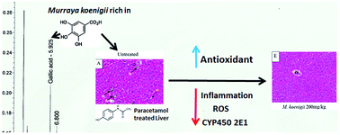
Murraya koenigii (L.) Spreng. (Rutaceae), a common spice, has been traditionally used to reduce inflammation and hepatitis. The present study aimed to reveal the antioxidant and anti-inflammatory activity as well as the regulation of cytochrome P450 levels elicited by aqueous extracts of M. koenigii leaves in response to paracetamol-induced liver toxicity in BALB/c mice. Liver toxicity was induced by an overdose of paracetamol followed by treatment with a M. koenigii leaf aqueous extract. The levels of serum liver markers, liver antioxidants, inflammatory markers and liver cytochrome P450 2E1 were quantified after 14 days of treatment. Histopathological analysis of the liver was also carried out. In vitro antioxidant levels and phenolic acid characterization were also performed. The extracts (50 and 200 mg kg−1 body weight) effectively restored the serum liver profiles (alanine transaminase, aspartate transaminase and alkaline phosphatase), liver antioxidant levels (superoxide dismutase, glutathione and ferric reducing ability of plasma) and inflammatory markers (tumor necrosis factor alpha, inducible nitric oxide synthase, nuclear factor kappa-light-chain-enhancer of activated B cells and nitric oxide) to healthy levels in a dosage dependent manner. The level of liver cytochrome P450 2E1 was also lowered in the extract treated groups. Histopathological assessment showed that treatment with 200 mg kg−1 of the M. koenigii aqueous extract was able to reduce liver necrosis in mice fed paracetamol. Gallic acid concentration was the highest among all the phenolic acids detected in the extract. These results suggested that the M. koenigii aqueous extract, which possessed antioxidant and anti-inflammatory effects, can be used as a potential treatment for liver diseases caused by oxidative stress.
1. Introduction
Plants, including spices, have been employed widely as food and dietary adjuncts. In addition, many are also used as medication in traditional therapy. Murraya koenigii (L.) Spreng., more commonly known as curry leaf tree, is a tropical plant in the Rutaceae family that can be found commonly in Asia including India, Sri Lanka, and Southeast Asia such as Malaysia.1 The leaves of M. koenigii are one of the most largely consumed flavoring ingredients in Indian cuisine. The spice is also utilized in Ayurveda medicine to treat inflammation, cuts, vomiting and dysentery. Nevertheless, many studies have been carried out to support these traditional applications and the collective findings showed that M. koenigii possessed anti-inflammatory, antioxidative and anti-diabetic effects in vitro and in vivo.2 Its leaves were found to be rich in polyphenols that contributed to its strong antioxidant activity.3
The liver is the major organ for lipid metabolism, protein synthesis and detoxification.4 However, prolonged exposure of the liver to xenobiotics and drugs including carbon tetrachloride (CCl4) and acetaminophen (APAP), commonly known as paracetamol, was found to induce high levels of reactive oxygen species (ROS) and inflammation, which subsequently damaged the liver.5 APAP is one of the commonly used over-the-counter (OTC) analgesic and antipyretic drugs. However, over-production of N-acetyl-p-benzoquinone imine (NAPQI), a by-product from the metabolization of APAP, can reduce the level of glutathione (GSH) and increase the level of cytochrome P450 which results in a significant increase of ROS in the liver. Thus, mice given an overdose of APAP have been widely used in an in vivo model for hepatoprotective studies of natural products.6 Enhancement of antioxidant levels especially the GSH level was found to potentially help the liver recover from damage induced by an overdose of APAP in rats.7 Many food and spices, such as pomegranate8 and nutmeg,9 were reported as potential antioxidant and hepatoprotective agents. Antioxidants in food have been proposed as ingredients that strengthen the body’s antioxidative status and subsequently help to alleviate degenerative diseases linked to oxidative stress.10 In relation to this, M. koenigii was reported to possess antioxidant and hepatoprotective effects in vitro and in vivo.1,5 These effects were also correlated to the high concentration of polyphenols in the leaf extract of M. koenigii.3,5 However, the major soluble phenolic acid content and the role of the M. koenigii leaf extract in alleviating inflammation and antioxidant levels in APAP-induced liver damage remains elusive. Our study therefore aimed to evaluate the effects of the M. koenigii leaf extract on antioxidant, inflammatory and cytochrome P450 levels in mice treated with an overdose of APAP. In addition, the total phenolic content, 1,1-diphenyl-2-picrylhydrazine (DPPH) scavenging activity, ferric reduction ability of plasma (FRAP) activity and the soluble phenolic acid content of the extract were also determined in this study.
2. Materials and methods
2.1. Plant material
M. koenigii leaves were obtained from a curry leaf plantation in Selangor, Malaysia in the months of April to June 2010. The plant was identified and deposited with the voucher number FRI 65673 by Science Officer Lim Chung Lu from the Forestry Division, Forest Research Institute Malaysia (FRIM) (Kepong, Malaysia). The leaves were air-dried, finely powdered using a grinder (HFM2413, Taiwan), heated with deionized water (1 g in 80 mL of water at 60 °C for 2 hours) and filtered through Whatman filter paper no. 1 (Millipore, Malaysia) and spray dried at an inlet temperature of 150 °C and outlet temperature of 100 °C (Buchi B-290, Switzerland). The native extract yield was 30% w/w with a moisture content <5%. The spray-dried M. koenigii aqueous extract was stored at 4 °C for the following in vivo and in vitro analyses.
2.2. In vivo hepatoprotective evaluation
APAP-induced hepatotoxicity in a mouse model was used to evaluate the hepatoprotective effect of the spray-dried M. koenigii aqueous extract. BALB/c mice (aged 5 weeks old, with an average body weight of 20 g) were purchased from the Animal House of Institute of Bioscience, Universiti Putra Malaysia. The procedures for this study were carried out according to the guidelines approved by the Animal Care and Use Committee, Faculty of Veterinary Medicine, Universiti Putra Malaysia (ref: UPM/FPV/PS/3.2.1.551/AUP-R168). Mice were acclimatized in plastic cages with 70% humidity and a temperature of 22 ± 3 °C for 7 days prior to the experiment and divided into 7 groups.
Group 1: mice were induced with 250 mg kg−1 APAP for 7 days followed by distilled water for another 14 days (untreated).
Group 2: mice were given distilled water for 7 days followed by 50 mg kg−1 of the M. koenigii aqueous extract treatment for another 14 days;
Group 3: mice were given distilled water for 7 days followed by 200 mg kg−1 of the M. koenigii aqueous extract treatment for another 14 days;
Group 4: mice were given distilled water only throughout the duration of the study (normal control);
Group 5: mice were induced with 250 mg kg−1 APAP for 7 days followed by a 50 mg kg−1 silybin treatment for another 14 days (positive control);
Group 6: mice were induced with 250 mg kg−1 APAP for 7 days followed by 50 mg kg−1 of the M. koenigii aqueous extract treatment for another 14 days;
Group 7: mice were induced with 250 mg kg−1 APAP for 7 days followed by 200 mg kg−1 of the M. koenigii aqueous extract treatment for another 14 days;
After 14 days of treatment, the mice were sacrificed, the serum was collected and the liver was harvested prior to performing the following assays.
2.2.1. Serum liver biomarkers and tumor necrosis factor alpha (TNF-α) quantification. Blood was centrifuged and the separated serum was used for several liver marker enzyme assays: alanine aminotransferase (ALT), alkaline phosphatase (ALP), and aspartate aminotransferase (AST). Assays were performed according to the manufacturer’s protocols (Roche Diagnostics GmbH, USA). TNF-α was quantified using a mouse TNF-α ELISA MAX (Biolegend, USA) according to the manufacturer’s instructions.
2.2.2. Liver homogenate preparation and antioxidant quantification. Liver was weighted and meshed using a 70 mm strainer (SPL, Korea) in cold phosphate buffer saline (PBS). The supernatant was separated from the pellet after centrifugation (8000 rpm, 15 minutes) and kept at −20 °C. Nitric oxide (NO), reduced glutathione and reactive oxygen species (ROS) levels were determined using the Griess assay kit (Invitrogen, USA), glutathione assay kit (Sigma, USA) and OxiSelect ROS assay kit (Cell Biolabs, USA), respectively. In addition, superoxide dismutase (SOD), malondialdehyde (MDA) and ferric reduction ability of plasma (FRAP) assays were performed according to the literature.11
2.2.3. Histology. An assay was performed according to Mohd Yusof et. al.11 Briefly, liver tissues were fixed in 10% neutral buffer formalin before paraffin embedding and stained with hematoxylin and eosin (H&E). The morphology of the liver samples were then observed using a Nikon Eclipse 90i microscope (New York, USA).
2.2.4. Quantitative real time reverse transcriptase PCR assay (qRT-PCR) of liver inducible nitric oxide synthase (iNOS) and nuclear factor kappa-light-chain-enhancer of activated B cell (NF-kB) gene expression. RNA was extracted from the liver of all groups using an RNeasy mini plus kit (Qiagen, Germany) and the extracted RNA was converted to cDNA using an iScript cDNA synthesis kit (Bio-Rad, USA). Expression of iNOS (iNOS forward primer: GCACCGAGATTGGAGTTC; iNOS reverse primer: GAGCACAGCCACATTGAT) and NF-kB (NF-kB forward primer: CATTCTGACCTTGCCTATCT; NF-kB reverse primer: CTGCTGTTCTGTCCATTCT) were evaluated using quantitative real time PCR (qRT-PCR) using a SYBR select master mix (Life Tech, USA) and an iQ5-Real Time PCR machine (Bio-Rad, USA). β-Actin (β-actin forward primer: TTCCAGCCTTCCTTCTTG; β-actin reverse primer: GGAGCCAGAGCAGTAATC) was used as the housekeeping control. Primer efficiency was determined by the standard curve of serially diluted cDNA samples isolated from the untreated control group. The PCR conditions were: 1 cycle of 50 °C/2 minutes for UDG activation, 1 cycle of 95 °C/2 minutes for DNA polymerase activation, 40 cycles of 95 °C/2 seconds for denaturation, and 52 °C for 30 seconds for annealing and extension. All samples were assayed in triplicate and the no-template controls were prepared for the specific assay. The relative expression of both genes in relation to housekeeping was calculated by the ΔΔCq method.12 The fold change between the treated and untreated control was presented in this study.
2.2.5. Western blot analysis of liver cytochrome P450. The liver from all groups was homogenized and the protein was quantified using the Bradford assay. 100 μg of the extracted protein were loaded onto a gel, subjected to SDS/PAGE (5% stacking gel and 10% running gel), and then electroblotted onto nitrocellulose membranes (Hybond-ECL, Amersham) using a semidry electroblotter (Transblot SD Semi-Dry Transfer Cell, BioRad). The membranes were blocked in a Tris-buffered saline (TBS)-Tween buffer, pH 7.5 (20 mM Tris/500 mM NaCl/0.05% Tween-20) containing 5% skimmed milk powder for 1 hour before exposure to an anti-beta actin antibody and anti-cytochrome P450 2E1 (Abcam, USA), at a dilution of 1/5000 in the TBS-Tween buffer, pH 7 for 1 hour. Membranes were then washed and incubated with Goat anti-Rabbit IgG H&L conjugated to alkaline phosphatase, diluted 1/5000 in the same buffer, for 1 hour. After a series of washes in the TBS-Tween buffer, protein bands were visualized by chemiluminescence with a CDP-STAR® reagent (NEB, UK) and visualized under a BioSpectrum system (UVP, US). The size of the protein bands were determined using electrophoresis colour markers. The protein level was normalized against β-actin to control for variance in the sample loading and transfer.
2.3. In vitro antioxidant assays
2.3.1. Total phenolic quantification. The total phenolic content was measured using the Folin–Ciocalteu method according to Ho et. al.13 with slight modification. Briefly, 1 mL of the sample, blank and gallic acid standard was placed in a test tube. Then, 5 mL of the Folin–Ciocalteu reagent was added and the mixture was vortexed and allowed to react for 5 minutes before adding 4 mL of 7.5% sodium carbonate. The mixture was then left at room temperature for 2 hours before being measured at 765 nm using a spectrophotometer (Beckman Coulter, USA). The results were expressed as gallic acid equivalents (GAE), using gallic acid as a standard.
2.3.2. 1,1-Diphenyl-2-picrylhydrazine (DPPH) radical scavenging test. The radical scavenging activity was measured using a modified DPPH method as described previously.14 A serial dilution was done for the sample and the standard. Trolox was used as a standard in this assay. Briefly, 50 μL of the sample was added to 250 μL of DPPH (0.04 mg mL−1) in a 96 well-plate. The mixture was allowed to react for 30 minutes in the dark. The absorbance was measured using an ELISA plate reader (BioTek Instrument, USA) at 515 nm.
2.3.3. Ferric reducing antioxidant power (FRAP) test. The FRAP assay was performed as previously described with slight modification.14 The FRAP reagent was prepared by mixing 300 mM acetate buffer, 10 mM TPTZ (in 40 mM HCl) and 20 mM of FeCl3·6H2O in a 10
:
1
:
1 ratio. The reagent was prewarmed at 37 °C before use. Briefly, 50 μL of the sample was loaded into a 96-well plate before adding 250 μL of the prewarmed FRAP reagent. The plate was incubated for 10 minutes in the dark at room temperature before an absorbance reading at 593 nm was taken. Results were calculated according to the calibration curve, using FeSO4·7H2O (100–1000 μM) as a standard.
2.3.4. Quantification of soluble phenolic acids. Soluble phenolic acids in the M. koenigii aqueous extract were quantified using reverse-phase high performance liquid chromatography.15 Concentrations of gallic acid, protocatechuic acid, 4-hydroxybenzoic acid, vanillic acid and syringic acid were determined via calibration with standards (external standard quantitation). The spray-dried aqueous extract of M. koenigii was solubilised in 2.5% methanol and filtered through a 0.2 μm membrane before injecting into a Chromolith Performance RP-18e (100 mm × 4.6 mm i.d.) column. Analysis was performed using a Waters Alliance 2695 Separation Module (Waters, Milford, MA) at 30 °C. The isocratic flow rate was set at 0.7 mL min−1 with 0.1% formic acid and methanol as the mobile phase.
2.4. Statistical analysis
All data were statistically analysed by one-way analysis of variance (ANOVA) using SPSS 15 software. Duncan’s multiple range tests were used for post-hoc analysis and p < 0.05 compared to the untreated control was regarded as significant.
3. Results
3.1. Effects of the M. koenigii aqueous extract on body weight, serum liver biochemical profiles, and TNF-α levels in APAP treated mice
Neither the sole treatment of APAP and M. koenigii on non-induced mice nor their post-treatment on APAP-induced mice resulted in significant changes in the body weight of the mice (Table 1). Blood glucose levels of the APAP-treated mice showed about a 1.5 fold increase as compared to normal control levels. Both 50 and 200 mg kg−1 of M. koenigii treated non-induced or APAP-induced mice were able to decrease serum glucose lower than that of the normal group mice. These mice also showed significantly higher serum levels of ALT, ALP, AST and TNF-α in comparison to the normal control. M. koenigii and silybin significantly (p < 0.05) reduced the elevated levels of AST, ALT, ALP and TNF-α in the APAP-induced mice. Reduction of ALP and TNF-α by a higher concentration of M. koenigii (200 mg kg−1 bw) was comparable to the effect of the positive control. Restoration to near normal levels was only observed for serum ALT in silybin treated mice.
Table 1 Effect of M. koenigii, silybin and APAP on the serum biochemical profiles of experimental micea
|
Body weight (g) |
Glucose (mmol L−1) |
ALT (U/L) |
ALP (U/L) |
AST (U/L) |
TNF-α (pg mL−1 protein) |
Different letters indicate a significant difference compared with the APAP untreated group, p < 0.05. |
Normal control |
22.41 ± 0.42a |
4.73 ± 1.14i |
42.32 ± 1.05y |
64.56 ± 1.12n |
143.76 ± 2.82β |
32.51 ± 28.13f |
M. koenigii (50 mg kg−1) |
22.03 ± 0.52a |
4.02 ± 1.56i |
41.58 ± 2.15y |
64.13 ± 1.52n |
141.46 ± 3.41β |
35.55 ± 21.99f |
M. koenigii (200 mg kg−1) |
22.18 ± 0.86a |
3.86 ± 0.89i,ii |
41.32 ± 1.77y |
61.39 ± 1.19n |
139.25 ± 4.56β |
30.18 ± 15.77f |
APAP |
21.93 ± 0.57a |
7.30 ± 1.75iii |
118.19 ± 2.75x |
83.83 ± 3.61m |
443.36 ± 9.99α |
585.96 ± 15.88e |
APAP + silybin (50 mg kg−1) |
21.99 ± 0.67a |
4.63 ± 1.11i |
46.23 ± 2.92y |
70.33 ± 1.49° |
205.00 ± 4.20γ |
220.14 ± 14.45g |
APAP + M. koenigii (50 mg kg−1) |
21.18 ± 1.66a |
4.18 ± 1.24i |
107.78 ± 2.46z |
75.42 ± 2.31p |
253.86 ± 5.61δ |
364.31 ± 23.42h |
APAP + M. koenigii (200 mg kg−1) |
22.53 ± 1.58a |
4.00 ± 1.40i |
67.79 ± 4.13zz |
71.00 ± 1.60° |
224.00 ± 3.35ε |
233.14 ± 20.75g |
3.2. Effects of the M. koenigii aqueous extract on liver antioxidant, oxidative stress and nitric oxide levels in APAP treated mice
The antioxidant capacity of the extract was evaluated via a few parameters, namely concentration of the liver antioxidant peptide GSH, antioxidant enzyme SOD, reduction of oxidative stress (MDA and ROS) and nitric oxide (NO) levels. In addition, the total antioxidant activity of the extract was also evaluated based on its ability to reduce the FeIII–TPTZ complex to FeII–TPTZ as indicated by the FRAP assay (Table 2). When compared to normal control, mice treated with 50 mg kg−1 of M. koenigii exhibited a slight increase in the total antioxidant activity with no significant difference in other parameters. On the other hand, a higher concentration (200 mg kg−1) of the extract not only enhanced the total antioxidant activity but also significantly increased GSH and SOD levels. Significant reduction of the NO level was observed in the 200 mg kg−1 group while there were insignificant changes in oxidative stress levels (MDA and ROS) in both M. koenigii-treated groups.
Table 2 Effect of M. koenigii, silybin and APAP on the liver antioxidant and NO levels of experimental micea
Group |
GSH (nM GSH per mg protein) |
SOD (U/mg per protein) |
FRAP (μM Fe(II) per mg protein) |
MDA (nM MDA per mg protein) |
ROS (DCF fluorescence intensity per mg of protein) |
NO (μM per mg protein) |
Different letters indicate a significant difference compared with the APAP untreated group, p < 0.05. |
Normal control |
6.42 ± 0.12a |
172.48 ± 5.06i |
30.55 ± 1.83° |
0.61 ± 0.18m |
431.93 ± 37.35α |
15.27 ± 1.99e |
M. koenigii (50 mg kg−1) |
6.88 ± 0.46a |
179.54 ± 4.61i |
33.89 ± 1.76p,q |
0.60 ± 0.25m |
400.15 ± 41.52α |
14.48 ± 1.44e |
M. koenigii (200 mg kg−1) |
9.15 ± 0.68b |
201.31 ± 6.88ii |
39.14 ± 1.93r |
0.55 ± 0.18m |
389.76 ± 29.64α |
11.85 ± 1.96f |
APAP |
1.88 ± 0.03c |
40.89 ± 1.92iii |
17.22 ± 1.30 s |
1.24 ± 0.19n |
2876.71 ± 247.01β |
33.16 ± 1.05g |
APAP + silybin (50 mg kg−1) |
4.33 ± 0.06d |
85.43 ± 5.54iv |
21.98 ± 2.21 t |
0.76 ± 0.21m |
1935.88 ± 166.29γ |
22.24 ± 1.38h |
APAP + M. koenigii (50 mg kg−1) |
2.91 ± 0.18e |
72.73 ± 2.36v |
19.62 ± 2.86 s,t |
0.76 ± 0.14m |
2315.14 ± 211.12δ |
30.78 ± 1.31g |
APAP + M. koenigii (200 mg kg−1) |
6.89 ± 0.32a |
174.16 ± 1.66i |
28.04 ± 1.76° |
0.66 ± 0.18m |
1148.35 ± 182.06ε |
13.37 ± 2.13e |
In contrast, APAP exposure significantly elevated (p < 0.05) oxidative stress (MDA, ROS, and NO) and impaired (p < 0.05) antioxidant activities (SOD, GSH, FRAP) in the mice. After treatment with M. koenigii, the total antioxidant activity and GSH and SOD levels were enhanced in the livers of APAP-induced mice. This effect was also associated with the reduction of oxidative stress (ROS), lipid peroxidation (MDA) and NO levels. Notably, 200 mg kg−1 of the extract resulted in a greater inhibition of oxidative stress and inflammation than the silybin group and was the only group that showed restoration of total antioxidant, SOD, GSH, MDA and NO levels to near normal.
3.3. Effects of the M. koenigii aqueous extract on liver histopathology in APAP treated mice
Histological sections (H&E staining) of normal control mice liver showed an intact centrilobular vein, healthy hepatocytes and a thin sinusoidal space (Fig. 1A). In contrast, mice exposed to APAP showed a non-uniform morphology of the hepatocytes, an enlargement of the sinusoidal space and necrosis (Fig. 1B). After treatment with 200 mg kg−1 of the M. koenigii aqueous extract (Fig. 1E), the liver tissue exhibited signs of recovery whereby intact hepatocytes were observed, with an integral centrilobular vein and marginal sinusoidal space similar to the normal control tissue (Fig. 1A) and silybin (Fig. 1C) treated mice. In addition, the group treated with 50 mg kg−1 of aqueous M. koenigii also showed a marginal recovery although some necrosis and a mild enlargement of the sinusoidal space could still be observed in the liver histological specimen (Fig. 1D).
 |
| Fig. 1 Representative histopathology of the liver of (A) normal healthy mice, (B) APAP treated only, (C) APAP + silybin treated mice, (D) APAP + 50 mg kg−1 M. koenigii, and (E) APAP + 200 mg kg−1 M. koenigii treated mice. (H&E staining, magnification 100×.) CV: centrilobular vein; SS: sinusoidal space; N: necrosis. | |
3.4. Effects of the M. koenigii aqueous extract on liver iNOS, NF-kB and cytochrome P450 levels in APAP treated mice
Quantitative real time reverse transcriptase PCR and Western blotting were used to evaluate the expression levels of inflammatory markers (iNOS and NF-kB) (Fig. 2) and cytochrome P450 (Fig. 3) in livers collected from the mice. Expression levels of iNOS and NF-kB were down-regulated in all groups except in the untreated mice (APAP-induced). The group treated with 200 mg kg−1 of the M. koenigii aqueous extract also showed the highest reduction of NF-kB levels among all the treated groups (Fig. 2). On the other hand, both the groups treated with 50 and 200 mg kg−1 of the M. koenigii aqueous extract also possessed similar significant (p < 0.05) reductions of cytochrome P450 expression as the positive control silybin treated group when compared to the untreated group by Western blot analysis (Fig. 3).
 |
| Fig. 2 Quantitative real time reverse transcriptase PCR evaluation of the expression of liver iNOS and NFkB in normal healthy, M. koenigii aqueous extract (50 and 200 mg kg−1) treated healthy, APAP treated only, APAP + silybin treated and APAP + M. koenigii aqueous extract (50 and 200 mg kg−1) treated mice. The statistical differences among all groups were assessed by one-way ANOVA followed by Duncan’s post-hoc. Means labelled with different letters are significantly different, p < 0.05. | |
 |
| Fig. 3 Western blot analysis of the levels of liver P450 protein of (a) APAP treated only, APAP + silybin treated and APAP + M. koenigii aqueous extract (50 and 200 mg kg−1) treated mice and (b) normal and M. koenigii aqueous extract (50 and 200 mg kg−1) treated healthy mice. The statistical differences among all groups were assessed by one-way ANOVA followed by Duncan’s post-hoc. Means labelled with different letters are significantly different, p < 0.05. | |
3.5. In vitro antioxidant capacity and soluble phenolic acid profiling of the M. koenigii aqueous extract
The in vitro antioxidant capacity and soluble phenolic acid profile of the M. koenigii aqueous extract are summarized in Table 3. The M. koenigii aqueous extract contained 304.10 μg ascorbic acid equivalents per mg sample and 193.97 μg gallic acid equivalents per mg sample as determined respectively by FRAP and total phenolic content assays. 300 μg mL−1 of the M. koenigii aqueous extract showed ∼26% inhibition to DPPH scavenging activity. Among the quantified soluble phenolic acids, gallic acid was most prevalent in the extract (674 μg mL−1), followed by vanillic acid, p-coumaric acid, syringic acid and protocatechuic acid. The concentration of 4-hydroxybenzoic acid was the lowest (90 μg mL−1) of them all.
Table 3 Total phenolic content, soluble phenolic acids content, DPPH and FRAP of the M. koenigii aqueous extracta
|
Murraya koenigii aqueous extract |
GAE: gallic acid equivalent. |
Total phenolic content (μg GAE per mg M. koenigii extract) |
193.97 ± 12.53 |
![[thin space (1/6-em)]](https://www.rsc.org/images/entities/char_2009.gif) |
Phenolic acid derivatives (HPLC) |
Gallic acid (μg mL−1) |
674.20 ± 5.81 |
Protocatechuic acid (μg mL−1) |
133.21 ± 3.84 |
4-Hydroxybenzoic Acid (μg mL−1) |
90.05 ± 2.33 |
Vanillic acid (μg mL−1) |
250.64 ± 7.28 |
Syringic acid (μg mL−1) |
219.06 ± 5.88 |
p-Coumaric acid (μg mL−1) |
247.60 ± 3.61 |
DPPH (percentage of inhibition at 300 μg mL−1) |
26.13 ± 1.66 |
FRAP (μM Fe(II) per mg M. koenigii extract) |
304.10 ± 3.88 |
4. Discussion
ALT is a cytosolic enzyme in hepatocytes, AST is a mitochondrial enzyme in liver parenchymal cells while ALP is an enzyme in the cells lining the biliary duct of the liver.16 Leakage of these enzymes in the blood could be promoted by the loss of hepatocyte membrane integrity, mitochondria damage in the liver tissue or large bile duct obstruction.16 Thus, an increase in enzyme levels can be conveniently used for measuring the extent of hepatic injury.13 Previous studies showed that hydroethanaolic and methaolic extracts of M. koenigii leaves were capable of reducing the levels of these 3 liver enzymes in rats treated with CCl4.5,17,18 A separate study on extracts of M. koenigii dried bark, extracted using different solvents including acetone, benzene, petroleum ether, chloroform, acetone and methanol, showed the significant ability of these extracts to reduce elevated ALP levels in CCl4-induced rats.19 Pre-treatment with the aqueous leaf extract of M. koenigii has also been shown to protect rats against liver damage induced by lead but its effect on the liver enzymes was not evaluated in the study.20 On the other hand, Sathaye et. al.21 reported that the ALP level was reduced in ethanol-induced mice that were co-treated with an aqueous leaf extract of M. koenigii. Cumulatively, these results suggested that M. koenigii is a potent hepatoprotective agent and it would be imperative to examine its effects on liver marker enzymes to assess the degree of recovery or protection against induced damage in liver cells.
APAP is the most widely used analgesic/antipyretic drug. However, an overdose of APAP has always been associated with acute hepatotoxicity. Thus, the drug is commonly used in studies for xenobiotic-induced hepatotoxicity22 and it is often associated with increased liver enzyme levels (ALT, AST and ALP), lipid peroxidation, cytochrome P450 2E1, and necrosis of hepatocytes.23,24 As observed in this study, mice which received APAP exhibited significantly higher levels of ALT, ALP and AST compared to the normal control (Table 1), signifying liver damage. These findings are also supported by H&E histopathological examination, which shows abnormalities including necrosis and enlargement of the sinusoidal space in the mouse livers (Fig. 1B). In contrast, both concentrations of the M. koenigii extract significantly reduced the serum levels of all 3 liver enzymes as compared to the levels in APAP-treated mice, demonstrating the protective properties against liver damage in a dosage dependent manner. A higher concentration of the extract exhibited greater reductions and more comparable effects to silybin. However, both concentrations did not recover the liver enzyme levels to near normal while silybin was able to restore the ALT level only to near normal. Histopathological examination of the liver tissues was also consistent with the observation whereby higher concentrations of M. koenigii and silybin resulted in more prominent signs of recovery than lower concentrations of M. koenigii (Fig. 1).
With preliminary evidence of recovery from APAP-induced damage, we were interested to know if the effect of M. koenigii is associated with the anti-inflammatory and anti-oxidant pathways that could alleviate liver damage. Necrosis caused by APAP would often lead to recruitment of macrophages and the release of pro-inflammatory factors such as cytokines and chemokines.25 This explains why liver inflammation is commonly observed in xenobiotic-induced hepatotoxicity. Among the regulators of inflammation, the NF-kB signalling pathway plays the most important role in the regulation of liver disease progression.26 Activation of NF-kB during acute inflammation caused by APAP-induced stress can eventually lead to fibrosis, cirrhosis and even increase the risk of progression to hepatocellular carcinoma.26 TNF-α, a proinflammatory cytokine, was found to be highly upregulated after treatment with APAP (Table 1). Apart from inducing neutrophil and macrophage accumulation and activation, high levels of TNF-α will also activate NF-kB and the subsequent production of iNOS in the liver.26,27 In this study, the activation of proinflammatory markers including iNOS, NF-kB, (Fig. 2) and NO (Table 2) was observed after exposure to an overdose of APAP (Table 1). After being treated with M. koenigii, the levels of TNF-α and NO in the liver reduced in a dosage dependent manner. The mRNA expression of iNOS and NF-kB was also down regulated in response to this. M. koenigii has been shown to potently scavenge nitric oxide in vitro in a previous study.28 In this study, both silybin and M. koenigii significantly reduced NO levels in the APAP-treated mice, and 200 mg kg−1 of M. koenigii was able to restore the NO level to near normal. Thus, along with the reduced mRNA expression of iNOS and NF-kB, it is suggested that M. koenigii possesses an anti-inflammatory effect over APAP-induced liver inflammation.
Besides TNF-α, the activation of the NF-kB proinflammatory signalling pathway could also be attributed to oxidative stress and lipid peroxidation. Consistent with other findings, APAP induced hyperglycemia (Table 1),29 oxidative stress (ROS), increased lipid peroxidation (MDA) while suppressing antioxidant levels (SOD, GSH and FRAP) in the liver (Table 2).27 APAP is catalysed into its metabolite, N-acetyl-p-benzoquinone imine (NAPQI), which can be detoxified at a nontoxic dose. When overdosed however, NAPQI leads to the depletion of GSH, exposing hepatocytes to the destructive effects of reactive oxygen species (ROS) and oxidative stress, which initiate necrosis.27 Besides GSH depletion, NAPQI was also reported to significantly deplete glycogen thus inducing hyperglycemia in mice.29 Both concentrations of M. koenigii were able to maintain the glucose level of non-induced and APAP-induced mice lower than that of normal mice, which may be contributable to its hypoglyemic action as previously reported by Khan et al., indicating that M. koenigii exhibited significant hypoglycemic action via an increase of hepatic glycogen and glycogenesis.30 ROS was increased by more than 6 fold by APAP while both concentrations of M. koenigii did not result in any significant change in the non APAP-treated mice. As indicated by both the non-induced and APAP-induced mice, 50 mg kg−1 of the M. koenigii extract possessed great hypoglycemic action to completely reverse the serum glucose level lower than that of the normal control, but the antioxidant effect of this treatment was not able to completely reverse the liver damage caused by APAP treatment. A significant reduction of ROS was observed in the group treated with 200 mg kg−1 of the M. koenigii extract as it exerted a greater antioxidant effect than treatment with 50 mg kg−1 of the extract or even silybin.
Strong antioxidants were shown to prevent APAP-induced cytochrome P450 2E1 expression in liver cells31 and could subsequently protect or recover the liver from APAP-induced hepatocellular damage. In the attempt to study the effect of M. koenigii in mediating APAP catalysis, we found that both concentrations of the extract down regulated the increased expression of P450 2E1 by APAP induction. Various extracts of the plant, including water, ethyl alcohol
:
water (1
:
1), methanol, ethanol, hexane, chloroform, acetone, ethyl acetate, benzene, petroleum ether, and methylene chloride had been previously tested for their antioxidant activities.3,32–36 Among them, the methanol extract of M. koenigii leaves was shown to possess greater antioxidant activity than the methanol extract of a few green leafy vegetables such as Amaranthus sp., Centella asiatica, and Trigonella foenum-graecum32 while its ethanolic leaf extract was reported to exhibit good antioxidant activity comparable to that of ascorbic acid.33 On the other hand, the antioxidant activity of the M. koenigii aqueous leaf extracts was claimed to confer significant protection against cadmium-induced oxidative stress in rat cardiac tissue.34 A separate study reported that 300 μg mL−1 of the extract scavenged ∼41% of DPPH radical activity3 while our study showed ∼26% DPPH scavenging activity with a lower concentration (100 μg mL−1) of the same extract. In addition, our study also evaluated antioxidant activity using the FRAP assay, which was reported to be more reliable in measuring antioxidant activity and correlates well with the total phenolic content.14 As seen in Table 3, the total antioxidant activity of the M. koenigii aqueous leaf extract was 304 μM Fe(II)/mg extract and the total phenolic content was 194 μg GAE/mg extract. Further analysis by HPLC identified the presence of a few common phenolic acid derivatives in the extract, with gallic acid (∼674 μg mL−1 extract) being the most abundant compound. As shown previously, phenolic acids, sesquiterpenes, rutinosides32 and alkaloids33 from M. koenigii had been reported to exert moderate hepatoprotective effects in vitro28 and anti-inflammatory effects in vivo.37 Among them, gallic acid had been reported to possess hepatoprotective effects against APAP-induced liver damage due to its antioxidant activity. The treatment was capable of restoring the depleted SOD and GSH levels and controlled inflammation induced by APAP in mice.37 The hepatoprotective effect of gallic acid was proposed to be correlated to its inhibitory effect on cytochrome P450 2E1 activity.38 For instance, gallic acid isolated from Orostachys japonicus was found to interfere with the increased hepatic activities of two cytochrome P450-dependent monooxygenases, namely aminopyrine N-demethylase (AMND) and aniline hydroxylase in mouse models.39 As this enzyme plays an important role in catalysing APAP to NAPQI, we propose that M. koenigii is capable of protecting the liver from damage by inhibiting APAP catalysis to its toxic metabolite NAPQI, and this could be attributed to the presence of gallic acid in the leaf extract.
5. Conclusion
In the present study, a M. koenigii aqueous extract demonstrated liver protective activities against the damage induced by APAP. In vitro antioxidant tests and HPLC profiling revealed the presence of various soluble phenolic acids, especially gallic acid, which may contribute to the hepatoprotective, antioxidative and anti-inflammatory effects of the M. koenigii aqueous leaf extract. As such, further studies are essential for elucidating the role of the individual phytochemicals in eliciting the anti-inflammatory and hepatoprotective effects of the M. koenigii leaves. This study could also serve as a lead to identify the standard target for regulating the M. koenigii aqueous extract as a functional food supplement for liver protection.
Acknowledgements
This project was funded by Research University Grants (RUGS) 91194, Universiti Putra Malaysia. We would like to thank Professor Tan Soon Guan for proof reading this manuscript.
References
- S. Sathaye, Y. Bagul, S. Gupta, H. Kaur and R. Redkar, Exp. Toxicol. Pathol., 2011, 63, 587–591 CrossRef CAS PubMed.
- Q. Ma, J. Tian, J. Yang, A. Wang, T. Ji, Y. Wang and Y. Su, Fitoterapia, 2013, 87, 1–6 CrossRef CAS PubMed.
- M. B. Ningappa, R. Dinesha and L. Srinivas, Food Chem., 2008, 106, 720–728 CrossRef CAS.
- S. Fu, L. Yang, P. Li, O. Hofmann, L. Dicker, W. Hide, X. Lin, S. M. Watkins, A. R. Ivanov and G. S. Hotamisligil, Nature, 2011, 473, 528–531 CrossRef CAS PubMed.
- S. N. Desai, D. K. Patel, R. V. Devkar, P. V. Patel and A. V. Ramachandran, Food Chem. Toxicol., 2012, 50, 310–314 CrossRef CAS PubMed.
- S. Bhanwra, J. Singh and P. Khosla, Indian J. Physiol. Pharmacol., 2000, 44, 64–68 CAS.
- R. Simeonova, V. Vitcheva, M. Kondeva-Burdina, I. Krasteva, V. Manov and M. Mitcheva, BioMed Res. Int., 2013, 757126, DOI:10.1155/2013/757126.
- I. S. Ashoush, O. I. El-Batawy and G. A. El-Shourbagy, Ann. Agric. Sci., 2013, 58, 27–32 Search PubMed.
- T. Morita, K. Jinno, H. Kawagishi, Y. Arimoto, H. Suganuma, T. Inakuma and K. Sugiyama, J. Agric. Food Chem., 2003, 51, 1560–1565 CrossRef CAS PubMed.
- V. Mani, K. Ramasamy, A. Ahmad, S. N. Wahab, S. M. Jaafar, T. L. Kek, M. Z. Salleh and A. B. A. Majeed, Phytother. Res., 2013, 27, 46–53 CrossRef CAS PubMed.
- H. Mohd Yusof, N. M. Ali, S. K. Yeap, W. Y. Ho, B. K. Beh, S. P. Koh, K. Long, S. Abdul Aziz and N. B. Alitheen, J. Evidence-Based Complementary Altern. Med., 2013, 274274, DOI:10.1155/2013/274274.
- M. W. Pfaffl, Nucleic Acids Res., 2001, 29, e45 CrossRef CAS PubMed.
- W. Y. Ho, S. K. Yeap, C. L. Ho, R. Abdul Rahim and N. B. Alitheen, J. Evidence-Based Complementary Altern. Med., 2012, 417953, DOI:10.1155/2012/417953.
- K. Thaipong, U. Boonprakob, K. Crosby, L. Cisneros-Zevallos and D. Hawkins Byrne, J. Food Compos. Anal., 2006, 19, 669–675 CrossRef CAS.
- V. Nagavani and T. Raghava Rao, Adv. Biol. Res., 2010, 4, 159–168 CAS.
- S. Rao, J. W. Einstein and K. Das, Int. Lett. Nat. Sci., 2014, 3, 30–43 CrossRef.
- R. Gupta and D. Singh, Pharmacologyonline, 2007, 1, 232–242 Search PubMed.
- B. N. Parimi, R. Mopuri and B. Meriga, J. Coastal Life Med., 2014, 2, 313–318 Search PubMed.
- M. Pande, S. Gupta and A. Pathak, J. Herb. Med. Toxicol., 2009, 3, 69–71 Search PubMed.
- D. Ghosh, S. B. Firdaus, E. Mitra, M. Dey, A. Chattopadhyay, S. K. Pattari, S. Dutta, K. Jana and D. Bandyopadhyay, Int. J. Pharm. Pharm. Sci., 2013, 5, 285–295 Search PubMed.
- S. Sathaye, P. D. Amin, V. B. Mehta and V. B. Zala, Int. J. Pharma Bio Sci., 2012, 3, 430–438 Search PubMed.
- K. C. D. Anantha, R. C. Siva and R. A. Manohar, J. Ayurveda Integr. Med., 2012, 3, 198–203 CrossRef PubMed.
- J. Das, J. Ghosh, P. Manna and P. C. Sil, Free Radical Res., 2010, 44, 340–355 CrossRef CAS PubMed.
- J. Hwang, Y.-H. Chang, J. H. Park, S. Y. Kim, H. Chung, E. Shim and H. J. Hwang, Lipids Health Dis., 2011, 10, 184 CrossRef CAS PubMed.
- H. Jaeschke, C. D. Williams, A. Ramachandran and M. L. Bajt, Liver Int., 2012, 32, 8–20 CrossRef CAS PubMed.
- B. Sun and M. Karin, Oncogene, 2008, 27, 6228–6244 CrossRef CAS PubMed.
- W.-T. Chen, C.-L. Yang and M.-C. Yin, Biomedicine, 2014, 4, 5 CrossRef PubMed.
- M. S. Baliga, G. C. Jagetia, S. K. Rao and K. Babu, Nahrung, 2003, 47, 261–264 CrossRef CAS PubMed.
- J. A. Hinson, J. B. Mays and A. M. Cameron, Biochem. Pharmacol., 1983, 32, 1979–1988 CrossRef CAS PubMed.
- B. A. Khan, A. Abraham and S. Leelamma, Indian J. Biochem. Biophys., 1995, 32, 106–108 CAS.
- S. C. Jeong, S. M. Kim, Y. T. Jeong and C. H. Song, Chin. Med., 2013, 8, 7 CrossRef PubMed.
- S. Gupta and J. Prakash, Plant Foods Hum. Nutr., 2009, 64, 39–45 CrossRef CAS PubMed.
- I. W. Kusuma, H. Kuspradini, E. T. Arung, F. Aryani, Y.-H. Min, J.-S. Kim and Y.-U. Kim, J. Acupunct. Meridian Stud., 2011, 4, 75–79 CrossRef PubMed.
- E. Mitra, A. K. Ghosh, D. Ghosh, D. Mukherjee, A. Chattopadhyay, S. Dutta, S. K. Pattari and D. Bandyopadhyay, Food Chem. Toxicol., 2012, 50, 1340–1353 CrossRef CAS PubMed.
- Y. Tachibana, H. Kikuzaki, N. H. Lajis and N. Nakatani, J. Agric. Food Chem., 2001, 49, 5589–5594 CrossRef CAS PubMed.
- M. Zahin, F. Aqil, F. M. Husain and I. Ahmad, BioMed Res. Int., 2013, 2013, 263509 Search PubMed.
- M. K. Rasool, E. P. Sabina, S. R. Ramya, P. Preety, S. Patel, N. Mandal, P. P. Mishra and J. Samuel, J. Pharm. Pharmacol., 2010, 62, 638–643 CrossRef CAS PubMed.
- K.-T. Ha, S.-J. Yoon, D.-Y. Choi, D.-W. Kim, J.-K. Kim and C.-H. Kim, J. Ethnopharmacol., 2005, 96, 529–535 CrossRef PubMed.
- J. C. Park, W. D. Han, J. R. Park, S. H. Choi and J. W. Choi, J. Ethnopharmacol., 2005, 102, 313–318 CrossRef CAS PubMed.
|
This journal is © The Royal Society of Chemistry 2015 |
Click here to see how this site uses Cookies. View our privacy policy here.