S. Rtimi
*a,
C. Pulgarin*a,
R. Sanjinesb and
J. Kiwia
aEcole Polytechnique Fédérale de Lausanne, EPFL-SB-ISIC-GPAO, Station 6, CH-1015, Lausanne, Switzerland. E-mail: sami.rtimi@epfl.ch; cesar.pulgarin@epfl.ch; Tel: +41 21 69 35150
bEcole Polytechnique Fédérale de Lausanne, EPFL-SB-IPMC-LPCM, Station 3, CH-1015, Lausanne, Switzerland
First published on 15th September 2015
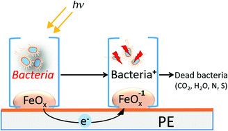
The first evidence for the synthesis of a uniform, adhesive polyethylene–FeOx (PE–FeOx) surface leading efficiently to bacterial inactivation is addressed in this study. PE was loaded with 0.04–0.08% Fe wt/wt PE after RF-plasma pretreatment was required to increase the active sites/polarity and roughness to adhere FeOx on PE. The repetitive bacterial inactivation proceeded in a stable way for several cycles. The oxidative radicals leading to bacterial inactivation under aerobic/anaerobic conditions were investigated by the use of appropriate scavengers. By X-ray photoelectron spectroscopy (XPS) and diffuse reflection spectroscopy (DRS) the changes on the PE–FeOx oxidation states and spectroscopic features during bacterial inactivation were monitored. The regeneration of the initial Fe-oxidation state and consequently of the initial Fe-oxidation state in the PE–FeOx was possible and followed by DRS. Inductive plasma coupled mass spectrometry (ICP-MS) indicated that only sub-ppb levels of Fe were released from the PE–FeOx surface within the reaction time.
In this study, we have selected polyethylene (PE) as substrate to sputter FeOx by direct current magnetron sputtering (DCMS) since PE it is a flexible low cost polymer resistant to corrosion and withstands 120 °C within the short times. The low surface energy of PE is the major reason for the poor particle adhesion leading to Fe–polymer composites. We focus on the binding of FeOx on PE as an innovative catalyst for bacterial disinfection. RF-pretreatment of PE has been carried out to fix a suitable amount of FeOx on the pretreated PE-hydrophilic sites.10 The FeOX was then deposited on PE by sputtering in a magnetron chamber11 and the amount of FeOx on the PE surface inactivated bacteria with acceptable kinetics.
During the last decades, iron oxide colloids have been reported as a photocatalyst in aqueous solutions to inactivate pollutants12 and also as a photocatalyst able to inactivate E. coli via photo-Fenton like processes in aqueous suspensions.13–15 When using Fe-oxides in suspension, separation and filtration of the catalyst at the end of the process is necessary for the catalyst reuse. For this reason our laboratory has reported degradation of pollutants on Fe-supported on PE–maleic anhydride films16,17 and also on Fe–sulfonic Nafion exchange membranes.18
Resistance of some bacteria to antibiotics is serious health problem leading to increased hospital-acquired infections (HAI).19,20 Antibiotic resistant bacteria induce healthcare-acquired infections (HCAI) with its associated high care costs.21 For this reason, there is a need at the present time to develop innovative composite surfaces and materials with faster antibacterial bacterial kinetics, like polymer films, rugs, curtains, medical devices and catheters.
The present study focuses on the preparation of innovative PE–FeOx uniform, adhesive, efficient and stable surfaces that can regenerated easily. We do not address the detailed mechanism of these Fe-oxide bacterial interaction surfaces but we address the photocatalysis presenting a systematic analysis of the surface properties. Therefore we focus on: (a) the optimization of the PE-surface pretreatment by RF-plasma to graft FeOx by reactive magnetron sputtering, (b) the evaluation of the E. coli inactivation kinetics and mechanism on PE–FeOx surfaces during the recycling of the catalyst and (c) the analysis/description of the PE–FeOx microstructure by surface science techniques to characterize the PE–FeOx films.
O, O–C
O, C–O–O–. This pretreatment modifies the PE surface by: (a) etching of the PE surface due to the residual O-radicals still present in the gas of the RF-plasma chamber at 1 Torr, (b) introducing hydrophilic groups on the PE-surface and (c) breaking the intermolecular PE- and the H–H bonds leading to a partial segmentation of PE. FeOx then binds the PE-negative functionalities by electrostatic attraction and chelation/complexation.10,11
The FeOx was sputtered from a 5 cm diameter target (Kurt Lesker, East Sussex, UK) by direct current magnetron sputtering (DC) on PE. Since Fe is paramagnetic, the target was modified by creating an artificial eroded zone by electro-erosion in order to let the magnetic field pass through the target forcing the electrons to follow a hopping trajectory along the target. The residual pressure Pr in the sputtering chamber was adjusted to Pr 10−4 Pa. The substrate to target distance was set at 10 cm. The FeOx films were deposited by reactive DC-magnetron sputtering (DC) in an O2 gas flow. The thickness of the FeOx sputtered layers was determined by means of a profilometer (Alphastep500, Tencor).
The polyethylene (PE) consists of highly branched low crystalline semi-transparent film with the formula H(CH2–CH2)nH. The (LDPE) 0.1 mm thick was obtained from Longfellow, UK (ET3112019), had a density of 0.92 g cm−3, and a flowing point of 185 °C. The PE was prepared by Blown Film Extrusion manufacturing.
The fluorescence stereomicroscopy was carried out on samples inoculated with 108 CFU of E. coli and incubated for 2 hours in a humidification chamber. This method uses a fluorochrome-based staining procedure from Filmtracer™ LIVE/DEAD® Biofilm Viability Kit (Molecular Probes, Invitrogen). The kit contains a combination of the SYTO® 9 green fluorescent nucleic acid stain and propidium iodide fluoro-chromes for the staining of live and dead cells, respectively. The sample fluorescence was monitored in a fluorescence stereomicroscope (Leica MZ16 FA, Leica Microsystems GmbH Wetzlar, Germany) and the images were processed using the LAS v.1.7.0 build 1240 software from Leica Microsystems CMS GmbH. Adhesion of bacteria to the sputtered PES was allowed for 2 min before washing the sample with sterile Milli-Q water to remove non-adherent bacteria.
The PE–FeOx films were irradiated with the Xe-400 W light in the Suntest solar simulator CPS (Atlas GmbH, Hanau, Germany) with a light dose of 52 mW cm−2 (∼0.8 × 10¹⁶ photons per s) and a cut-off filter was added in the Suntest cavity to block the light <310 nm.
The local pH-changes during the bacterial inactivation was followed by a Jenco 6230N (pH/mV/Temp meter) provided for with a hand held microprocessor in splash proof case with 3 points calibration. The device can be monitored via RS-232-C IBM compatible communication interface and BNC, pH/ORP connector with 8-pin DIN ATC connector.
The Finnigan™ ICPS used was equipped with a double focusing reverse geometry mass spectrometer with an extremely low background signal and a high ion-transmission coefficient. The FeOx of the samples was digested with nitric acid 69% (1
:
1 HNO3 + H2O) to remove the organics in the solution and to guarantee that there were no remaining ions adhered to the flacon wall. The samples droplets are introduced to the ICP-MS trough a peristaltic pump to the nebulizer chamber at ∼7700 °C allowing the sample components evaporation and ionization. The Fe found in the nebulizer droplets was subsequently quantified by mass spectrometry (MS).
![]() | ||
| Fig. 1 Diffuse reflectance spectroscopy (DRS) spectra of RF-plasma pre-treated polyethylene (PE) surfaces at a pressure of 1 Torr. | ||
In the presence of residual oxygen, RF-plasma leads to the build-up of C–O groups on PE surface such as: C
O (n → Π* 280 nm ε = 20 M−1 cm−1); C
C–C
O (n → Π* 220 nm ε = 2 × 10⁵ M−1 cm−1) and C
C–C
O (n → Π* 290 nm ε = 30 M−1 cm−1).26 RF-plasma at 1 Torr enhances the capture length of the electrons generated by the applied electric field leading to excited O* and atomic O, singlet 1O2, anion-radicals O− and cation-radicals O+.27 These species react with the PE surface introducing oxidative C–O type functionalities, breaking H–H and C–C bonds segmenting the PE-film.28 Concomitantly oxidative functionalities like: –C–O−, –COO−, –COH–, O–C
O−, –COOH, percarboxylates, epoxides, phenolic and lactam and these negative polar groups by electrostatic attraction bind positive Fe3+-cation (FeOx) leading to surface coordination/chelation.29 The residual H2O-vapor still present in the chamber being decomposes during the sputtering being an additional source of O-atoms and O-ions.
The changes in the concentration of the oxidative functional groups C–OH, C
O and O
C–OH and the ratio to the C–C, C
C and –CH2 reductive species obtained by XPS are shown in Fig. 2a. Fig. 2b shows the C 1s deconvolution before and after RF-plasma pretreatment supporting the results found in Fig. 2a.
The deconvoluted peaks centred at BE of 285.9, 286.6 and 288.1 eV were ascribed respectively to the C–OH, C
O and O
C–OH according to their binding energy (BE) values.22–24 The binding energy (BE) at 285.0 eV was assigned to the C–C, C
C and CH (reduced functionalities) to estimate the values in the y-axis in Fig. 2a. The ratio of oxidized to reduced functionalities (COOH + COH/CH + C–C) remained constant beyond 20 min RF-pretreatment, at the time the highest polarity was reached on the PE-film.30–32
Fig. 3 shows the roughness of the PE-films as a function of the RF-plasma pretreatment times. The root mean square (rms) of the samples pretreated at times (a) zero, (b) 5 min and (c) 10 min led to Rg values of 3 nm, 7 nm and 13 nm respectively. The increased non-uniformity in the topmost PE fibers introduced at an RF-plasma pretreatment time of 10 min is consistent with the time necessary to introduce the highest polarity on the PE–FeOx as shown in Fig. 2.
![]() | ||
| Fig. 3 Roughness and topography of pre-treated PE films by RF plasma: (a) time zero, (b) 5 min and (c) 10 min. For other details see Ms text. | ||
![]() | ||
Fig. 4 E. coli adhesion on PE–FeOx sputtered for 60 s: ( ) RF-plasma pre-treated surface for 15 min and ( ) PE–FeOx sputtered for 60 s non-pre-treated surface. | ||
This trend was also similar to the trend reported in Fig. 3 for Rg values as a function of the RF pretreatment time suggesting that the surface roughness had a significant effect on bacterial adhesion. The diffusion of the generated ROS by the PE–FeOx film under light on the bacterial load (100 μL samples) allows to kill also the non-adhered or the weakly adhered bacteria. The weakly adhered bacteria could be removed by sonication.
Fig. 5a presents the E. coli inactivation under solar simulated light and in the dark on 10 min RF pretreated PE–FeOx with different Fe-loadings (Table 1).
| Conc.% Fe2O3 | Error% | |
|---|---|---|
| FeOx–PE sputtered for 30 s | 0.0401 | 0.006 |
| FeOx–PE sputtered for 60 s | 0.0607 | 0.007 |
| FeOx–PE sputtered for 120 s | 0.0842 | 0.007 |
PE–FeOx sputtered for 60 s led to the faster bacterial inactivation time (Fig. 5a, trace 2).
Table 2 shows that the half time for bacterial inactivation (E50%) depends on the time of sputtering of Fe on PE leading to PE–FeOx films.
| E50% (min) | |
|---|---|
| FeOx–PE sputtered for 30 s | 52.7 |
| FeOx–PE sputtered for 60 s | 21.7 |
| FeOx–PE sputtered for 120 s | 28.2 |
| FeOx–PE sputtered for 150 s | 28.0 |
The thickness determined by profilometry of the FeOx layer of 42 nm. This layer thickness seems to induce the most suitable size/surface loading of the FeOx clusters on the PE. If one atomic layer is ∼0.2 nm thick and including 1015 atoms per cm2, the Fe deposition rate can be estimated as 3.5 × 10¹⁵ atoms per cm2 s.11 The number of Fe-layers sputtered within a thickness of 42 nm is ∼210 layers.10,22 Sputtering for 30 s (trace 4) did not deposit enough FeOx on the PE (Table 1). The bacterial inactivation time shown in Fig. 5a, traces 3 and 4 reveal longer bacterial inactivation times for sputtering times of 120 and 150 s, due to two effects: (a) the increase in layer thickness leading to bulk inward diffusion of the charge carriers2,10 and (b) the size of the FeOx-clusters increased with the sputtering time. This leads to FeOx agglomerates, but the catalytic activity per exposed atom would consequently decreases due to agglomeration.2,6,9 Fig. 5a, trace 5 also shows that the bacterial inactivation does not take place in the dark on PE–FeOx films nor does it proceed on PE alone under simulated solar irradiation.
Fig. 5b shows the live and dead E. coli bacteria by using the dye fluorochrome that enters the cell and stains the DNA when the cell wall membranes are damaged by PE–FeOx showing an abnormal high permeability.2,3
The time sequence for E. coli cells wall destabilization/damage leading ultimately to cell dead is shown in Fig. 5b the increasing population of red dots. The red dots are indicative of cell membrane damage. On the other hand, bacterial cells incubated on PE alone kept alive up to 120 min light exposure remaining green.
![]() | ||
| Fig. 6 Recycling of PE–FeOx sputtered samples for 60 s under low intensity solar simulated light (52 mW cm−2). | ||
Fig. 7a shows the diffuse reflectance spectroscopy (DRS) of PE–FeOx samples up to the 5th disinfection cycle in Kubelka–Munk units. The spectrum of Fe2O3 between 250 and 500 nm has been reported and the amplitude of the Fe2O3 spectral peaks observed at 350, 300 and 265 nm are a function of the film thickness and Fe2O3 grain size.35,36 The peak around 220 nm in Fig. 7a is due to the PE optical absorption (Fig. 1). The stability of the PE–FeOx DRS spectra up to the 5th recycling shown in Fig. 7a is consistent with sample cycling shown in Fig. 6. Fig. 7b, trace 1 reports the spectrum of PE–FeOx before bacterial inactivation. Fig. 7b, trace 2, reports the spectrum of PE–FeOx after bacterial inactivation. The regeneration of the initial state of Fe2O3 (on the PE–FeOx surface) was possible by the use of a 1 M NaOH solution35 as it can be seen in Fig. 7b trace (3). Fe2O3 is an n-type semiconductor with a band-gap of ∼2.3 eV able to activate charge separation on at a potential ∼0.1 eV and holes at +2.2 eV.35,36
The visual appearance of PE unsputtered samples and for PE–FeOx sputtered samples for 30 s, 60 s and 120 s are shown in Fig. 8. The blue line in Fig. 8 was added for clarity of presentation due to the transparent nature the FeOx–PE film. The orange-brown colour in the sample shown in Fig. 8c is characteristic of Fe2O3 in samples sputtered for 60 s figures in Fig. 8e shows the decrease in colour after 2 bacterial inactivation cycles and further discoloration is shown after five bacterial inactivation cycles in Fig. 8f. The discoloration was due to the accumulation of the colourless FeO after in the PE–FeOx samples after bacterial inactivation. The molar absorption coefficient of the Fe(II) band at 250–265 nm is 20 M−1 cm−1 is much smaller compared to molar absorption coefficient of an ε = 1000 M−1 cm−1 (ref. 16 and 17) at 366–375 nm as shown in Fig. 8c for the PE–FeOx samples.
Scavenging the OH-radicals with DMSO slows down bacterial inactivation but this effect seems not to be significant since the bacterial inactivation is slowed down in a very limited way. More important is the almost complete suppression of the bacterial inactivation shown in Fig. 9 by EDTA-2Na to detect the vbh+. The addition of SOD was used to detect the HO2/O2−. Both scavengers preclude almost entirely the bacterial inactivation as shown in Fig. 9.
The intervention of HO2˙/O2−, OH˙ radicals and vbh+ during the PE–FeOx mediated bacterial inactivation is suggested below:
| PE–FeOx + hν → h+ + e− | (1) |
| e− + O2 + H+ → HO2˙, E0 − 0.05NHE9 | (2) |
| HO2˙/O2−˙, E0 0.75NHE9pKa = 4.8 | (3) |
| e− + O2(ads) → O2−(ads), E0 − 0.16NHE10 | (4) |
| h+ + OHads → OH˙, E0 − 1.90NHE40 | (5) |
| h+ + H2Oads → OHads + H+ | (6) |
To investigate further the nature of the oxidative radicals leading to bacterial inactivation, we measured the local pH-changes occurring during bacterial inactivation. The inset in Fig. 10 shows the pH changes at the outer cell wall within 120 min, decreasing stepwise from 6.6 to 4.8. This pH drop is equivalent to an increase of almost two orders (one hundred fold) in the (H+) concentration. In anaerobic conditions the pH decreases from a pH 6.6 to 6.4, an increase of about two times in the (H+) concentration. The decrease of pH under aerobic conditions can be suggested by eqn (7)–(10) below:
| Bact + [PE–FeOx] + hν → [Bact*⋯FeOx]PE → [Bact+˙ + FeOx]PE + cbe− | (7) |
| FeOx + hν → FeOx (vbh+) | (8) |
| vbh+ + H2Oads → OH˙ads + H+ | (9) |
| cbe− + O2ads → O2− | (10) |
![]() | ||
| Fig. 11 XPS investigation of iron oxide oxidation states on the PE–FeOx surfaces sputtered for 60 s before and after bacterial inactivation under solar simulated light (52 mW cm−2). | ||
This changes in the surface atomic concentration percentages is accompanied by redox processes shifting the peaks position of the FeOx photocatalyst under Suntest simulated light irradiation. After bacterial inactivation the XPS peak positions for the Fe(III), FeO(II/III) and Fe(II) peaks were: 711.4 eV; 711.4 eV; 708.6 eV and 713.8 eV respectively. The three Fe-oxides in Fig. 11 offer different potentials to catalyze the bacterial reduction inherent to its own oxidation states. No accumulation of residual intermediates on the catalyst after bacterial inactivation as shown by the decrease of the C-content on the catalyst surface in Table 3.
| Before bacterial inactivation | After bacterial inactivation | |
|---|---|---|
| Fe 2p | 7.87 | 7.39 |
| O 1s | 31.11 | 35.27 |
| C 1s | 61.02 | 57.34 |
| N 2p | 0.9 | 1.19 |
| HO2˙ + Fe3+ → Fe2+ + O2 + H+ | (11) |
![]() | ||
| Fig. 12 Mechanism suggested for the bacterial inactivation mechanism under solar simulated light on PE–FeOx sputtered surfaces. | ||
The Fe2O3 is reduced in the initial stages while oxidizing bacteria.
This is shown in Fig. 8 when Fe(III) transforms to Fe(II) and further confirmed by the XPS experiments reported in Fig. 11. The Fe3O4/FeO produced in this first step in Fig. 11 subsequently re-oxidizes to Fe2O3 in aerobic media. The conduction band of Fe2O3 at −0.1 V is at a more positive potential than the potential required for the one electron oxygen reduction O2 + H+ + e− → HO2˙ (−0.22 V).2 This precludes the consumption of H+ allowing the H+ to drive the local pH towards more acidic values as shown in the insert in Fig. 10.
One of most important finding in this study relates to the PE–FeOx mediated bacterial reduction in Fig. 4a, trace 1 showing the bacterial inactivation for PE–FeOx sputtered for 60 s. The amount of Fe-released within the inactivation time is shown in Table 4.
| Fe (ppb) | |
|---|---|
| FeOx sputtered for 30 s | 0.5 |
| FeOx sputtered for 60 s | 0.6 |
| FeOx sputtered for 120 s | 0.9 |
The released FeOx was <1 ppb, far below the 200–400 ppb Fe-concentration reported to be toxic to mammalian cells.37–39 The PE–FeOx mediated disinfection would proceed with practically no loss of Fe.40,41
The cytotoxic effect of PE–FeOx on bacteria could be rationalized by the formation of a Fe–C composite at the PE–FeOx film interface. This composite at the interface would be highly toxic even at low levels of Fe.42 In the petrochemical industry C-metal interfaces present an increase catalytic activity compared to metal NP's alone.43 Our findings in this study are very different to the subjects reported in many recent studies where Fe has been reported to be released from Fe-heterogeneous catalysts during degradation of pollutants/bacteria in solution leading to Fenton reactions, but in the presence of Fe in the ppm-range.16–18,33,34
| This journal is © The Royal Society of Chemistry 2015 |