
A miniature room temperature formaldehyde sensor with high sensitivity and selectivity using CdSO4 modified ZnO nanoparticles
Xinghua Changab,
Mi Penga,
Junfeng Yanga,
Teng Wanga,
Yu liuc,
Jie Zheng*a and
Xingguo Li*a
aBeijing National Laboratory for Molecular Sciences (BNLMS), The State Key Laboratory of Rare Earth Materials Chemistry and Applications, College of Chemistry and Molecular Engineering, Peking University, Beijing 100871, China. E-mail: zhengjie@pku.edu.cn; xgli@pku.edu.cn
bAcademy for Advanced Interdisciplinary Studies, Peking University, Beijing 100871, China
cSharp Laboratory of China CO., LTD, Beijing Research Center, Phoenix Palace No. A5, Shuguangxili, Chaoyang District, Beijing 100028, China
First published on 26th August 2015
Abstract
A light activated miniature formaldehyde sensor working at room temperature is fabricated by CdSO4 modified ZnO nanoparticles. The CdSO4 is deposited on the surface of the ZnO nanoparticles as a separated phase rather than doping into the lattice of ZnO. The Cd2+ and SO42− on the surface play a synergic effect for the high sensitivity to formaldehyde. The sensor shows high sensitivity to formaldehyde, with detection limit lower than 1 ppm while shows no response to ethanol and very weak response to acetone. With engineering efforts, a highly compact prototype formaldehyde sensor is obtained, which is very convenient for portable formaldehyde specific detection.
Introduction
Formaldehyde is associated with many health risk factors and has been identified as a major cause of sick building syndrome.1,2 The World Health Organization has set a 30 min exposure limit of 0.08 ppm. As formaldehyde is widely found in indoor environment, it is highly desirable to monitor the ambient formaldehyde concentration in real time. Although formaldehyde can be readily determined by well-established spectroscopic or chromatographic methods with sufficient sensitivity, these methods require expensive instrumentation with well-trained operators.3 Therefore, developing portable, cheaper and easier operating formaldehyde gas sensors with high sensitivity and selectivity is necessary to meet the requirement of real time monitoring the formaldehyde concentration in residential environment.The electronic properties of metal oxide semiconductor (MOS) nanostructures are very sensitive to the molecules adsorbed on the surface.4–6 This property, together with the wide availability of highly sensitive conductivity measurement devices, makes MOS nanostructures very attractive for high sensitivity gas sensors.7 Formaldehyde detectors using SnO2 nanostructures have been extensively reported.8–10 One drawback of the SnO2 based detector is the high working temperature (typically above 200 °C).11–13 Such high temperature detectors usually exhibit responses to many volatile organic compounds (VOCs) that can be oxidized, such as ethanol and acetone which are also frequently found in indoor environment. This poor selectivity also limits its application for formaldehyde specific detection, as less harmful VOCs will cause false signals of the detector. Alternatively, the MOS nanostructures can be activated by light. In this case, the sensor can work at room temperature. ZnO is one of the most important electronic and photonic semiconductor materials, which is considered to be a good candidate for formaldehyde sensor.14–17 But the sensitivity to formaldehyde of undoped ZnO are poorer compared to that of thermal activated SnO2 nanostructures.
Here we improve the formaldehyde sensing performance of ZnO nanoparticles by surface CdSO4 modification. It is found that the Cd2+ cation and the SO42− anion exhibit a synergic effect in promoting the sensitivity of formaldehyde detection. A miniature UV light assisted formaldehyde detector is fabricated using CdSO4 modified ZnO nanoparticles, which shows high sensitivity up to 20% response to low formaldehyde concentration of 1 ppm and good selectivity for formaldehyde over other frequently encountered VOCs in indoor environments.
Experiment methodology
Materials
Analytical-grade chemicals zinc sulphate heptahydrate (ZnSO4·7H2O), ammonium bicarbonate (NH4HCO3), cadmium sulphate (3CdSO4·8H2O), formaldehyde solution (formaldehyde, 37%) were purchased from Beijing Chemical Works (China) and used without further purification.Synthesis of CdSO4–ZnO nanoparticles
NH4HCO3 solution (50 mL, 1.26 mol L−1) is added to ZnSO4 solution (25 mL, 1.5 mol L−1) dropwisely under vigorous stirring. The mixture is stirred at 40 °C for 1 h and the precipitate is separated by filtration. The solid product is thoroughly washed with deionized water for three times and is dried in an oven at 80 °C for 12 h and then 120 °C for 2 h. Finally, the dried precursor was calcinated at 500 °C for 2 h to give the ZnO nanoparticles.The obtained ZnO nanoparticles (0.400 g) are dispersed in aqueous CdSO4 solution with different concentration (100 mL) by sonicating for 10 min. The resulting suspension was heated to 120 °C under vigorous stirring to remove the water. The obtained solid was dried in an oven at 80 °C for 12 h and then 120 °C for 2 h. At last, the sample was calcinated at 450 °C for 4 h to give the surface CdSO4 modified ZnO nanoparticles. The samples are designated as ZnO–xCdSO4, where x is the molar ratio of Cd/Zn.
Material characterization
The structure and morphology of products was characterized by X-ray diffraction (XRD, Rigaku D/max 2000 diffractometer, Cu Kα), scanning electron microscopy (SEM, Hitachi S4800, 10 kV) with an energy dispersive X-ray spectroscopy (EDS) analyzer, The X-ray photoelectron spectroscopy (XPS) analysis is performed on an AXIS-Ultra spectrometer (Kratos Analytical) using monochromatic Al Kα radiation (225 W, 15 mA, 15 kV).Fabrication of the gas sensor
A schematic illustration of the structure of the sensor is shown in Fig. 1. A printed circuit board (PCB) with two interpolated Au electrode arrays is used as the substrate. A layer of ZnO–xCdSO4 nanoparticles are coated onto the substrate by drop casting ethanol suspension of the nanoparticles. After evaporation of the ethanol at room temperature, a semiconductor layer with total material mass loading of 20 mg cm−2 is formed. A UV LED (3 mW) is attached to the semiconductor layer to activate the ZnO–xCdSO4 nanoparticles during operation. | ||
| Fig. 1 (a) The schematic structure of the fabricated sensor; (b) and (c) photographs of the prototype sensor. | ||
Measurement
To measure the gas sensing properties of the materials, a miniature formaldehyde sensor is fabricated with the lateral dimension of only about 1 cm (Fig. 1). The nanoparticle covered PCB is sealed inside a quartz container with calibrated internal volume, and The current passing through the two Au electrode arrays is measured at a constant voltage of 10 V by an electrochemical workstation (model: CHI 760b). The background current under UV irradiation i0 is measured at a relative humidity (RH) of 30 ± 5%. The RH is created by injecting 0.5 μL deionized water using a microdosing syringe into the container. 0.5 μL aqueous formaldehyde solution with different concentration was injected into the quartz container to give the desired formaldehyde concentration. The formaldehyde containing air is removed from the chamber by pumping. The UV LED is turned on and off with the irradiation time lasting for 100 s in each on–off cycle. The sensor response (R) was defined as| R = (ig − i0)/i0, | (1) |
Result and discussion
Structural characterization of the CdSO4 modified ZnO nanocomposite
Fig. 2 shows the X-ray diffraction patterns of ZnO–xCdSO4 nanoparticles with different x value. For the undoped ZnO sample, all the peaks can be indexed to the wurtzite ZnO (JCPDS card 36-1451). The samples are highly crystallized, as indicated by the sharp diffraction peaks. The ZnO–2%CdSO4 sample shows very similar pattern to the undoped sample. However, when the CdSO4 reaches 5 mol%, additional peaks attributed to CdSO4 (JCPDS no. 14-0352) start to appear, as indicated by the solid squares in Fig. 1. Therefore, at high CdSO4 concentration, the excessive CdSO4 starts to form a separated phase. Interestingly, there is no shift of the diffraction peaks with the increase of CdSO4/ZnO molar ratio, indicating that the CdSO4 modification causes little Cd substitution in the lattice of ZnO. The XRD results suggest that the introduced CdSO4 prefer to exist as a separated phase rather than homogeneously doped into the lattice of the ZnO phase. | ||
| Fig. 2 XRD patterns of ZnO–xCdSO4 samples. | ||
Fig. 3 shows the SEM images of the ZnO–xCdSO4 samples. The undoped ZnO sample is composed of interconnected nanoparticles around 50 nm in size. The aggregation of the particles is resulted from the slight sintering effect during the calcination process. Such interconnected particles creates pathway for electron transport, which is favourable to obtain good sensitivity for formaldehyde. Modification by different amount of CdSO4 shows little effect on the particle size and morphology (Fig. 3b–d). Elemental analysis of ZnO–2%CdSO4 suggests that the nanocomposite is composed of Zn, O, Cd, S. The atomic ratio of Cd and S is about 1![[thin space (1/6-em)]](https://www.rsc.org/images/entities/char_2009.gif) :1 (Fig. 3e), which matches well to the stoichiometric ratio of CdSO4.
:1 (Fig. 3e), which matches well to the stoichiometric ratio of CdSO4.
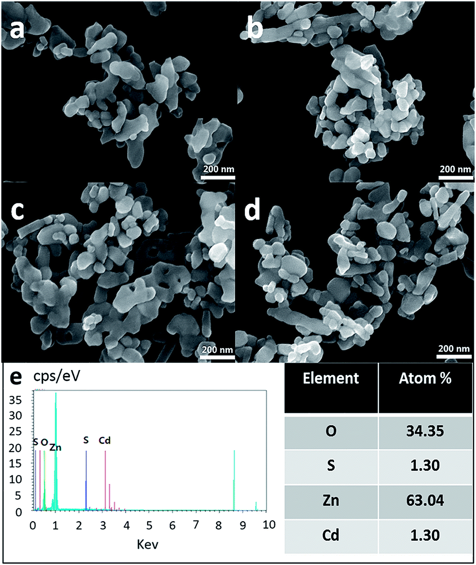 | ||
| Fig. 3 SEM images of ZnO–xCdSO4 samples: (a) pure ZnO; (b) ZnO–2%CdSO4; (c) ZnO–5%CdSO4; (d) ZnO–10%CdSO4. (e) The EDS spectrum and the corresponding elemental composition of ZnO–2%CdSO4 sample. | ||
The surface state of the ZnO–2%CdSO4 sample is further investigated by XPS spectra, as shown in Fig. 4. The O 1s spectrum can be deconvoluted into two components (Fig. 4b). The main peak at 530.2 eV is assigned to the lattice oxygen in ZnO crystals.18 The shoulder peak centred at 531.6 eV can be attributed to the chemisorbed oxygen species (O2−) and Zn–OH groups.19 The existence of the functional groups and chemisorbed oxygen species indicates an active surface of the CdSO4 modified ZnO nanoparticles. In the Cd 3d3/2 spectrum (Fig. 4c), the peak is located at 412.4 eV, which is attributed to divalent Cd.20 Besides, the S 2p spectrum (Fig. 4d) can be deconvoluted into two peaks which are centred at 168.6 eV and 169.7 eV, respectively, which can be assigned to sulphate species.21 Composition analysis from XPS gives Cd/Zn molar ratio of 1:8.4 and Cd/S molar ratio of 1.05:1 (Fig. 4a). The Cd/Zn ratio is much higher than the doping level while the Cd/S ratio remains close to that in CdSO4, indicating surface enrichment of the CdSO4 species. Therefore, both the XRD and XPS results indicate that CdSO4 modification with molar ratio less than 2 mol% yields ZnO nanoparticles with surface decorated CdSO4. There is no apparent Cd doping into the ZnO lattice.
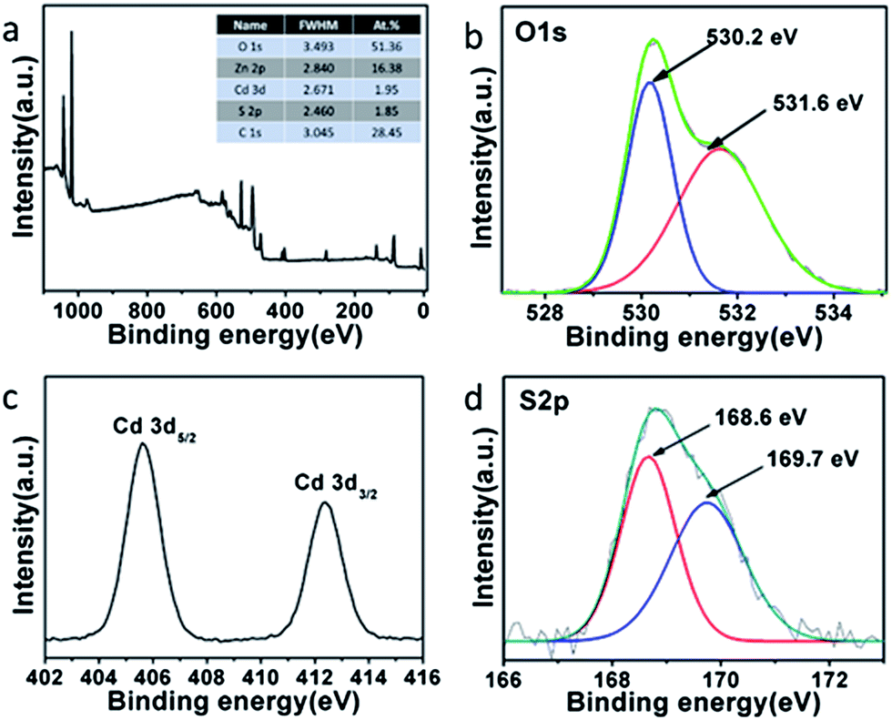 | ||
| Fig. 4 XPS spectra of the ZnO–2%CdSO4 sample: (a) a full range spectrum and the corresponding surface elemental composition; (b) O 1s; (c) Cd 3d; (d) S 2p. | ||
Gas sensing studies
Fig. 5 is the dynamic conductivity response of the formaldehyde sensor fabricated by the ZnO–2%CdSO4 sample to UV irradiation in different atmosphere. Under UV irradiation, the conductivity of the ZnO–2%CdSO4 sample rapidly increases even without ambient formaldehyde. The photo current in the steady state is more than 25 times higher than the dark current. After turning off the UV lamp, the conductivity reversibly decrease to the original level. This is a typical phenomenon of photon generation of carriers in semiconductors. With low concentration of formaldehyde of 3 ppm, the photo-current becomes even higher. Calculated using eqn (1), the sensor shows a positive response of about 36% to 3 ppm formaldehyde. | ||
| Fig. 5 Current signal of the ZnO–2%CdSO4 sample to 3 ppm formaldehyde at room temperature. | ||
Fig. 6 shows the sensor response versus different formaldehyde concentration from 1 to 30 ppm with UV irradiation. The sensor's responses increases with the ambient formaldehyde concentration. There remains 20% response for 1 ppm formaldehyde, indicating that the sensor has sufficient sensitivity to detect low concentration of formaldehyde to 1 ppm. The response increases more rapidly for formaldehyde concentration lower than 10 ppm while shows saturation trend at higher concentration, which is favourable for monitoring the low formaldehyde concentration on a quantitative basis. Table 1 compares the sensitivity of some MOS based formaldehyde sensors reported in literature. It clearly shows that our light activated sensor based on CdSO4 modified ZnO nanoparticles is one of the most sensitive formaldehyde sensor based on light activated MOS nanostructure.
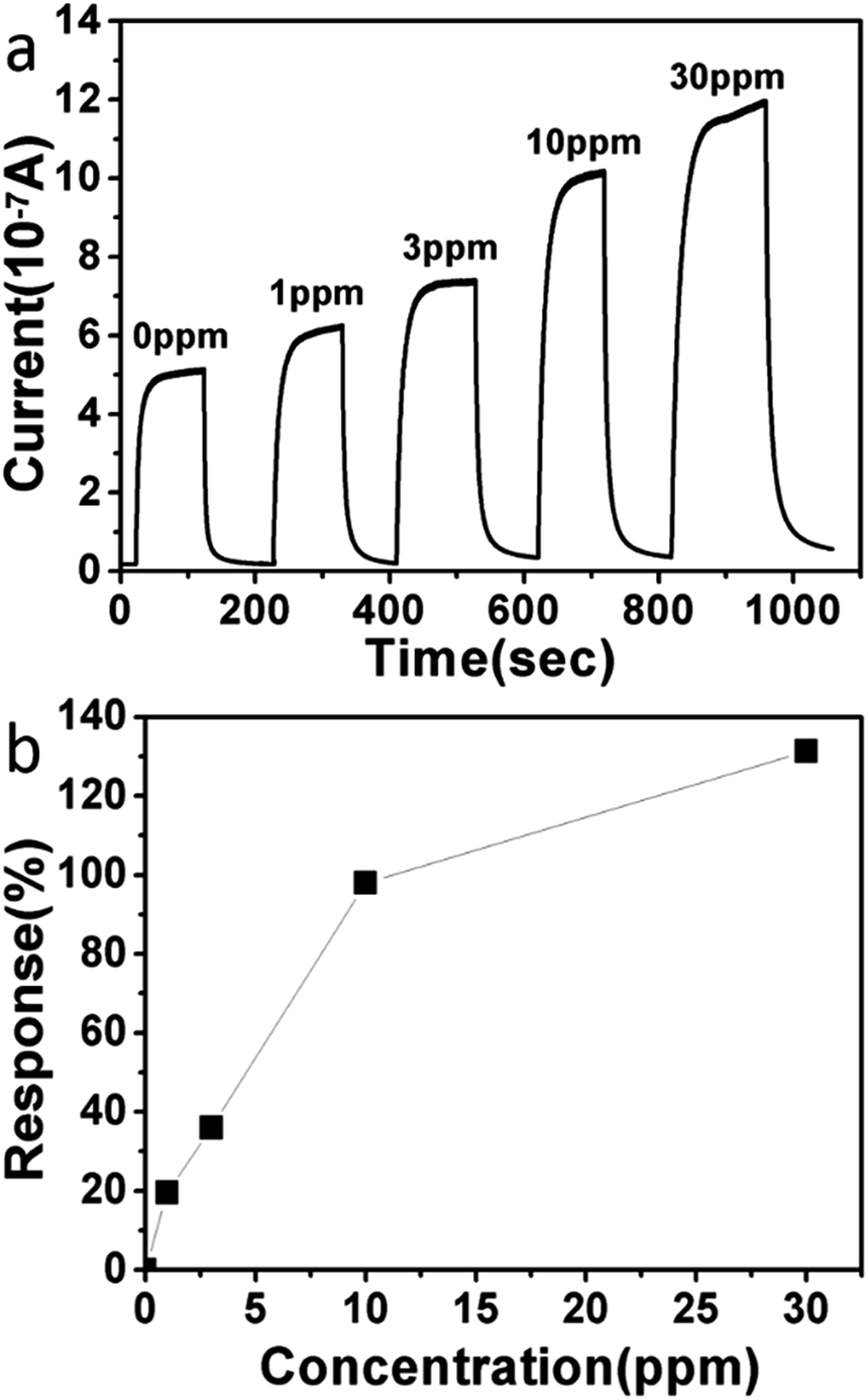 | ||
| Fig. 6 (a) Current signal of the formaldehyde sensor based on ZnO–2%CdSO4 nanoparticles to different formaldehyde concentration. (b) The response for the sensor at different formaldehyde concentration. | ||
| Materials | Operation temperature | Formaldehyde concentration | Response | Reference |
|---|---|---|---|---|
| CdSO4–ZnO | RT | 1 ppm | 20% | This work |
| ITO | RT | 10 ppm | Very weak | 22 |
| Ag–ZnO | RT | 5 ppm | 9.4% | 23 |
| Au–SnO2 | RT | 20 ppm | 1% | 16 |
| P3HT–ZnO | RT | 4 ppm | <1% | 24 |
| NiO | 340 °C | 5 ppm | 12.6% | 25 |
| SnO2 | 280–300 °C | 1 ppm | 7.7% | 9 |
| Li–In2O3 | 300 °C | 20 ppm | 50% | 26 |
| ZIF67 | 150 °C | 5 ppm | Very weak | 27 |
| Zn–NiO | 360 °C | 100 ppm | 15% | 28 |
| Fe2O3 | 325 °C | 25 ppm | 10% | 29 |
The real-time response-recovery characteristics of the ZnO–2%CdSO4 sensor to 3 ppm formaldehyde at room temperature with UV irradiation is given in Fig. 7. It is observed that the current rapidly increases to stable value when 3 ppm formaldehyde is injected, and reversibly decrease to the baseline when the formaldehyde is removed. Calculated using eqn (1), the sensor shows a positive response of about 36% to 3 ppm formaldehyde, in excellent agreement with the results shown in Fig. 5 and 6. The sensor also shows rapid response to the concentration change. The response time of the ZnO–2%CdSO4 sensor, defined as the time for reaching 90% (t90) of the full response of the sensor, is about 33 s, and the 90% recovery time (tr90) is about 40 s. The above results demonstrate that the sensor based on ZnO–2%CdSO4 nanoparticles is highly suited for real time monitoring the change of ambient formaldehyde with low concentration.
 | ||
| Fig. 7 Real-time response-recovery characteristics of the ZnO–2%CdSO4 sensor to 3 ppm formaldehyde. | ||
Selectivity
Ethanol and acetone are commonly encountered volatile organic compounds (VOCs) in indoor environment. A good formaldehyde sensor should exhibit little response to these VOCs other than formaldehyde to avoid false signal. MOS based sensors operating at high temperature also exhibit response for these VOCs, which results in strong interference to the detection of formaldehyde. However, our light activated sensor fabricated from CdSO4 modified ZnO nanoparticles exhibits excellent selectivity to formaldehyde. As shown in Fig. 8, the sensor barely shows any response to ethanol and only shows very weak response to acetone (6% with concentration of 3 ppm). Therefore, our light activated sensor will not give false signals to other commonly seen, but far less harmful VOCs in indoor environment. This is a distinct advantage over the high temperature sensors.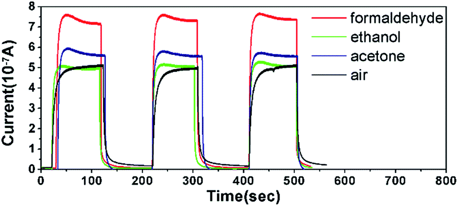 | ||
| Fig. 8 Current signal of the ZnO–2%CdSO4 to 3 ppm various VOCs at room temperature. | ||
The ZnO–2%CdSO4 sensor is further tested under different relative humidity conditions at room temperature (Fig. 9). The photocurrent is 61 μA in dry air, which monotonically decreases with RH to 44 μA at RH = 90 ± 5%. The negative response to water might be attributed to the occupation of the adsorption sites for oxygen by the water molecules. According to Meyer et al.30 and Dulub et al.,31 when two water molecules absorbed on the neighbouring lattice sites, dissociation of the water molecules may occur. The dissociated water molecules could capture electrons and holes generated during the UV light irradiation, decreasing the carrier density of the ZnO–CdSO4 samples. The response to water causes little interference on formaldehyde sensing as the response to formaldehyde and water is completely reverse.32,33 It also indicates that the sensitivity to formaldehyde can be further enhanced when the sensor is calibrated with the ambient humidity. In practical application, this can be easily achieved by coupling a humidity detector which is widely available.
 | ||
| Fig. 9 Current signal of the ZnO–2%CdSO4 sample to 3 ppm formaldehyde in different RH condition. | ||
Mechanism
The enhanced photocurrent due to ambient formaldehyde is attributed to the oxidation of the adsorbed formaldehyde molecules on the surface. During the UV light irradiation, electron–hole pairs are generated, which causes a sudden increase of the carrier density.34 This explains the photocurrent observed both with and without ambient formaldehyde. The ZnO nanostructures in ambient environment have surface adsorbed oxygen species which decreases the density and mobility of the current carriers by a creating depletion layer near the surface.35–37 With the presence of the surface adsorbed formaldehyde molecules, the chemisorbed oxygen will react with formaldehyde and release the immobilized electrons, which further increases the photocurrent. This explains the even higher conductivity with the presence of ambient formaldehyde, as ZnO is intrinsically n-type, higher electron density will lead to higher conductivity.38–40 It also explains the excellent selectivity for formaldehyde with respect to ethanol and acetone, as ethanol and acetone are more difficult to be oxidized compared to formaldehyde.According to this mechanism, the response will increase with the surface density of the adsorbed formaldehyde molecules. At low formaldehyde concentration when there is sufficient surface adsorption sites on the surface, the response will be nearly proportional to the formaldehyde concentration in the vapour phase. When the formaldehyde concentration further increases, however, the chemisorbed oxygen consumption rate due to formaldehyde oxidation is limited by the availability of the surface adsorption sites for formaldehyde. A gradually saturation trend in the conductivity is expected. Such concentration dependence is indeed observed (Fig. 6b).
To study the surface modification effect on the sensitivity, we first consider the effect of CdSO4 doping level. As shown in Fig. 10a, the response shows a volcano type of curve with the CdSO4 doping level. The optimal response is obtained for the ZnO–2%CdSO4 sample. Further raising the CdSO4 doping level results in decrease of the response. Interestingly, in the ZnO–2%CdSO4 sample there is no detectable CdSO4 phase in XRD. The above results suggest that the CdSO4 phase is unfavourable for formaldehyde sensing.
 | ||
| Fig. 10 (a) Responses of sensors fabricated from ZnO–xCdSO4 samples with different CdSO4 modification level x. (b) Responses of sensors fabricated by modified ZnO nanoparticles using different reagents with concentration of 2 mol%. The formaldehyde concentration. | ||
Other Cd sources are also studied. As shown in Fig. 10b, by replacing CdSO4 with Cd(NO3)2, the sensor response is reduced by almost 50% at the same formaldehyde concentration, which indicates SO42− as the anion is more favourable. However, if only (NH4)2SO4 is used in the surface modification process, the response becomes very weak. The sulphate anion alone, therefore, also cannot explain the high sensitivity. We conclude that the Cd2+ and SO42− play a synergic effect in promoting the sensitivity for formaldehyde. To the best of our knowledge, such synergism has never been reported previously. A possible reason is that surface modification by CdSO4 may either enhance the surface adsorption of the formaldehyde molecules or favour the formaldehyde oxidation process (or both). The detailed mechanism is under investigation and will be reported in future.
Conclusions
In summary, a light activated miniature formaldehyde sensor with high sensitivity and selectivity is fabricated by using CdSO4 modified ZnO nanoparticles. The sensor shows sufficient sensitivity with detection limit lower than 1 ppm formaldehyde. The sensor shows no response to ethanol and very weak response to acetone, which is highly favourable for formaldehyde specific detection. Structural characterization suggests that CdSO4 modification results in the CdSO4 phase deposited on the surface of the ZnO nanoparticles. The Cd2+ and SO42− on the surface play a synergic effect for the high sensitivity to formaldehyde.Acknowledgements
This study is supported by National Natural Science Foundation of China (No. U1201241, 11375020, 51431001 and 21321001) and Sharp Laboratory of China CO., LTD.Notes and references
- D. Farmanzadeh and L. Tabari, Comput. Theor. Chem., 2013, 1016, 1–7 CrossRef CAS PubMed.
- W. Hu, S. Chen, L. Liu, B. Ding and H. Wang, Sens. Actuators, B, 2011, 157, 554–559 CrossRef CAS PubMed.
- M. Grutter, E. Flores, G. Andraca-Ayala and A. Báez, Atmos. Environ., 2005, 39, 1027–1034 CrossRef CAS PubMed.
- D. E. Williams, Sens. Actuators, B, 1999, 57, 1–16 CrossRef CAS.
- J. Watson, Sens. Rev., 1994, 14, 20–23 CrossRef.
- G. Korotcenkov, Sens. Actuators, B, 2005, 107, 209–232 CrossRef CAS PubMed.
- N. Yamazoe, Sens. Actuators, B, 2005, 108, 2–14 CrossRef CAS PubMed.
- Q. He, W. Zeng, M. Wu and Y. Wang, J. Mater. Sci., 2013, 24, 2390–2397 CAS.
- J. Huang, L. Wang, C. Gu, Z. Wang, Y. Sun and J. J. Shim, Sens. Actuators, B, 2015, 207, 782–790 CrossRef CAS PubMed.
- N. Barsan, M. Schweizer-Berberich and W. Göpel, Fresenius' J. Anal. Chem., 1999, 365, 287–304 CrossRef CAS.
- Y. Zhang, X. He, J. Li, Z. Miao and F. Huang, Sens. Actuators, B, 2008, 132, 67–73 CrossRef CAS PubMed.
- Q. Wan and T. H. Wang, Chem. Commun., 2005, 3841–3843 RSC.
- J. Zhang, J. Guo, H. Xu and B. Cao, ACS Appl. Mater. Interfaces, 2013, 5, 7893–7898 CAS.
- C.-Y. Lu, S.-J. Chang, S.-P. Chang, C.-T. Lee, C.-F. Kuo and H.-M. Chang, Appl. Phys. Lett., 2006, 89, 153101–153103 CrossRef PubMed.
- X. Chu, T. Chen, W. Zhang, B. Zheng and H. Shui, Sens. Actuators, B, 2009, 142, 49–54 CrossRef CAS PubMed.
- F.-C. Chung, Z. Zhu, P.-Y. Luo, R.-J. Wu and W. Li, Sens. Actuators, B, 2014, 199, 314–319 CrossRef CAS PubMed.
- J. Gong, Y. Li, X. Chai, Z. Hu and Y. Deng, J. Phys. Chem. C, 2010, 114, 1293–1298 CAS.
- A. Kolmakov, S. Potluri, A. Barinov, T. O. Mentes, L. Gregoratti, M. A. Nino, A. Locatelli and M. Kiskinova, ACS Nano, 2008, 2, 1993–2000 CrossRef CAS PubMed.
- M. Batzill, K. Katsiev, J. M. Burst and U. Diebold, Phys. Rev. B: Condens. Matter Mater. Phys., 2005, 72, 1–19 CrossRef.
- P. Thakur, S. S. Joshi and K. R. Patil, Appl. Surf. Sci., 2010, 257, 1390–1394 CrossRef CAS PubMed.
- D. Mandrino, D. Vrbanic, M. Jenko, D. Mihailovic and S. Pejovnik, Surf. Interface Anal., 2008, 40, 1289–1293 CrossRef CAS PubMed.
- V. S. Vaishnav, S. G. Patel and J. N. Panchal, Sens. Actuators, B, 2014, 202, 1002–1009 CrossRef CAS PubMed.
- J. Cui, D. Wang, T. Xie and Y. Lin, Sens. Actuators, B, 2013, 186, 165–171 CrossRef CAS PubMed.
- H. Tai, X. Li, Y. Jiang, G. Xie and X. Du, Sensors, 2015, 15, 2086–2103 CrossRef CAS PubMed.
- I. Castro-Hurtado, C. Malagu, S. Morandi, N. Perez, G. G. Mandayo and E. Castano, Acta Mater., 2013, 61, 1146–1153 CrossRef CAS PubMed.
- N. G. Pramod and S. N. Pandey, Ceram. Int., 2015, 41, 527–532 CrossRef CAS PubMed.
- E.-X. Chen, H. Yang and J. Zhang, Inorg. Chem., 2014, 53, 5411–5413 CrossRef CAS PubMed.
- S. Shijiu, Z. Kailaing, W. Fang, Y. Yujie, H. Yemei, Z. Tiantian, W. Meng and L. Chunjing, ECS Trans., 2014, 60, 1115–1120 CrossRef PubMed.
- P. Das, B. Mondal and K. Mukherjee, RSC Adv., 2014, 4, 31879–31886 RSC.
- B. Meyer, D. Marx, O. Dulub, U. Diebold, M. Kunat, D. Langenberg and C. Woll, Angew. Chem., Int. Ed., 2004, 43, 6642–6645 CrossRef PubMed.
- O. Dulub, B. Meyer and U. Diebold, Phys. Rev. Lett., 2005, 95, 136101 CrossRef.
- Q. H. Li, T. Gao, Y. G. Wang and T. H. Wang, Appl. Phys. Lett., 2005, 86, 123117 CrossRef PubMed.
- Y. Wang, M. Muhler and C. Will, Phys. Chem. Chem. Phys., 2006, 8, 1521–1524 RSC.
- Y. Li, F. Della Valle, M. Simonnet, I. Yamada and J.-J. Delaunay, Appl. Phys. Lett., 2009, 94, 023110–023113 CrossRef PubMed.
- S. Lenaerts, J. Roggen and G. Maes, Spectrochim. Acta, Part A, 1995, 51, 883–894 CrossRef.
- N. Barsan and U. Weimar, J. Electroceram., 2001, 7, 143–167 CrossRef CAS.
- Z. Yang, M. Wang, X. Song, G. Yan, Y. Ding and J. Bai, J. Mater. Chem. C, 2014, 2, 4312–4319 RSC.
- C. Xie, L. Xiao, M. Hu, Z. Bai, X. Xia and D. Zeng, Sens. Actuators, B, 2010, 145, 457–463 CrossRef CAS PubMed.
- C. Soci, A. Zhang, B. Xiang, S. A. Dayeh, D. P. R. Aplin, J. Park, X. Y. Bao, Y. H. Lo and D. Wang, Nano Lett., 2007, 7, 1003–1009 CrossRef CAS PubMed.
- H. W. Ra, K. S. Choi, J. H. Kim, Y. B. Hahn and Y. H. Im, Small, 2008, 4, 1105–1109 CrossRef CAS PubMed.
| This journal is © The Royal Society of Chemistry 2015 |