Physically crosslinked poly(vinyl alcohol)–carrageenan composite hydrogels: pore structure stability and cell adhesive ability

Yabin Zhanga, Lei Yea, Man Cuib, Boguang Yanga, Junjie Lic, Hong Sun*ab and Fanglian Yao*a
aSchool of Chemical Engineering and Technology, Key Laboratory of Systems Bioengineering of Ministry of Education, Tianjin University, Tianjin 300072, China. E-mail: yaofanglian@tju.edu.cn; Fax: +86-22-27403389; Tel: +86-22-27402893
bDepartment of Basic Medical Sciences, North China University of Science and Technology, Tangshan 063000, China. E-mail: xsun1@hotmail.com; Fax: +86-315-3726552; Tel: +86-315-3725740
cDepartment of Advanced Interdisciplinary Studies, Institute of Basic Medical Sciences and Tissue Engineering Research Center, Academy of Military Medical Science, Beijing 100850, China

Received 14th June 2015 , Accepted 9th September 2015

First published on 9th September 2015


Abstract

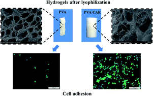
Poly(vinyl alcohol) (PVA) hydrogels have gained comprehensive attention in the biomedical area. However, their resistance to cell adhesion is a drawback for applications such as tissue engineering. Besides, the controllability of the porous structure of PVA-based hydrogels during lyophilization needs to be further improved. Herein, we prepared PVA–carrageenan (CAR) composite hydrogels as tissue engineering scaffolds using the freeze–thaw technique. The hydrogels were found to possess deformation resistance, preserving their shape during the lyophilization process without shrinkage. Besides, ATDC5 cells showed good adherence and proliferation activity on the composite hydrogels. In addition, the PVA–CAR composite hydrogels possess good hemocompatibility and did not cause any adverse effects in the inflammatory response from RAW 264.7 macrophage cells. Overall, the results obtained indicate that the PVA–CAR composite hydrogels show potential applications in the field of tissue engineering based on their good structural stability, excellent biocompatibility and mild fabrication process.


1. Introduction

Macroporous scaffolds provide the necessary architecture for the three-dimensional organization of cells and the open pore morphology to allow the supply of nutrients and the removal of waste metabolites.1,2 Recently, cryogelation technology, which has emerged as a potential approach to prepare cryogels at temperatures below the freezing point of the solvent (e.g. water), has attracted considerable attention because it allows the production of macroporous materials by utilizing frozen solvent crystals as the interconnecting porogen without using toxic organic solvents.3 In particular, the hydrogels based on poly(vinyl alcohol) (PVA) formed via the cryogenic route have several useful properties including a high water content, non-toxicity and biocompatibility.4,5

Generally, PVA in aqueous solution can gelatinize through cryogelation and the structure is imprinted by formation of ice crystals within a not yet structured water/PVA liquid mixture, during the freezing step.6 When subjected to freeze–thaw cycles, the crystallization of PVA leads to the formation of a porous network structure where the crystallites serve as junction points and the polymer segments ensure connectivity all over the macroscopic gel sample filling with free water in the pores.7,8 The water acted as a porogen in the hydrogel-forming solution and needed to be removed after gelation to create the desired pores. Water removal is usually achieved by lyophilization, which has been widely used to prepare porous materials for tissue engineering and biological applications. In recent years, the method of lyophilization has been explored as a unique route to produce highly porous materials.9 However, the pure PVA hydrogel after lyophilization will be accompanied by the shrinkage of the polymer network and the collapse of the pores,10 leading to the inhomogeneous performance of the material even in the state of re-swelling. Besides, the pores present within the physically crosslinked PVA hydrogels are less than 10 μm in size and that will be further decreased due to the shrinkage phenomenon after lyophilization, which adversely affects the cell growth and rapid nutrient diffusion required for cell survival and growth when the PVA hydrogels are used as tissue engineering scaffolds.3 A similar result is found in physically crosslinked gelatin/PVA, with either insufficient pore sizes or a reliance on chemical crosslinking for stability.11 Therefore, obtaining PVA-based hydrogels with a stable pore structure will be significant.

There is a general requirement that cells adhere to the surface of scaffolds and proliferate to eventually form a specific extracellular matrix (ECM) in tissue engineering applications.12 The ideal scaffold for tissue engineering should have good cell affinity and enough mechanical strength to serve as an initial support.13 Regretfully, the suitability of the pure PVA hydrogel as a tissue engineering scaffold still is a debatable topic because of its resistance to protein adsorption and cell adhesion.14,15 Polysaccharides have attracted much interest due to their inherently desirable biocompatibility and interaction with biological systems, being widely proposed as scaffold materials in tissue engineering applications as well as carriers for drug delivery systems. Therefore, in order to expand their application range, PVA is often combined with polysaccharides such as chitosan,16 starch,17 gelatin18 and bacterial cellulose19 etc.

Carrageenan (CAR) is a naturally occurring polysaccharide extracted from marine red algae (Rhodophyceae), and possesses a basic linear primary structure based on an alternating disaccharide repeating unit of 1,4-linked α-D-galactose and 1,3-linked β-D-galactose, with varying degrees of sulfation.20 CAR is biocompatible, biodegradable, non-toxic, and gel-forming. Most importantly, due to its backbone composition of sulphated disaccharides, this hydrophilic polysaccharide resembles the naturally occurring glycosaminoglycans (GAGs) which are an important component of connective tissues.21 Thus, CAR has been investigated for a range of biomedical applications, such as wound dressing22 and tissue engineering.23,24

In this paper, we used a facile, freeze–thaw approach for the fabrication of PVA–CAR composite hydrogels. The aims of this study were to improve the pore stability during lyophilization and enhance cell adhesion in comparison to the pure PVA hydrogel. The physiochemical properties of the PVA–CAR composite hydrogels (morphology, stability, water absorption and mechanical strength) were evaluated. In addition, the hemolysis test and the in vitro cellular behaviors of both ATDC5 cells and RAW 264.7 macrophage cells were examined to determine their suitability as tissue engineering scaffolds. In conclusion, the resultant composite hydrogels possess good collapse resistance, enhance cell adhesion and induce almost no adverse effects in inflammatory response in vitro.

2. Materials and methods

2.1. Materials

PVA was purchased from Guangfu Fine Chemical Co., Ltd (Tianjin, China) with a saponification degree of 97% and a number-average polymerization degree of 1750 ± 50. ι-Carrageenan was provided by Kasei Kogyo Co., Ltd (Tokyo, Japan). Other chemical agents were all of analytical purity and used as received.

2.2. Preparation of PVA–CAR composite hydrogels

Definite amounts of PVA and CAR were dissolved into distilled water and heated at 90 °C with vigorous stirring under reflux. The total solid content was 10 wt%. Subsequently, bubbles were removed by refluxing the solution with a lower stirring speed for 1 h at 70 °C, obtaining a homogeneous solution. The resulting solution was transferred into a tubular mold and subjected to freezing temperatures at −20 °C for 24 h and subsequently thawed at room temperature over 6 h. This freeze–thaw cycle was repeated up to 5 times to provide the hydrogels for further experiments.

The composite hydrogels were coded as CP5, CP10, CP20 and CP30 corresponding to a CAR content (based on the total solid content) of 5, 10, 20 and 30 wt%, respectively. The as-prepared hydrogels were lyophilized for 24 hours before analysis.

2.3. Microstructure characterization

The morphologies of the PVA–CAR composite hydrogels were observed by scanning electronic microscopy (SEM) to evaluate their micro- and macrostructures. Cubic samples were cut from the hydrogels and were swollen to equilibrium in distilled water at room temperature for 24 h, then frozen in liquid nitrogen immediately, and lyophilized before SEM observation. Prior to examination, the samples were sputter coated with a thin layer of gold. A Hitachi S-4800 scanning electron microscope (Tokyo, Japan) was used to perform the image analysis. The porosity of the samples was estimated according to previously reported liquid displacement methods.25

2.4. FT-IR spectral analysis

The samples were cut into small pieces and then ground into powder. The infrared spectral analysis was performed on a WQF-510A FT-IR spectrophotometer (Rayleigh, China) by recording the IR spectra of the powder samples in KBr discs. The absorbance measurements were carried out in the range of 400–4000 cm−1, with 16 scans and a resolution of 4 cm−1.

2.5. X-ray diffraction analysis

The PVA/CAR composite materials were characterized by an X-ray diffractometer (D8-Focus, Bruker, Germany) using CuKα radiation generated at 36 kV and 200 mA; the range of the diffraction angle (2θ) was 10–60° at a scanning speed of 6° min−1.

2.6. Differential scanning calorimetry

The differential scanning calorimetry (DSC) analyses were carried out using a NETZSCH STA 449 F3 (Netzsch, Germany) under an argon gas flow rate of 50 mL min−1 and at a heating rate of 10 °C min−1 from room temperature up to 250 °C.

2.7. Swelling characterization

To investigate the swelling ratio, the dry samples were immersed directly into distilled water at room temperature until their weight became constant. The swollen samples were freed of surface water using filter paper to remove excess water and then weighed. The swelling ratio was determined according to the following equation:
 
image file: c5ra11331h-t1.tif(1)
where W0 and W are the weight of the PVA–CAR composite materials before and after immersing in water respectively. The tests were conducted in quintuplicate to minimize error and reported as a mean value.

2.8. Mechanical behavior

The hydrogels were first swollen in distilled water to equilibrium and then subjected to a compression strength test using mechanical test equipment (M350-2208, Testometric, U.K.) at room temperature with a crosshead speed of 5 mm min−1. The cylindrical samples were cut to an appropriate size (10 mm in diameter and 10 mm in thickness). The diameter and the thickness of the samples were checked with an electric digital caliper. Five specimens were tested for each material.

2.9. Proliferation and adhesion of ATDC5 cells on the PVA–CAR composite hydrogels

Prior to cell culture studies, the samples were sterilized by 60Co radiation and placed into a 24-well tissue culture plate. ATDC5 cells, a cell line established from chondrocytic cells of a mouse embryonal carcinoma, were seeded at a density of 2 × 10⁴ cells per well onto the surfaces of the PVA–CAR composite hydrogels. The ATDC5 cells were then cultured for 7 days in maintenance medium consisting of a 1[thin space (1/6-em)]:[thin space (1/6-em)]1 mixture of Dulbecco’s modified Eagle’s medium and Ham’s F-12 medium (DMEM-F12, Gibco, U.K.) supplemented with 10% fetal bovine serum (BI, Israel), and 1% antibiotics (100 U mL−1 penicillin, 100 mg mL−1 streptomycin, Sigma-Aldrich, U.S.A.) at 37 °C in a humidified incubator with 5% CO2.
2.9.1. Cell adhesion. After each of the selected time points of culture, the samples were removed from the culture media and washed three times with PBS (2 min each), then fixed with 4% paraformaldehyde for 12 h. After being permeabilized with 0.1% Triton X-100, the samples were blocked with bovine serum albumin for 20 min at 37 °C. Then the ATDC5 cells were incubated with anti-type II collagen at 4 °C overnight. After washing with PBS three times (2 min each), the samples were incubated with a secondary antibody (FITC-conjugated) for 2 h in the dark, followed by multiple washing with PBS. Then the nuclei were counterstained by 4′,6-diamidino-2-phenylindole (DAPI, Sigma-Aldrich, U.S.A.) for 10 min in the dark, followed by fluorescence imaging.
2.9.2. Cell proliferation. At each time interval, cell proliferation was quantitatively analyzed by 3-[4,5-dimethylthiazol-2-yl]-2,5-diphenyl tetrazolium bromide (MTT, Sigma-Aldrich, U.S.A.) assay. Briefly, at corresponding time points 20 μL of MTT solution (5 mg mL−1 in Dulbecco’s PBS) was added to each well, followed by 4 h of incubation at 37 °C. After removal of the medium, 100 μL of dimethyl sulfoxide solution (Sigma-Aldrich, U.S.A.) was added to each well. Then a solution (100 μL) of each sample was transferred to another 96-well plate. The absorbance of the converted dye is measured at a wavelength of 570 nm using a microplate reader (Bio-Rad, Berkeley, CA).
2.9.3. Cell morphology. After the ATDC5 cells had cultured for 7 days, the samples were fixed with 2.5% glutaraldehyde in 0.1 M PBS (pH 7.4) and then dehydrated through a graded series of ethanol, vacuum dried and gold-sputtered prior to SEM observation.

2.10. Hemolysis assay

Fresh human blood stabilized with heparin was kindly provided by Second Affiliated Hospital of Tianjin University of Traditional Chinese Medicine (Tianjin, China). The protocol used to isolate and purify the red blood cells (RBCs) and to quantitate hemolysis is described in the literature. Briefly, fresh human blood was centrifuged at 2000 rpm for 10 min to collect RBCs. 0.2 mL of the RBCs was diluted to 4 mL with sterile PBS buffer (5% hematocrit) after successively washing five times. Each sample was mixed with 1.0 mL dilute RBC suspension. The diluted RBC suspension incubated with distilled water and PBS was employed as the positive and negative control, respectively. After being incubated at 37 °C for 4 h, the mixtures were centrifuged at 2000 rpm for 5 min, and the supernatant of the erythrocyte suspension was collected and detected on an ultraviolet spectrophotometer (UV spectrophotometer 1201, Shimadzu Co., Ltd Japan) at a wavelength of 542 nm. The percentage of hemolysis was calculated using the following equation:
 
image file: c5ra11331h-t2.tif(2)

2.11. Inflammation study

2.11.1. Culture of RAW 264.7 cell. RAW 264.7 (murine macrophage cell line) cells were kindly gifted to us by Tianjin University of Traditional Chinese Medicine and then cultured in Dulbecco’s modified Eagle’s medium (DMEM, Gibco, U.K.) supplemented with 10% FBS (BI, Israel), antibiotics (1% (v/v) mixture of penicillin and streptomycin), 4.5 g L−1 D-glucose (Gibco, U.K.), 300 mg L−1 L-glutamine (Gibco, U.K.), and 110 mg L−1 sodium pyruvate (Gibco, U.K.).
2.11.2. RAW 264.7 cellular response. To analyze the immune response, the murine macrophage cell line RAW 264.7 was employed to observe the macrophage response to the foreign body. The nitric oxide (NO) concentration released from the RAW 264.7 cells was measured to investigate the effect of the composite hydrogels on the macrophage activation. RAW 264.7 cells were seeded into a tissue culture grade 24-well plate (2 × 10⁵ cells per well) and incubated for 2 h at 37 °C, with 5% CO2 for cell attachment. Then, the cells were stimulated with LPS (1 μg mL−1), PBS and composite hydrogels. PBS and LPS served as the negative (0% activation) and positive (100% activation) controls, respectively. The cells were further incubated at 37 °C, with 5% CO2 for 24 h. The culture supernatant was subjected to Griess assay for nitrite determination. The percentage activation was calculated according to the following equation:
 
image file: c5ra11331h-t3.tif(3)
where Ct, Cn, and Cp are the NO concentrations obtained from the test, PBS and LPS, respectively. The assay was repeated six times for each sample (n = 6).

2.12. Statistical analysis

Statistical analysis was performed with Origin 8.0 (Graph Pad Inc., San Diego, CA). All quantitative data were expressed as the mean ± standard error. Analysis of variance was used to analyze the experimental data from all the experiments. Differences were considered significant at p values < 0.05.

3. Results and discussion

3.1. Morphological observation

Generally, the microarchitecture of the scaffold plays a key role in regulating cellular aggregation and function.26 The pores present within the physically crosslinked PVA hydrogels are less than 10 μm in size and that will be further decreased due to pore structure shrinkage after lyophilization. Thus, the pore sizes are considered to be too small for tissue engineering applications. Herein our primary goal was to increase the pore size and enhance the porous structure stability of PVA-based hydrogels. With the addition of CAR, the pore structure stability of the PVA–CAR composite hydrogel was improved significantly (Fig. 1). The PVA hydrogel after lyophilization presents a serious shrinking state, the same as CP5 and CP10. Besides, the shrinkage problem still exists even after undergoing the treatment of re-swelling, which means that the polymer network structure is distorted during the lyophilization process and can’t reconstruct by re-swelling. However, with an increase of the CAR feeding ratio, the original morphology of the composite hydrogels (CP20 and CP30) basically remains. Thus, the pore structure and the anti-shrinkage performance of the PVA–CAR composite hydrogels have strong relationships with the CAR concentration.
image file: c5ra11331h-f1.tif
Fig. 1 The photographs of PVA and PVA–CAR composite hydrogels in different states. After lyophilization, PVA, CP5 and CP10 were found to be in different degrees of morphology shrinkage; however, CP20 and CP30 can maintain the original morphology. The shrinkage problem still exists even after re-swelling.

We speculate that the key role of the CAR content to overcome the shrinkage of the PVA–CAR composite hydrogels can be summarized as the following two points: (i) the crystallization behavior of PVA may be affected by the addition of CAR content, which changes the physical crosslinking density and the pore size of the composite hydrogels formed through freeze–thaw cycles; and (ii) when water is removed by lyophilization, the non-volatile CAR content can fill within the pores, preventing the collapse of the pore structure.

Cyclic freeze–thaw processing produces PVA-based hydrogels that are physically crosslinked by the presence of hydrogen bonding interactions and crystalline regions. Fig. 2 shows the formation of porous structures of the pure PVA and PVA–CAR composite hydrogels. The polymer was dissolved into distilled water at 90 °C to obtain a homogeneous solution (Fig. 2A and E). Microphase separation of PVA from water is a crucial and unique mechanism that occurs during freeze–thaw PVA-based hydrogel gelation. PVA in aqueous solution can gelatinize because the viscosity increases quickly and the motion of PVA segments in the solution ceases along with the water freezing. With a decreasing temperature, the size of the ice increases and the PVA polymer chains come into close contact with each other. Then the regular pendant hydroxyl groups of the PVA polymer chains participate in hydrogen bonding and crystallites form. Consequently, the porous structure is imprinted by the formation of ice crystals in zones placed inside the hydrogel during the freezing step. In the hydrogel network, the PVA polymer chains are restricted to limited regions because of the presence of permanent elastic constraints and the segments exhibit limited thermal fluctuation around fixed average positions (Fig. 2B). Upon thawing at room temperature, some PVA chain segments can be activated and thus have the ability to rearrange upon freezing once again, leading to an improved crystallization. The gelation arises from the hydrogen bonding followed by crystallization, which leads to the formation of a porous network structure in which PVA crystallites serve as junction points.


image file: c5ra11331h-f2.tif
Fig. 2 Schematic illustration for the formation of the shrinkage and stable porous structure in pure PVA and PVA–CAR composite hydrogels, respectively.

However, for the PVA–CAR composite system, CAR molecules enter the PVA hydrogel network and become entangled with PVA polymer chains, which may affect the motion of the PVA polymer chains. Addition of CAR to the PVA system prevents the PVA polymer chains from contacting each other. Thus the interaction of hydrogen bonding is weakened and the physical crosslinking density is decreased, resulting in a loose structure (Fig. 2F).

Removal of the ice crystals from the PVA-based hydrogels generates materials with a porous structure at the stage of lyophilization. For the pure PVA hydrogel, because of the flexible character of the PVA polymer chains, they could further contact each other again with the sublimation of the ice from the dense network, leading to the pore collapse and structure shrinkage (Fig. 2C and D). As for the PVA–CAR composite hydrogels, however, due to the space steric hindrance caused by the bulky sugar ring structure of CAR, the motion and association of the PVA polymer chains during lyophilization is relatively difficult. Besides, part of the CAR content is dispersed in the pores together with the ice crystals inside the composite hydrogels during the freezing process (Fig. 2F) while the pure PVA hydrogel contained water only (Fig. 2B). The rigid CAR chains stuck in the pore may support the pore structure through the strong hydrogen bonding interactions with PVA chains and inhibit their collapse during lyophilization, resulting in a structure with less shrinkage (Fig. 2G and H). Therefore, the CAR content in the PVA–CAR composite hydrogels plays an important role as a pore-forming agent, leading to the significant difference in morphology of the PVA–CAR composite hydrogels compared with the pure PVA hydrogel after lyophilization. As shown in Fig. 1, the shrinkage phenomenon occurs in the PVA system while the PVA–CAR composite hydrogels can maintain the original morphology with a relatively higher CAR content after lyophilization.

The porous structures of the PVA–CAR hydrogels can be clearly observed from their SEM micrographs (Fig. 3). All of the samples exhibited a homogeneous porous structure. According to these micrographs, the pure PVA (Fig. 3A1) provides a surface with pore diameters in the range of 1–2 μm. The pore sizes increased significantly to provide pores with 5–10 μm range diameters for CP5 (Fig. 3B1) and 8–15 μm for CP10 (Fig. 3C1). However, irregular surface morphologies with some pore sizes of more than 40 μm were observed for CP20 and CP30 (Fig. 3D1 and E1).


image file: c5ra11331h-f3.tif
Fig. 3 SEM micrographs of the PVA and PVA–CAR composite hydrogels investigated from the surface (A1–E1) and the cross-section (A2–E2): PVA (A1 and A2), CP5 (B1 and B2), CP10 (C1 and C2), CP20 (D1 and D2), and CP30 (E1 and E2). Magnification: 500×.

In the cross-section, CP30 (Fig. 3E2) has a large pore size of more than 50 μm. Interestingly, with insight into the internal pore structure of CP20, CP30 and pure PVA (Fig. 3D2, E2 and A2), a significant difference could be observed. Cobweb-like fiber networks appeared inside the pores in the PVA–CAR composite hydrogels, while the inner space of the pores was completely empty for the pure PVA hydrogel (Fig. 2H). It has been reported that the structure of physically crosslinked PVA hydrogel in terms of a porous polymer network is where the crystals act as knots and the polymer segments ensure connectivity all over the macroscopic gel sample filling with free water in the pores. Namely, the inside of the PVA hydrogel pore after lyophilization is empty.27 Thus, this special porous structure of the PVA–CAR composite hydrogels is conducive to protect the pore from collapse, leading to the higher stability of the network structure. Besides, the porosity of the samples is 64.34% ± 2.35% for PVA, 66.59% ± 3.57% for CP5, 69.12% ± 1.25% for CP10, 86.88% ± 3.88% for CP20, and 87.52% ± 4.25% for CP30. Evidently, the addition of CAR loosens the structure of the hydrogel and makes the pores larger. This is because CAR molecules enter the PVA hydrogel network and become entangled with the PVA chains. Thus, it is revealed that the rigid CAR content plays an important role in improving the structural stability of the composite hydrogel in the process of lyophilization, which is in accordance with the aforementioned mechanism analysis.

3.2. FT-IR spectral analysis

Through the preceding research and analysis, it is found that addition of CAR to the PVA system can prevent the pore collapse and morphology shrinkage during lyophilization. Furthermore, it is also of interest to have a more in-depth and detailed understanding of the molecular interaction based on hydrogen bonding between PVA and CAR, as well as the crystallization behavior of PVA when adding CAR content. Therefore, the FT-IR spectrum is firstly used to investigate the hydrogen bonding interaction. As shown in Fig. 4A, the absorption band which has appeared at 3300–3600 cm−1 is attributed to –OH stretching vibrations due to the intermolecular and intramolecular hydrogen bonds. In comparison with the pure PVA sample, this band becomes broader with the addition of CAR content, due to the presence of diversified OH groups in both PVA and CAR. Meanwhile, this characteristic band of 3441 cm−1 (pure PVA) is shifted to lower wavenumbers, as a consequence of the intermolecular hydrogen bond interactions between PVA and CAR.
image file: c5ra11331h-f4.tif
Fig. 4 (A) FT-IR spectra, (B) X-ray diffraction profiles and (C) DSC curves of the PVA and PVA–CAR composite hydrogels.

Previous study has shown that an absorption peak at 1141 cm−1 arising from a C–C stretching vibration is indicative of PVA crystallinity and will intensify with an increased degree of crystallinity.28 The appearance of the band at 1143 cm−1 in the PVA–CAR composite hydrogels indicates that a crystalline effect has occurred. The band around 1078 cm−1 can be attributed to the vibration absorption of C–O in the amorphous area of PVA.29 Relative to the absorption at 1143 cm−1 corresponding to the crystalline region, the intensity of the absorption of the amorphous area at 1078 cm−1 of PVA increases significantly along with the increased amount of CAR, indicating an increase of the amorphous region. It means that the crosslinking networks of the PVA–CAR composite hydrogels become looser as a result of disordered arrangement of the PVA polymer chains.

3.3. X-ray diffraction analysis

The degree of crystallinity of the PVA–CAR samples was characterized by XRD analysis and the results are shown in Fig. 4B. The peaks at 2θ = 19.4° and 41° belong to the characteristic crystalline peaks of PVA. The sharp peak at 2θ = 19.4° caused by the diffraction of the (101) crystal plane of PVA is attributed to the intermolecular interference between the PVA chains in the direction of intermolecular hydrogen bonding.30 The X-ray diffractogram of CAR reveals diffuse diffraction characteristics, with a weak peak at 2θ = 21.9°. This peak is broad, having a steamed bread peak type, indicating that CAR is amorphous as a result of possessing an orderly short chain and disorderly long chain structure.31 It can be visualized that the peak around 2θ = 19.4°, i.e., the (101) diffraction of the PVA crystal, has a clear difference in its intensity with the increase of the CAR content.

The degrees of crystallinity (Xc) were approximately evaluated from Fig. 4B as the ratio between the area of the crystalline reflection in the 2θ range 18–21° (A1) and the area subtending the whole diffraction profile of the samples (A2):

 
image file: c5ra11331h-t4.tif(4)

The Xc of the PVA–CAR samples decreases with the increasing amount of CAR; they are 35.6% (PVA), 24.3% (CP5), 22.1% (CP10), 16.3% (CP20) and 14.6% (CP30), respectively. The XRD analysis results indicate that the PVA–CAR composite hydrogels are semicrystalline. The formation of the hydrogen bonds between the hydroxyl groups of the two polymers hindered the crystallization of PVA, resulting in the decrease of the physical crosslinking density in the composite hydrogels.

3.4. Differential scanning calorimetry analysis

It’s well known that the melting point of a polymer composite has a close relationship with the miscibility of the composite components. The multiplying peaks of the melting process indicate that there may be two distinct phases which are partially miscible. All the peaks (Fig. 4C) corresponding to the melting process of the PVA–CAR samples are present as a single peak, demonstrating the excellent miscibility of PVA and CAR.

The degree of crystallinity (Xc) of PVA in the composites can be calculated as the ratio between the enthalpy under the melting peak over the range 200–240 °C (ΔH) and the enthalpy of melting for a 100% crystalline PVA sample (ΔH0) (138.6 J g−1):

 
image file: c5ra11331h-t5.tif(5)
where WPVA is the PVA weight fraction in the PVA–CAR composite hydrogels.

The thermal parameters (Tm and ΔH) obtained from the DSC curves, for both the PVA and PVA–CAR composite hydrogels, are shown in Table 1. The decrease both of the Tm value and the calculated percentage of crystallinity with the increased CAR content were found. In other words, the crystallinity of the samples depends on the composition, and this is consistent with the XRD analysis results. Increasing the content of CAR is associated with increased hydrogen bonding between PVA and CAR, which renders the PVA chains more rigid with more constrained chain movement. Therefore, this interaction between the components can reduce the accessibility of the PVA chains. That’s the main reason for the PVA–CAR composite hydrogels preserving their shape during the lyophilization process without shrinkage compared with pure PVA.

Table 1 Thermal characteristics of PVA and PVA–CAR composite hydrogelsa
Samples ΔH (J g−1) Xc (%) Tm (°C)
a Tm: melting temperature; ΔH: enthalpy of fusion; Xc: crystallinity.
PVA 48.6 35.1 232.5
CP5 30.2 22.9 229.8
CP10 23.5 18.5 227.8
CP20 18.6 15.0 224.9
CP30 15.5 13.3 223.8


3.5. Swelling measurements

The swelling degree of the materials is closely related to their structure and composition and the results for the swelling behavior of the PVA–CAR samples are shown in Fig. 5A. The equilibrium degree of the PVA–CAR samples was higher than that of the pure PVA sample, which is associated with the high hydrophilicity of the CAR content. Moreover, a lower crosslinking degree or larger pore size of the composite hydrogels also affects the swelling degree.
image file: c5ra11331h-f5.tif
Fig. 5 (A) Equilibrium swelling degree of PVA and PVA–CAR composite hydrogels and (B) stress–strain curves of PVA and PVA–CAR composite hydrogels. *Statistically significant difference compared to PVA (*p < 0.05).

3.6. Mechanical behavior

Fig. 5B shows that the compression behaviors of the composite hydrogels are non-linear and take on the general shape of curving up towards the stress axis. The behavior of hydrogels under stress significantly depends on the polymer structure and the interstitial water. When applying compression to the composite hydrogel network, the interstitial water begins to squeeze out and the position of the polymer chains is changed. So at the very beginning, a relatively small force will result in a significant strain. A three-dimensional network structure in the composite hydrogel holds lots of free spaces. These free spaces enable the polymer chains to respond rapidly to the external force and rearrange simultaneously, which ultimately results in the relatively high compressive strain ratio of the composite hydrogels. So there will be little interstitial water left and the polymer chains tend to be uniform as the compression continues, and the friction caused by the flow of interstitial water begins to produce a hardening effect on the hydrogels.32 Therefore, higher stress is required to deform the hydrogel. The non-linear compression features of the PVA–CAR composite hydrogels indicate their elastic characteristics. Compared to pure PVA hydrogels, the PVA–CAR composite hydrogels possess a large pore structure leading to the increase of interstitial water in the network.

3.7. ATDC5 cell adhesion and proliferation behaviors

The cell attachment and proliferation ability in vitro is often employed as an important initial evaluation of cellular compatibility of the materials. PVA hydrogels show high mechanical properties and biological safety and are widely used as articular cartilage scaffolds, but are in lack of cell adhesion.33 Natural polysaccharides usually can play important roles in providing appropriate micro-environments that will modulate the cell attachment and proliferation.34

Fig. 6 and 7 summarize the fluorescence images of the ATDC5 cells on the surface of the PVA and PVA–CAR composite hydrogels for 3 and 7 days. The attachment and proliferation of the ATDC5 cells seeding on the hydrogels can be visualized, showing the good compatibility of the cell with the samples. The collagen II immunohistochemical positive stain indicates that the ATDC5 cells cultured with the PVA–CAR samples in vitro maintained the specific chondrocytes phenotype. The adhesion of ATDC5 cells was the least on the pure PVA hydrogel (Fig. 6A3), and there was almost no proliferation of the ATDC5 cells with the increase of incubation time (Fig. 7A3). However, cells on the PVA–CAR composite hydrogels showed a higher density and a well-spread morphology, indicating that the addition of CAR greatly promoted the ATDC5 cell adhesion and proliferation. In sharp contrast, the samples with 20% and 30% of CAR had a higher cell density and a more evenly spread morphology than that of the 5% and 10% samples.


image file: c5ra11331h-f6.tif
Fig. 6 Fluorescence images of ATDC5 cells cultured on PVA (A1–A3), CP5 (B1–B3), CP10 (C1–C3), CP20 (D1–D3) and CP30 (E1–E3) for 3 days and stained with DAPI (nucleus, blue) and collagen II (green). Magnification: 200×.

image file: c5ra11331h-f7.tif
Fig. 7 Fluorescence images of ATDC5 cells cultured on PVA (A1–A3), CP5 (B1–B3), CP10 (C1–C3), CP20 (D1–D3) and CP30 (E1–E3) for 7 days and stained with DAPI (nucleus, blue) and collagen II (green). Magnification: 200×.

Apart from the adhesive proteins, many other ECM molecules, including GAGs, have also been used to improve the cell attachment of biomaterials.19,35 CAR has been proposed as a potential candidate for tissue engineering applications, due to its resemblance to natural glycosaminoglycans (GAGs).23 Therefore, the cell proliferation activity was significantly enhanced in the composite hydrogels compared with the PVA hydrogel (shown in Fig. 8).


image file: c5ra11331h-f8.tif
Fig. 8 Cell proliferation on the PVA and PVA–CAR composite hydrogels. Cells were seeded initially at a density of 2.0 × 10⁴ cells per well. *p < 0.05 was observed on the PVA–CAR composite hydrogels with respect to the PVA hydrogel.

SEM images of ATDC5 cells seeded on the PVA and PVA–CAR composite hydrogels after 7 days are presented in Fig. 9. The cells seeded on the surface of the PVA hydrogel possessed an oval morphology. However, the morphology of the cells residing on CP5 and CP10 was changed to polygonal in shape. Moreover, cells cultured on CP20 and CP30 were almost short shuttle-like or polygonal in shape. The morphology of the ATDC5 cells on CP20 and CP30 showed high motility with filopodia, which indicated that the cells spread much more.


image file: c5ra11331h-f9.tif
Fig. 9 SEM images depicting the morphologies of ATDC5 cells after 7 days cultured on PVA and PVA–CAR composite hydrogels. Magnification: 500×.

3.8. Hemolysis test in vitro

The materials used for tissue engineering should have good biocompatibility, which is a central criterion for ultimately deciding the feasibility of implantation in the body. For materials that come into contact with the living body for the intended period, hemolysis is the most undesirable but frequently occurring event that restricts clinical application of the biomaterial. The membrane disruption of RBCs was estimated to determine the blood compatibility of the PVA–CAR composite hydrogels. Fig. 10 shows that, compared to the positive control (distilled water) which registers ≈100% hemolysis, the sample groups treated RBCs showed very little hemolysis (less than 5%), which suggests good blood compatibility of the PVA–CAR composites. This is also evident from the optical images (Fig. 10, inset) of the blood samples where the positive control shows the leakage of hemoglobin into the supernatant, but the sample groups show a clear supernatant and the undamaged RBCs settled down to the bottom of the tube.
image file: c5ra11331h-f10.tif
Fig. 10 Assessment of hemolytic potential of red blood cells (RBCs) treated with both PVA and PVA–CAR samples. Inset: representative optical photographs showing no significant hemoglobin leakage from sample-treated RBCs compared to the positive control (distilled water).

3.9. Inflammation analysis

Implantation of biomaterials is nearly always associated with an inflammation, which is a complex biological response mediated by activated inflammatory cells such as macrophages. The primary role of inflammation is to both directly combat the infection and to govern the magnitude and type of the subsequent adaptive immune response. When macrophages encounter an infection they release toxic molecules such as nitrite, produce a broad array of pro-inflammatory and immunomodulatory molecules (cytokines), and express various co-stimulatory molecules. The inflammatory response to the PVA–CAR composite hydrogel was investigated by evaluating the NO production which represents one of the inflammation markers. An increase of NO production can be induced by LPS-stimulated treatment of RAW 264.7 macrophages in vitro. As compared to cells growing on the PVA hydrogel, RAW cells cultured on the PVA–CAR hydrogels had no significantly increased NO production (Fig. 11). Previous study shows that a PVA implant produced only mild inflammation.36 Elena G. Popa’s study has demonstrated that there is no necrosis at the carrageenan hydrogel implantation sites within a context of a moderate inflammatory process.37 In this study, the PVA–CAR composite hydrogels had a slight additional effect on NO production, which indicated that the materials did not result in any adverse effects, thus demonstrating the suitability of the PVA–CAR composite hydrogels as implantable scaffolds.
image file: c5ra11331h-f11.tif
Fig. 11 NO produced from activated RAW 264.7 cells after treatment with LPS, PBS and PVA–CAR composite hydrogels. PBS and LPS served as the negative (0% activation) and positive controls (100% activation), respectively.

4. Conclusions

In summary, the PVA–CAR composite hydrogels exhibit desirable and tunable scaffold characteristics that could make them suitable for a range of tissue engineering applications. The hydrogels were prepared via a facile, freeze–thaw approach. Surprisingly, compared to a pure PVA hydrogel after lyophilization, the resultant PVA–CAR composite hydrogels possess a larger pore size and highly interconnected porous structures while maintaining their original shape. Furthermore, ATDC5 cells cultured in vitro confirmed that these composite hydrogels were capable of promoting cell attachment and proliferation significantly. In addition, RAW 264.7 cell responses towards the PVA–CAR composite hydrogels were benign, demonstrating their potential suitability for implantation purposes. With such positive results, the PVA–CAR composite hydrogel could serve as an excellent scaffold for tissue engineering.

Acknowledgements

This work is supported by National Nature Science Foundation of China (grant no. 51073119, 31271016 and 81101448), Ministry of Science and Technology of China (2013DFG52040) and Natural Science Foundation of Hebei Province (grant no. H2012401017).

References

  1. L. H. Han, J. H. Lai, S. Yu and F. Yang, Biomaterials, 2013, 34, 4251–4258 CrossRef CAS PubMed.
  2. M. C. Xu, D. Zhai, J. Chang and C. T. Wu, Acta Biomater., 2014, 10, 463–476 CrossRef CAS PubMed.
  3. T. M. A. Henderson, K. Ladewig, D. N. Haylock, K. M. McLean and A. J. O’Connor, J. Mater. Chem. B, 2013, 1, 2682–2695 RSC.
  4. M. H. Alves, B. E. B. Jensen, A. A. A. Smith and A. N. Zelikin, Macromol. Biosci., 2011, 11, 1293–1313 CrossRef CAS PubMed.
  5. N. A. Peppas, Makromol. Chem., 1975, 176, 3433–3440 CrossRef CAS PubMed.
  6. F. A. Annamaria Tedeschi, R. Ricciardi, G. Mangiapia, M. Trifuoggi, L. Franco, C. de Rosa, R. K. Heenan, L. Paduano and G. D’Errico, J. Phys. Chem. B, 2006, 110, 23031–23040 CrossRef PubMed.
  7. J. L. Holloway, A. M. Lowman and G. R. Palmese, Acta Biomater., 2013, 9, 5013–5021 CrossRef CAS PubMed.
  8. J. K. Wu, X. L. Gong, Y. C. Fan, H. S. Xia and S. Aarrass, Soft Matter, 2011, 7, 6205–6212 RSC.
  9. L. Qian and H. F. Zhang, J. Chem. Technol. Biotechnol., 2011, 86, 172–184 CrossRef CAS PubMed.
  10. K. Kudo, J. Ishida, G. Syuu, Y. Sekine and T. Ikeda-Fukazawa, J. Chem. Phys., 2014, 140, 044909 CrossRef PubMed.
  11. N. E. Vrana, P. A. Cahill and G. B. McGuinness, J. Biomed. Mater. Res., Part A, 2010, 94, 1080–1090 Search PubMed.
  12. V. M. Gun’ko, I. N. Savina and S. V. Mikhalovsky, Adv. Colloid Interface Sci., 2013, 187, 1–46 CrossRef PubMed.
  13. Y. Tanaka, H. Yamaoka, S. Nishizawa, S. Nagata, T. Ogasawara, Y. Asawa, Y. Fujihara, T. Takato and K. Hoshi, Biomaterials, 2010, 31, 4506–4516 CrossRef CAS PubMed.
  14. S. Gupta, T. J. Webster and A. Sinha, J. Mater. Sci.: Mater. Med., 2011, 22, 1763–1772 CrossRef CAS PubMed.
  15. C. R. Nuttelman, S. M. Henry and K. S. Anseth, Biomaterials, 2002, 23, 3617–3626 CrossRef CAS.
  16. N. E. Vrana, Y. R. Liu, G. B. McGuinness and P. A. Cahill, Macromol. Symp., 2008, 269, 106–110 CrossRef CAS PubMed.
  17. Y. X. Wang, C. Y. Chang and L. N. Zhang, Macromol. Mater. Eng., 2010, 295, 137–145 CrossRef CAS PubMed.
  18. S. Moscato, L. Mattii, D. D’Alessandro, M. G. Cascone, L. Lazzeri, L. P. Serino, A. Dolfi and N. Bernardini, Micron, 2008, 39, 569–579 CrossRef CAS PubMed.
  19. R. A. Perez, J. E. Won, J. C. Knowles and H. W. Kim, Adv. Drug Delivery Rev., 2013, 65, 471–496 CrossRef CAS PubMed.
  20. A. Bartkowiak and D. Hunkeler, Colloids Surf., B, 2001, 21, 285–298 CrossRef CAS.
  21. E. G. Popa, M. E. Gomes and R. L. Reis, Biomacromolecules, 2011, 12, 3952–3961 CrossRef CAS PubMed.
  22. M. Sen and E. N. Avci, J. Biomed. Mater. Res., Part A, 2005, 74, 187–196 CrossRef PubMed.
  23. S. M. Mihaila, A. K. Gaharwar, R. L. Reis, A. P. Marques, M. E. Gomes and A. Khademhosseini, Adv. Healthcare Mater., 2013, 2, 895–907 CrossRef CAS PubMed.
  24. N. Almeida, A. Mueller, S. Hirschi and L. Rakesh, J. Biomed. Mater. Res., Part A, 2014, 102, 1510–1517 CrossRef PubMed.
  25. M. Mour, D. Das, T. Winkler, E. Hoenig, G. Mielke, M. M. Morlock and A. F. Schilling, Materials, 2010, 3, 2947–2974 CrossRef CAS PubMed.
  26. A. Khademhosseini and R. Langer, Biomaterials, 2007, 28, 5087–5092 CrossRef CAS PubMed.
  27. M. He, Z. G. Wang, Y. Cao, Y. T. Zhao, B. Duan, Y. Chen, M. Xu and L. N. Zhang, Biomacromolecules, 2014, 15, 3358–3365 CrossRef CAS PubMed.
  28. N. A. Peppas, Hydrogels in medicine in pharmacy, CRC Press, Boca Raton, FL, 1987, pp. 1–48 Search PubMed.
  29. E. Fathi, N. Atyabi, M. Imani and Z. Alinejad, Carbohydr. Polym., 2011, 84, 145–152 CrossRef CAS PubMed.
  30. Z. Lian and L. Ye, J. Appl. Polym. Sci., 2013, 128, 3325–3329 CrossRef CAS PubMed.
  31. R. Meena, K. Prasad and A. K. Siddhanta, Int. J. Biol. Macromol., 2007, 41, 94–101 CrossRef CAS PubMed.
  32. A. S. Maiolo, M. N. Amado, J. S. Gonzalez and V. A. Alvarez, Mater. Sci. Eng., C, 2010, 32, 1490–1495 CrossRef PubMed.
  33. R. X. Hou, G. H. Zhang, G. L. Du, D. X. Zhan, Y. Cong, Y. J. Cheng and J. Fu, Colloids Surf., B, 2013, 103, 318–325 CrossRef CAS PubMed.
  34. T. Segura, B. C. Anderson, P. H. Chung, R. E. Webber, K. R. Shull and L. D. Shea, Biomaterials, 2005, 26, 359–371 CrossRef CAS PubMed.
  35. X. H. Hu, D. Li, F. Zhou and C. Y. Gao, Acta Biomater., 2011, 7, 1618–1626 CrossRef CAS PubMed.
  36. N. Alexandre, J. Ribeiro, A. Gärtner, T. Pereira, I. Amorim, J. Fragoso, A. Lopes, J. Fernandes, E. Costa, A. Santos-Silva, M. Rodrigues, J. D. Santos, A. C. Maurício and A. L. Luís, J. Biomed. Mater. Res., Part A, 2014, 102, 4262–4275 Search PubMed.
  37. E. G. Popa, P. P. Carvalho, A. F. Dias, T. C. Santos, V. E. Santo, A. P. Marques, C. A. Viegas, I. R. Dias, M. E. Gomes and R. L. Reis, J. Biomed. Mater. Res., Part A, 2014, 102, 4087–4097 CrossRef PubMed.

This journal is © The Royal Society of Chemistry 2015
Click here to see how this site uses Cookies. View our privacy policy here.