
Concave gold nanoparticle-based highly sensitive electrochemical IgG immunobiosensor for the detection of antibody–antigen interactions
Youju Huang†
*ab,
Palanisamy Kannan†c,
Lei Zhanga,
Tao Chen*a and
Dong-Hwan Kim*b
aDivision of Polymer and Composite Materials, Ningbo Institute of Material Technology and Engineering, Chinese Academy of Science, 1219 Zhongguan West Road, Ningbo 315201, China. E-mail: yjhuang@nimte.ac.cn; tao.chen@nimte.ac.cn
bSchool of Chemical and Biomedical Engineering (SCBE), Nanyang Technological University, Singapore. E-mail: dhkim@ntu.edu.sg
cSingapore Centre on Environmental Life Science Engineering (SCELSE), Nanyang Technological University, Singapore
First published on 25th June 2015
Abstract
In this work, we report a novel electrochemical sensor for the label-free, real time and highly sensitive detection of antibody–antigen interactions based on concave gold nanocuboids (CAuNCs). In contrast to low-index facet gold nanoparticles (AuNPs) such as gold nanorods (AuNRs), the CAuNCs provide higher surface atoms with enhanced chemical activities, which can efficiently catalyse the oxidation reaction of amino groups on antibodies (anti-bovine IgG produced in rabbit). This leads to an obvious redox current response being observed in cyclic voltammetry (CV) measurements. Upon the introduction of an IgG antigen, a notable decrease of the anodic peak current was observed, which is attributed to the formation of an antigen–antibody complex between the IgG antigen and the antibody on the CAuNCs. The unique electrocatalytic property of the CAuNCs allows easy detection of the rabbit IgG antigen in a wide range of concentrations (from 10 to 200 ng mL−1), with a limit of detection (LOD) to 5 ng mL−1 (signal to noise ratio 3 (S/N = 3)) by using a CV method.
1. Introduction
Accurate and sensitive detection of proteins is of great significance in both biomedical research and clinical diagnosis.1–3 Numerous immunoassay methods such as fluorescence,4 chemiluminescence,5 surface plasmon,6,7 and mass spectrometric immunoassays8 have been developed for the detection of different proteins.9–11 However, these methods often suffer from considerable time consumption, high cost and tediously professional operation, which make them imperfect candidates for practical applications and demand a more convenient detection method with a comparable or even higher sensitivity.12–14 In this sense, an electrochemical label-free immunoassay is ideal for the detection of proteins and has recently aroused extensive interest due to its rapid recognition, simple instrumentation and easy operation.15,16Antibodies are large Y-shaped protein molecules produced by the immune system, which are typically used to identify and neutralize foreign objects such as bacteria and viruses.17–20 Their excellent reorganization and binding properties provide numerous opportunities in various applications such as biological research, biotechnology, and medicine.20,21 The characterization of antibody–antigen interactions has therefore long been a hot topic of research. Generally, there are two typical detection approaches (electrochemical and optical techniques) to this kind of immunological sensing.20,22 Compared with optical techniques such as fluorescence fluctuation spectroscopy (FFS)23 and colloidal Au-enhanced localized surface plasmon resonance (LSPR),24 an electrochemical method offers some advantages such as simple design and fabrication, small dimensions, low power requirements and low cost.25
Among the various electrochemical techniques, electrochemical impedance spectroscopy is a common method to detect antibody–antigen interactions.26,27 In this case, after the introduction of an antigen, a thick antibody–antigen complex layer should be formed on the electrode surface, which leads to a change of resistance. This allows detection of antibody–antigen interactions by monitoring the electrochemical impedance response. Although cyclic voltammetry (CV) is the most widely used technique for acquiring qualitative information about electrochemical reactions based on the detection of antibody–antigen interactions.28,29 This is mainly because the direct electron transfer (DET) between an electrode and immobilized proteins is rarely produced, especially without redox mediators in the system.
Recent developments in nanotechnology offer excellent opportunities for the sensible design of functional nanomaterials with unique surface and electronic properties for electrochemical immunoassay.30,31 The large surface-to-volume ratio of nanomaterials ensures the immobilization of a sufficient amount of recognition elements, while the high electronic conductivity, excellent catalytic activity and biocompatibility provide significantly improved signal transductions.32–35 As a result, high sensitivity, good selectivity, a low detection limit, fast response, and miniaturization of the sensing devices can be achieved.32,33 Therefore, nanomaterials can facilitate electron transfer or act as an electrocatalyst for the oxidation reaction with antibodies, and are important and necessary modifications on electrodes. In this view point, anisotropic gold nanoparticles exhibit high chemical and plasmonic sensitivity as well as multiple plasmon resonance bands36–39 along with their individual axes. This enables tuning of the plasmon resonance from the visible to near-IR spectral regions.40,41 Inspired by such appealing optical and chemical properties of anisotropic AuNPs, we have synthesized concave Au rectangular nanocuboids (CAuNCs) by a seeded growth method.42 It is very important to mention that the CAuNCs are not common nanostructures, and generally possess high-index facets {720}, which exhibit much higher chemical activities40,41,43 and superior electrocatalytic performance compared with conventional AuNPs enclosed by low-index facets such as {111}, {100}, and {110}.44–47 Despite a potentially marked influence of these concave AuNPs, to the best of our knowledge there is no report on the applications of concave AuNPs in electrochemical sensors, probably due to challenges in synthesis, which arise from their high surface energy. In this paper, we report a simple route based on seeded growth to the synthesis of Au concave nanocuboids bounded by high index {720} facets (CAuNCs) and then used to develop a highly sensitive label-free electrochemical immunobiosensor. Due to the higher electrocatalytic activity of the CAuNCs, the as-proposed sensor shows great potential towards the detection of rabbit IgG antigen. The observed result indicates that the as-prepared CAuNCs are very promising candidates for practical clinical purposes.
2. Experimental section
2.1. Chemical and regents
Hydrogen tetrachloroaurate trihydrate (HAuCl4·3H2O) was purchased from Sigma-Aldrich, Singapore. Trisodium citrate, sodium borohydride (NaBH4), cetyltrimethylammonium bromide (CTAB), silver nitrate (AgNO3), and ascorbic acid (AA) were purchased from Merck (Singapore) and were used as received. All other chemicals used in this investigation were of analytical grade and were used without further purification. The 0.2 M phosphate buffered (PB) solution was prepared from Na2HPO4 and NaH2PO4, and a freshly prepared solution of AA was used in all the experiments. Double-distilled water was used to prepare all the experimental solutions.2.2. Instrumentation
UV-visible spectra were recorded with a PerkinElmer Lamda 35 spectrophotometer. Scanning electron microscopic measurements were performed with a JEOL JSM6700F field emission scanning electron microscope. Electrochemical measurements were performed in a conventional two-compartment three-electrode cell with mirror polished 3 mm glassy carbon (GC) as the working electrode, Pt wire as the counter electrode, and a NaCl saturated Ag/AgCl as the reference electrode. The electrochemical measurements were carried out with a CHI model 670D electrochemical workstation (Austin, TX, USA). All the electrochemical experiments were carried out under a nitrogen atmosphere. The immunobiosensor design and detection principle is shown in Fig. 1. Herein, a sandwich-type immunobiosensing approach was adopted. Prior to modification, the glassy carbon electrode was pre-treated with polishing and ultrasonic cleaning. First, 3 μL of CAuNCs were deposited on the glass carbon electrode, and allowed to dry for 3 h under nitrogen atmosphere. After that, the electrode was incubated in an anti-rabbit IgG solution for 12 h at 4 °C. As a result, the anti-IgG was immobilized on the electrode through electrostatic interaction. In order to prevent nonspecific adsorption, the unreacted sites were blocked with 2% bovine serum albumin (BSA) in PBS (pH 7.4) for 1 h at 4 °C. Subsequently, the modified electrode was used as a probe to detect rabbit IgG. | ||
| Fig. 1 Schematic representation of the immunobiosensor design and the detection principle. | ||
2.3. Preparation of AuNR seeds
AuNR seed preparation includes two subsequent experimental steps based on our previous report42 i.e., the formation of spherical Au seeds (∼3 nm in diameter) followed by the growth of AuNR seeds (∼61 nm in length and ∼18 nm in width). Briefly, the spherical Au seeds was prepared by the addition of a freshly prepared, ice-cold, aqueous NaBH4 solution (0.01 M, 0.6 mL) into an aqueous mixture composed of HAuCl4 (0.01 M, 0.25 mL) and CTAB (0.1 M, 9.75 mL). The resultant solution was mixed by rapid inversion for 2 min and then kept at room temperature for at least 2 h before use. To fabricate AuNR seeds, a growth solution was prepared by mixing an aqueous solution of HAuCl4 (0.01 M, 4 mL), AgNO3 (0.01 M, 0.8 mL), and CTAB (0.1 M, 80 mL) together. Then, a freshly prepared, aqueous ascorbic acid solution (0.1 M, 0.64 mL) was added to the above mixture, followed by the addition of an aqueous HCl solution (1.0 M, 1.6 mL). The resultant solution was mixed by rapid inversion, followed by the addition of the spherical Au seed solution (0.02 mL). The above mixture was subjected to gentle inversion for 10 s and then left undisturbed for at least 6 h, resulting in the formation of AuNR seeds.2.4. Preparation of concave Au nanocuboids
The Au nanocuboids was prepared according to our previous report.42 7.5 mL of a growth solution containing a mixture of 2.5 × 10⁻⁴ M HAuCl4 and 0.01 M CTAB solutions were added to five 20 mL conical flasks. Then, 41 μL of 0.1 M freshly prepared ascorbic acid was added into each flask followed by gentle stirring for 2 min. Finally, 0.2 mL of the AuNR seed solutions at different concentrations (0.2C0, 0.5C0, C0, 2C0 and 5C0, where C0 represents concentration of the AuNR seed stock solution) were added into each flask and the mixtures were kept at 30 °C in a water bath for at least 6 hours. The concentrated AuNR seed solutions (i.e., 5C0 and 2C0) were prepared by re-dispersing the centrifuged precipitation of the AuNR seeds into 0.1 M CTAB solution.3. Results and discussion
3.1. Morphological characterization of the CAuNCs
In a typical synthesis, an aqueous solution of ascorbic acid was injected (with a pipette) into a mixture containing HAuCl4 and CTAB solutions under magnetic stirring. Then the AuNR seeds were added to the above reaction mixture, and kept at 30 °C in a water bath for at least 6 h (vide supra). A closer examination indicates that a small portion of the cubes were somewhat elongated along one of the axes to form rectangular bars with aspect ratios (length to width) slightly larger than one (Fig. 2a). Since both the cubes and bars were enclosed by {001} facets, they are collectively called “nanocuboids” for easiness. We have discussed that the creation of concave structures is not thermodynamically favourable, particularly for the case of seeded growth,48 the growth rate of atoms on the seeds can be manipulated by kinetic control.48 As the concentration of the AuNR seeds plays an essential role in the control of nanoparticle growth, we have optimized various seed concentrations ranging from 5 × C0 to 0.2 × C0 (C0 represents the concentration of the AuNR seed stock solution) to obtain concave nanoparticles, and the resultant products are designated as AuNP-5C0, AuNP-2C0, AuNP-C0, AuNP-0.5C0, and AuNP-0.2C0.42 It is known that the higher seed concentration of 5 × C0 has been frequently used for AuNP synthesis.49–51 Such higher seed concentrations normally lead to the isotropic overgrowth of Au seeds, resulting in the formation of a rod-like nanostructure with flat surfaces.42 When the seed concentration is reduced to 2 × C0, the grown AuNPs begin to exhibit facets with irregular trapezoidal and triangular structures on the surface.42 The concave nanocuboids can be obtained in a moderate seed concentration between 0.5 × C0 and to 1 × C0 (Fig. 2a). The AuNPs synthesized at a concentration of 1 × C0 (AuNP-C0) were used as representative seeds for the concave nanocuboids throughout the experiment. | ||
| Fig. 2 Representative FE-SEM images of the Au nanocuboids (a), synthesized at the AuNR seed concentrations of 1 × C0. The close examination of the edges of the concave structure (b and c) focused from the single Au nanocuboids. The corresponding HR-TEM image of the CAuNCs (d). | ||
The nanoparticle products were extensively characterized by FE-SEM (Fig. 2b and c). As seen from the side view or top view, the nanoparticles appear to have a clear concave morphology. The FE-SEM images show that the faces of the caves each have contrast lines in the shape of a wide “X” (Fig. 2b and c). The size of the concave nanocuboids, as determined by the edge length, can be adjusted from tens to hundreds of nanometers by varying the volume of seed particles added to the growth solution. Therefore, concave cuboids with edge lengths of 50 ± 5 nm (Fig. 2b), and 100 ± 5 nm (Fig. 2c) can be synthesized by our optimized protocol. The FE-SEM images of these nanoparticles show that the concave faces of the cuboids are maintained regardless of particle size, although the facets are better defined or more easily assignable for most of the larger particles. To completely characterize the structure of the concave nanocuboids, the facets on its surface are indexed. The high-index facets of an Au nanocuboid can be theoretically obtained by the interfacial angles.52 The average measured angle is 17 ± 1°, indicating that the facets of the concave cubes are indeed high-index {720} facets, which have a calculated angle of 16.0°. Note that a few facets appear to have a slightly larger angle such as 18–19°, which can be attributed to different high-index facets such as {510}, {830}, and {310}.42 From the angles of several concave nanocubes, we could conclude that the surface of the Au concave nanocuboids was mainly enclosed by {720} facets, together with a few other high-index facets such as {510}, {830}, and {310}. A similar structure was recently reported by Mirkin and co-workers for concave nanocubes of Au, the surface of which was enclosed by mostly {720} and some {310} planes.52 Overall, we have found that the facets of the concave cubes are mostly {720} planes, mixed with a few {510}, {830}, and {310} planes. A similar observation has also been made for the electrochemically prepared Pt tetrahexahedral nanoparticles, where, in addition to the most prevalent {730} planes, a few other planes such as {520}, {210}, and {310} planes were also identified.53 The morphological and structural characterizations were further confirmed by HR-TEM image (Fig. 2d), and the concave nanocuboids were enclosed by 24 high-index {720} facets. The sizes of these concave nanocubes could be controlled in the range of 50–100 nm by simply using Au nanorods with different edge lengths as the seeds. We found that a low volume of Au nanorods was beneficial to the formation of Au concave nanocuboids by preferential overgrowth at the corner and edge sites of a rod seed.
3.2. Electrochemical characterization of CAuNCs
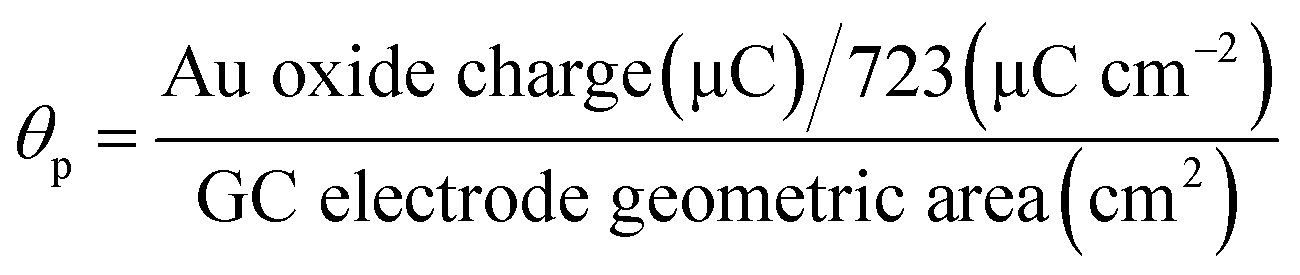
The deposition of CAuNCs on the GC electrode was characterized by a cyclic voltammetry (CV) method. The cyclic voltammograms obtained for the AuNRs, and the CAuNCs modified GC electrodes in 0.1 M H2SO4 solution are shown in Fig. 3. The AuNR modified GC electrode shows an oxidation peak at 1.26 V and a corresponding reduction peak at 0.9 V (Fig. 3a). The redox peaks are attributed to the oxidation and subsequent reduction of surface gold oxide (AuOx) formation of the AuNRs, as a result of the positive potential polarization of the AuNR modified GC electrode. On the other hand, the CAuNC modified GC electrode shows an oxidation peak at 1.15 V, which is a 0.11 V less positive potential than the AuNRs, and a corresponding distinguished reduction peak at 0.9 V (Fig. 3b). The amount of Au oxide exposed to the solution was calculated by integrating the charge under the Au oxide reduction peak corrected for the baseline, and it was found to be 33.84 μC.54 We have calculated the particle coverage (θp) of the CAuNC GC electrode using eqn (1). The particle coverage is defined as the ratio between the electrochemically accessible CAuNC area and the geometric area of the GC electrode in contact with the electrolyte solution.55 The particle coverage of the CAuNC modified GC electrode was calculated by using the charge involved in the reduction of the electrochemically formed Au oxide from the recorded cyclic voltammogram, between the potential window from 1.0 to 0.5 V in 0.1 M H2SO4 solution, by assuming that the charge density for the reduction peak of the Au oxide is 723 μC cm−2.55
 | (1) |
 | ||
| Fig. 3 CVs obtained for the AuNR (a), and CAuNC (b) modified GC electrodes in 0.1 M H2SO4 solution at a scan rate of 50 mV s−1. | ||
The CAuNC particle coverage on the GC electrode was found to be 19.79%. However, the AuNR deposited electrode showed a particle coverage of only 1.45%. Therefore, the CAuNCs showed a very higher particle coverage on the GC electrode, which is effectively utilized for immobilizing large amounts of IgG antibody for the detection of IgG proteins.
3.3. Electrochemical IgG immunobiosensor
To study the potential utilization of the IgG antibody/CAuNC modified electrode, the electrocatalytic oxidation and electrochemical determination of the IgG antigen were carried out in a 0.2 M PB solution (pH 7.2). The AuNR/GC electrode did not give any electrochemical response for the IgG antibody and the IgG antigen–antibody immobilized on the AuNRs, as shown in Fig. 4A, curve a and b. This is due to the fact that the surface atoms of the AuNRs, composed of low index facets (111), cannot catalyze this reaction. Moreover, a lower coverage of nanorods (1.45%) may not allow large amounts of antibodies and antigens on its surface. On the other hand, the CAuNC electrode showed a broad oxidation peak at ∼0.45 V, and a corresponding reduction peak at 0.01 V (Fig. 4B, curve a). The above oxidation is mainly because of the functional amino groups present in the IgG antibody, which are involved in the electrochemical oxidation in the PB solution. The observed result is closely matched with the oxidation of a gold nanoparticle based polymer film nanocomposite electrode.56 From the CV results, it can be seen that the CAuNC modified electrode has a clear response in the presence of IgG antibodies and antibody–antigen interactions. The CAuNCs possess high index facets {720}, which can provide higher electrochemically active atoms/sites for this oxidation reaction. However, the electrochemical response disappeared at ∼0.45 V and 0.01 V (Fig. 4B, curve b) and a new peak was observed at ∼0.25 V due to the interaction and the complex formation between the IgG antigen and IgG antibody. Moreover, the catalytic current response decreases due to the high coverage of IgG antigen on the IgG antibody/CAuNC surface, which results in a resistive layer on the electrode. In the presence of an IgG antigen molecule, the IgG antibody incorporated CAuNC electrode showed a broad oxidation peak for IgG antigen (∼0.25 V; vide supra), which clearly indicates that the IgG antibody-CAuNC modified electrode effectively detects the IgG antigen due to the specific interaction of the IgG-incorporated CAuNCs with the IgG antigen. The immobilized CAuNCs provide an efficient catalysis platform for the present IgG immunobiosensor. In other words, the deposition of the whole IgG antibodies on the CAuNC surface relies on electrostatic interactions (vide supra). Simultaneously, the physical adsorption is one of the protein binding processes, although rather uncontrollable. Random orientation of the assembled antibodies and close proximity between adsorptive surface and the antigen-binding site could enable the detection through an impeding CV response. Moreover, the CAuNCs greatly increase the active surface area for loading large amounts of IgG antibodies. Furthermore, no reduction peak is seen for the IgG antigen during interaction with the IgG antibody modified CAuNC electrode (Fig. 4B, curve a). These results reveal that the oxidation process of the IgG antigen at the IgG antibody/CAuNC electrode is faster than the AuNR electrode and also that the IgG antibody-incorporated CAuNC electrode possesses higher electrocatalytic activity towards the IgG oxidation. The electrochemical response of the IgG antibody/CAuNC electrode has been investigated as a function of the IgG antigen concentration (10–200 ng mL−1) using a CV technique (Fig. 5A). The current increase on each addition of IgG is attributed to the formation of an antigen–antibody complex between the IgG antigen and the IgG antibody on the CAuNCs. | ||
| Fig. 4 Cyclic voltammograms obtained for the IgG antibody (a) and IgG antigen–antibody complex (b) on the AuNR (A) and CAuNC (B) modified GC electrodes in a 0.2 M PB solution at a scan rate of 50 mV s−1. | ||
 | ||
| Fig. 5 (A) Cyclic voltammograms recorded for the IgG antibody on the CAuNC-modified immunoelectrode as a function of varying concentrations of IgG antigen (a–h); 10, 25, 50, 100, 150, 200, 250 and 300 ng mL−1, respectively, in a 0.2 M PB solution (pH 7.2) at a scan rate of 50 mV s−1. (B) Calibration plot obtained for the current response vs. function of conc. of IgG antigen at a scan rate of 50 mV s−1. | ||
Fig. 5B shows the calibration curve obtained as a function of the IgG antigen concentration. A linear relationship between the magnitude of current and IgG concentration can be fitted to the experimental points from 10 to 200 ng mL−1 with a correlation coefficient of 0.9913. The value of the association constant (Ka) was calculated to be 5.32 × 10¹¹ L mol−1 using a Lineweaver–Burk like plot,57 indicating a high affinity of the IgG antibody towards the IgG antigen, attributed to prevalent electrostatic interactions. The increased activity of the IgG antibody due to favourable electrostatic interactions with the CAuNCs results in a high value of Ka. The detection limit of the IgG antibody/CAuNC electrode towards the IgG antigen was calculated from the CV method, and it was 5 ng mL−1 (S/N = 3).
4. Conclusions
We described a novel electrochemical sensor for the label-free, real time and highly sensitive detection of antibody–antigen interactions based on CAuNCs. The CAuNCs provide higher surface atoms with enhanced chemical activities, which can efficiently catalyse the oxidation reaction of amino groups on antibodies (anti-bovine IgG produced in rabbit). An obvious redox current response was observed in the CV response, and for the introduction of IgG antigens, a notable decrease of anodic peak current was observed, which reduces the protein mobility so less amine can participate in the electrochemical oxidation reaction after the antibody–antigen interactions. The electrocatalytic properties of the CAuNCs showed an excellent sensing platform towards the rabbit IgG antigen in a wide range of concentrations (from 10 to 200 ng mL−1) with a limit of detection (LOD) of 5 ng mL−1 (S/N = 3) by using a CV method.Acknowledgements
We gratefully acknowledge Defense Acquisition Program Administration and Agency for Defense Development under the contract UD140080GD and the Natural Science Foundation of China (21404110 and 51473179).Notes and references
- A. Hucknall, S. Rangarajan and A. Chilkoti, In Pursuit of Zero: Polymer Brushes that Resist the Adsorption of Proteins, Adv. Mater., 2009, 21, 2441–2446 CrossRef CAS PubMed.
- D. Khatayevich, T. Page, C. Gresswell, Y. Hayamizu, W. Grady and M. Sarikaya, Selective Detection of Target Proteins by Peptide-Enabled Graphene Biosensor, Small, 2014, 10, 1505–1513 CrossRef CAS PubMed.
- N. Wang, C. Gao, Y. Han, X. Huang, Y. Xu and X. Cao, Detection of human immunoglobulin G by label-free electrochemical immunoassay modified with ultralong CuS nanowires, J. Mater. Chem. B, 2015, 3, 3254–3259 RSC.
- Q. Yu, X. Wang and Y. Duan, Capillary-Based Three-Dimensional Immunosensor Assembly for High-Performance Detection of Carcinoembryonic Antigen Using Laser-Induced Fluorescence Spectrometry, Anal. Chem., 2014, 86, 1518–1524 CrossRef CAS PubMed.
- F. Ma, Y. Yang and C.-y. Zhang, Ultrasensitive Detection of Transcription Factors Using Transcription-Mediated Isothermally Exponential Amplification-Induced Chemiluminescence, Anal. Chem., 2014, 86, 6006–6011 CrossRef CAS PubMed.
- J. S. Lane, J. L. Richens, K.-A. Vere and P. O’Shea, Rational Targeting of Subclasses of Intermolecular Interactions: Elimination of Nonspecific Binding for Analyte Sensing, Langmuir, 2014, 30, 9457–9465 CrossRef CAS PubMed.
- Y. Zhang, N. Islam, R. G. Carbonell and O. J. Rojas, Specific Binding of Immunoglobulin G with Bioactive Short Peptides Supported on Antifouling Copolymer Layers for Detection in Quartz Crystal Microgravimetry and Surface Plasmon Resonance, Anal. Chem., 2013, 85, 1106–1113 CrossRef CAS PubMed.
- K. Meyer and P. M. Ueland, Targeted Quantification of C-Reactive Protein and Cystatin C and Its Variants by Immuno-MALDI-MS, Anal. Chem., 2014, 86, 5807–5814 CrossRef CAS PubMed.
- K. Maehashi, T. Katsura, K. Kerman, Y. Takamura, K. Matsumoto and E. Tamiya, Label-Free Protein Biosensor Based on Aptamer-Modified Carbon Nanotube Field-Effect Transistors, Anal. Chem., 2007, 79, 782–787 CrossRef CAS PubMed.
- J. S. Lee, H.-A. Joung, M.-G. Kim and C. B. Park, Graphene-Based Chemiluminescence Resonance Energy Transfer for Homogeneous Immunoassay, ACS Nano, 2012, 6, 2978–2983 CrossRef CAS PubMed.
- T. Li, E.-J. Jo and M.-G. Kim, A label-free fluorescence immunoassay system for the sensitive detection of the mycotoxin, ochratoxin A, Chem. Commun., 2012, 48, 2304–2306 RSC.
- X. Yu, B. Munge, V. Patel, G. Jensen, A. Bhirde, J. D. Gong, S. N. Kim, J. Gillespie, J. S. Gutkind, F. Papadimitrakopoulos and J. F. Rusling, Carbon Nanotube Amplification Strategies for Highly Sensitive Immunodetection of Cancer Biomarkers, J. Am. Chem. Soc., 2006, 128, 11199–11205 CrossRef CAS PubMed.
- G. Wang, H. Huang, G. Zhang, X. Zhang, B. Fang and L. Wang, Dual Amplification Strategy for the Fabrication of Highly Sensitive Interleukin-6 Amperometric Immunosensor Based on Poly-Dopamine, Langmuir, 2011, 27, 1224–1231 CrossRef CAS PubMed.
- M. Campàs and J.-L. Marty, Highly sensitive amperometric immunosensors for microcystin detection in algae, Biosens. Bioelectron., 2007, 22, 1034–1040 CrossRef PubMed.
- L. Yang, H. Zhao, S. Fan, S. Deng, Q. Lv, J. Lin and C.-P. Li, Label-free electrochemical immunosensor based on gold–silicon carbide nanocomposites for sensitive detection of human chorionic gonadotrophin, Biosens. Bioelectron., 2014, 57, 199–206 CrossRef CAS PubMed.
- J. Zhou, M. Xu, D. Tang, Z. Gao, J. Tang and G. Chen, Nanogold-based bio-bar codes for label-free immunosensing of proteins coupling with an in situ DNA-based hybridization chain reaction, Chem. Commun., 2012, 48, 12207–12209 RSC.
- G. W. Litman, J. P. Rast, M. J. Shamblott, R. N. Haire, M. Hulst, W. Roess, R. T. Litman, K. R. Hinds-Frey, A. Zilch and C. T. Amemiya, Phylogenetic diversification of immunoglobulin genes and the antibody repertoire, Mol. Biol. Evol., 1993, 10, 60–72 CAS.
- M. Diaz and P. Casali, Somatic immunoglobulin hypermutation, Curr. Opin. Immunol., 2002, 14, 235–240 CrossRef CAS.
- J. J. Wilson, R. Burgess, Y.-Q. Mao, S. Luo, H. Tang, V. S. Jones, B. Weisheng, R.-Y. Huang, X. Chen and R.-P. Huang, Chapter Seven - Antibody Arrays in Biomarker Discovery, in Advances in Clinical Chemistry, ed. S. M. Gregory, Elsevier, 2015, vol. 69, pp. 255–324 Search PubMed.
- T. R. J. Holford, F. Davis and S. P. J. Higson, Recent trends in antibody based sensors, Biosens. Bioelectron., 2012, 34, 12–24 CrossRef CAS PubMed.
- D. C. T. Parker, Cell-Dependent B Cell Activation, Annu. Rev. Immunol., 1993, 11, 331–360 CrossRef CAS PubMed.
- C. I. L. Justino, A. C. Freitas, R. Pereira, A. C. Duarte and T. A. P. Rocha Santos, Recent developments in recognition elements for chemical sensors and biosensors, TrAC, Trends Anal. Chem., 2015, 68, 2–17 CrossRef CAS PubMed.
- Q. Ruan and S. Y. Tetin, Applications of dual-color fluorescence cross-correlation spectroscopy in antibody binding studies, Anal. Biochem., 2008, 374, 182–195 CrossRef CAS PubMed.
- A. C. Crisostomo, L. Dang, J. L. Digambaranath, A. C. Klaver, D. A. Loeffler, J. J. Payne, L. M. Smith, A. L. Yokom and J. M. Finke, Kinetic analysis of IgG antibodies to beta-amyloid oligomers with surface plasmon resonance, Anal. Biochem., 2015, 481, 43–54 CrossRef CAS PubMed.
- J. Gao, Z. Guo, F. Su, L. Gao, X. Pang, W. Cao, B. Du and Q. Wei, Ultrasensitive electrochemical immunoassay for CEA through host–guest interaction of β-cyclodextrin functionalized graphene and Cu@Ag core–shell nanoparticles with adamantine-modified antibody, Biosens. Bioelectron., 2015, 63, 465–471 CrossRef CAS PubMed.
- A. Sargent and O. A. Sadik, Monitoring antibody–antigen reactions at conducting polymer-based immunosensors using impedance spectroscopy, Electrochim. Acta, 1999, 44, 4667–4675 CrossRef CAS.
- Y. Xiao, C. M. Li and Y. Liu, Electrochemical impedance characterization of antibody–antigen interaction with signal amplification based on polypyrrole–streptavidin, Biosens. Bioelectron., 2007, 22, 3161–3166 CrossRef CAS PubMed.
- F. Darain, S.-U. Park and Y.-B. Shim, Disposable amperometric immunosensor system for rabbit IgG using a conducting polymer modified screen-printed electrode, Biosens. Bioelectron., 2003, 18, 773–780 CrossRef CAS.
- K.-J. Huang, D.-J. Niu, W.-Z. Xie and W. Wang, A disposable electrochemical immunosensor for carcinoembryonic antigen based on nano-Au/multi-walled carbon nanotubes–chitosans nanocomposite film modified glassy carbon electrode, Anal. Chim. Acta, 2010, 659, 102–108 CrossRef CAS PubMed.
- A. P. F. Turner, Biosensors: sense and sensibility, Chem. Soc. Rev., 2013, 42, 3184–3196 RSC.
- S. Dong and B. Wang, Electrochemical Biosensing in Extreme Environment, Electroanalysis, 2002, 14, 7–16 CrossRef CAS.
- Z. Ren and P.-X. Gao, A review of helical nanostructures: growth theories, synthesis strategies and properties, Nanoscale, 2014, 6, 9366–9400 RSC.
- C. Zhu, G. Yang, H. Li, D. Du and Y. Lin, Electrochemical Sensors and Biosensors Based on Nanomaterials and Nanostructures, Anal. Chem., 2015, 87, 230–249 CrossRef CAS PubMed.
- N. Li, X. Su and Y. Lu, Nanomaterial-based biosensors using dual transducing elements for solution phase detection, Analyst, 2015, 140, 2916–2943 RSC.
- B. Pérez-López and A. Merkoçi, Nanomaterials based biosensors for food analysis applications, Trends Food Sci. Technol., 2011, 22, 625–639 CrossRef PubMed.
- L. Chen, B. Wu, L. Guo, R. Tey, Y. Huang and D.-H. Kim, A single-nanoparticle NO2 gas sensor constructed using active molecular plasmonics, Chem. Commun., 2015, 51, 1326–1329 RSC.
- L. Guo, Y. Huang, Y. Kikutani, Y. Tanaka, T. Kitamori and D.-H. Kim, In situ assembly, regeneration and plasmonic immunosensing of a Au nanorod monolayer in a closed-surface flow channel, Lab Chip, 2011, 11, 3299–3304 RSC.
- Y. Huang, A. R. Ferhan, Y. Gao, A. Dandapat and D.-H. Kim, High-yield synthesis of triangular gold nanoplates with improved shape uniformity, tunable edge length and thickness, Nanoscale, 2014, 6, 6496–6500 RSC.
- Y. Huang, A. R. Ferhan and D.-H. Kim, Tunable scattered colors over a wide spectrum from a single nanoparticle, Nanoscale, 2013, 5, 7772–7775 RSC.
- Y. Yu, Q. Zhang, X. Lu and J. Y. Lee, Seed-Mediated Synthesis of Monodisperse Concave Trisoctahedral Gold Nanocrystals with Controllable Sizes, J. Phys. Chem. C, 2010, 114, 11119–11126 CAS.
- C. J. DeSantis, A. A. Peverly, D. G. Peters and S. E. Skrabalak, Octopods versus Concave Nanocrystals: Control of Morphology by Manipulating the Kinetics of Seeded Growth via Co-Reduction, Nano Lett., 2011, 11, 2164–2168 CrossRef CAS PubMed.
- Y. Huang, L. Wu, X. Chen, P. Bai and D.-H. Kim, Synthesis of Anisotropic Concave Gold Nanocuboids with Distinctive Plasmonic Properties, Chem. Mater., 2013, 25, 2470–2475 CrossRef CAS.
- C.-L. Lu, K. S. Prasad, H.-L. Wu, J.-a. A. Ho and M. H. Huang, Au Nanocube-Directed Fabrication of Au–Pd Core–Shell Nanocrystals with Tetrahexahedral, Concave Octahedral, and Octahedral Structures and Their Electrocatalytic Activity, J. Am. Chem. Soc., 2010, 132, 14546–14553 CrossRef CAS PubMed.
- P. Kannan, S. Sampath and S. A. John, Direct Growth of Gold Nanorods on Gold and Indium Tin Oxide Surfaces: Spectral, Electrochemical, and Electrocatalytic Studies, J. Phys. Chem. C, 2010, 114, 21114–21122 CAS.
- P. Kannan, H. Y. Tiong and D.-H. Kim, Highly sensitive electrochemical determination of neutrophil gelatinase-associated lipocalin for acute kidney injury, Biosens. Bioelectron., 2012, 31, 32–36 CrossRef CAS PubMed.
- P. Kannan and S. A. John, Highly sensitive determination of hydroxylamine using fused gold nanoparticles immobilized on sol–gel film modified gold electrode, Anal. Chim. Acta, 2010, 663, 158–164 CrossRef CAS PubMed.
- P. Kannan and S. A. John, Highly sensitive electrochemical determination of nitric oxide using fused spherical gold nanoparticles modified ITO electrode, Electrochim. Acta, 2010, 55, 3497–3503 CrossRef CAS PubMed.
- H. Zhang, M. Jin and Y. Xia, Noble-Metal Nanocrystals with Concave Surfaces: Synthesis and Applications, Angew. Chem., Int. Ed., 2012, 51, 7656–7673 CrossRef CAS PubMed.
- K. Sohn, F. Kim, K. C. Pradel, J. Wu, Y. Peng, F. Zhou and J. Huang, Construction of Evolutionary Tree for Morphological Engineering of Nanoparticles, ACS Nano, 2009, 3, 2191–2198 CrossRef CAS PubMed.
- Y. Huang and D.-H. Kim, Dark-field microscopy studies of polarization-dependent plasmonic resonance of single gold nanorods: rainbow nanoparticles, Nanoscale, 2011, 3, 3228–3232 RSC.
- H. A. Keul, M. Möller and M. R. Bockstaller, Structural Evolution of Gold Nanorods during Controlled Secondary Growth, Langmuir, 2007, 23, 10307–10315 CrossRef CAS PubMed.
- J. Zhang, M. R. Langille, M. L. Personick, K. Zhang, S. Li and C. A. Mirkin, Concave Cubic Gold Nanocrystals with High-Index Facets, J. Am. Chem. Soc., 2010, 132, 14012–14014 CrossRef CAS PubMed.
- N. Tian, Z.-Y. Zhou, S.-G. Sun, Y. Ding and Z. L. Wang, Synthesis of Tetrahexahedral Platinum Nanocrystals with High-Index Facets and High Electro-Oxidation Activity, Science, 2007, 316, 732–735 CrossRef CAS PubMed.
- N. Alexeyeva and K. Tammeveski, Electroreduction of oxygen on gold nanoparticle/PDDA-MWCNT nanocomposites in acid solution, Anal. Chim. Acta, 2008, 618, 140–146 CrossRef CAS PubMed.
- S. Kumar and S. Zou, Electrooxidation of Carbon Monoxide on Gold Nanoparticle Ensemble Electrodes:
![[thin space (1/6-em)]](https://www.rsc.org/images/entities/char_2009.gif) Effects of Particle Coverage, J. Phys. Chem. B, 2005, 109, 15707–15713 CrossRef CAS PubMed.
Effects of Particle Coverage, J. Phys. Chem. B, 2005, 109, 15707–15713 CrossRef CAS PubMed. - P. Kannan and S. A. John, Ultrasensitive detection of l-cysteine using gold–5-amino-2-mercapto-1,3,4-thiadiazole core–shell nanoparticles film modified electrode, Biosens. Bioelectron., 2011, 30, 276–281 CrossRef CAS PubMed.
- H. Lineweaver and D. Burk, The Determination of Enzyme Dissociation Constants, J. Am. Chem. Soc., 1934, 56, 658–666 CrossRef CAS.
Footnote |
| † These authors contribute equally to this work. |
| This journal is © The Royal Society of Chemistry 2015 |