Extending pharmacological dose-response curves for salsalate with natural deep eutectic solvents

E. Rozema a, A. D. van Damb, H. C. M. Sipsb, R. Verpoortea, O. C. Meijerb, S. Kooijman§ *b and Y. H. Choi§*a
aNatural Products Laboratory, Institute of Biology, Leiden University, 2333 BE Leiden, The Netherlands. E-mail: y.h.choi@biology.leidenuniv.nl
bDepartment of Medicine, Division of Endocrinology, Post Zone C7Q. Leiden University Medical Center, 2300 RC Leiden, The Netherlands. E-mail: s.kooijman@lumc.nl

Received 29th May 2015 , Accepted 8th July 2015

First published on 8th July 2015


Abstract

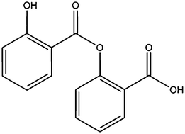
Natural deep eutectic solvents (NADES) are recently developed green solvents that are attractive for their great solubilising power and intrinsic lack of toxicity. In this paper we report the results of a study on the feasibility and benefits of dissolving the poorly water-soluble salsalate in a NADES composed of 1,2-propanediol–choline–water as an alternative to DMSO for functional in vitro assays. The increase in solubility allows an extension of the range of the dose-response curve of salsalate-induced activation of brown adipocytes.


The solubility in aqueous media of a drug is one of the most critical features to be considered when designing in vitro and in vivo bioactivity tests. The solubility influences the bioavailability, transport and uptake of a drug in cells.1,2 Often, hydrophobic compounds only dissolve in non-aqueous, organic or toxic solvents, which are therefore not suitable for pharmacological studies.

Deep eutectic solvents are a class of solvents obtained by mixing solid compounds, generating a eutectic mixture with a melting point that is much lower than that of the individual components. Particularly natural deep eutectic solvents (NADES), which comprise components from natural origin, hold great promise as solvents for pharmaceutical applications because of their biodegradability, low toxicity levels and ability to dissolve chemically diverse compounds, including relatively non-water soluble compounds (e.g. rutin, cinnamic acid, taxol) as well as macromolecules (e.g. DNA and starch).3,4 These solvents are combinations of two or more naturally occurring compounds, mainly primary plant metabolites, which form a liquid when mixed in certain molar ratios. This liquid is formed due to the establishment of strong hydrogen bonding between their components. This hydrogen bonding also occurs between the NADES and solutes, with a probable stabilising effect of solutions of even scarcely soluble compounds. Furthermore, the NADES that is appropriate for each case can potentially be tailored to the needs of the application by choosing the right combination and ratio of its components. Additionally, besides functioning as solvent, NADES may also exhibit intrinsic effects.5

Salicylates are compounds that occur in plants6 and play a role in the plant defence system against pathogen attack and environmental stress.7 Non-acetylated (e.g. salsalate, sodium salicylate) and acetylated salicylates (e.g. aspirin) (Fig. 1) are well known to have anti-inflammatory and anti-diabetic activities in humans.8 Recently, we identified salsalate as an inducer of brown adipose tissue (BAT) activity.9 BAT activity allows large amounts of glucose and triglycerides to be converted into heat through uncoupled respiration due to the expression of uncoupling protein1 (UCP1). Therefore, BAT is considered to be a promising target in the treatment of obesity and associated diseases, including type II diabetes and atherosclerosis.10


image file: c5ra10196d-f1.tif
Fig. 1 Structures of salsalate and salicylate.

Salsalate has the advantage over its metabolite sodium salicylate of being insoluble at the acid pH of the stomach, ensuring that it passes straight through it without direct contact with the gastric mucosa. Undesirable gastric side-effects are thus avoided.8,11,12 However, salsalate is only soluble in a few organic solvents such as dimethyl sulfoxide (DMSO). This limits the possibilities of in vivo and in vitro investigation of salsalate bioactivities. In this study, we first assessed the possibility of dissolving salsalate in a NADES and then the in vitro effects of this solution on cell viability and the function of brown adipocytes.

To select the NADES in which salsalate had the highest solubility, we tested a range of sugar-based NADES composed of mixtures of sugars with alcohols, acids or bases with an acid in given molar ratios.4

These included malic acid–choline chloride–water (MCH, molar ratio 1[thin space (1/6-em)]:[thin space (1/6-em)]1[thin space (1/6-em)]:[thin space (1/6-em)]2), glucose–choline chloride–water (GCH, 2[thin space (1/6-em)]:[thin space (1/6-em)]5[thin space (1/6-em)]:[thin space (1/6-em)]5), lactic acid–glucose–water (LGH, 5[thin space (1/6-em)]:[thin space (1/6-em)]1[thin space (1/6-em)]:[thin space (1/6-em)]3), 1,2-propanediol–choline chloride–water (PCH, 1[thin space (1/6-em)]:[thin space (1/6-em)]1[thin space (1/6-em)]:[thin space (1/6-em)]1), 1,2-propanediol–malic acid–water (PMH, 1[thin space (1/6-em)]:[thin space (1/6-em)]1[thin space (1/6-em)]:[thin space (1/6-em)]3), fructose–glucose–sucrose–water (FGSH, 1[thin space (1/6-em)]:[thin space (1/6-em)]1[thin space (1/6-em)]:[thin space (1/6-em)]1[thin space (1/6-em)]:[thin space (1/6-em)]11) and sucrose–choline chloride–water (SCH, 1[thin space (1/6-em)]:[thin space (1/6-em)]4[thin space (1/6-em)]:[thin space (1/6-em)]4). Among these, the highest solubility was obtained with PCH. The solubility of salsalate at room temperature as determined by HPLC/DAD was 85 mg mL−1 (0.33 M) against 3.3 g mL−1 in DMSO. The stability of a PCH solution of salsalate was monitored over a three-month period by HPLC/DAD. For this, samples were prepared and analysed by HPLC directly after preparation (T0) and 30 and 90 days later. The HPLC system consisted of a reversed phase C18 column and isocratic elution with methanol-1% acetic acid (60[thin space (1/6-em)]:[thin space (1/6-em)]40). The chromatogram of salsalate in both PCH (Fig. 2a) and DMSO (Fig. S1) at T0 showed one peak with a retention time of approximately 7.14 min. After 30 days, two additional small peaks with retention times of 4.0 and 5.7 minutes (Fig. 2d) appeared in the PCH salsalate solution. Both had a similar spectrum to salsalate with UV max at 238 nm and 306–308 nm (Fig. 2e–f). The retention times of the peaks did not correspond with that of salicylic acid nor methyl salicylate reference compounds (Fig. S2), indicating some degradation of salsalate. On the other hand, the DMSO solutions of salsalate were more stable and no additional peaks were detected after one or three months (Fig. S1).


image file: c5ra10196d-f2.tif
Fig. 2 HPLC chromatogram of freshly prepared (T0) PCH salsalate solution with detection at 220 nm (red) and 280 nm (blue) (a) and UV spectra of the peak with Tr = 2.1 min (UVmax = 216 nm) (b) and Tr = 7.1 min with UVmax = 238 and 308 nm (c) detected in the salsalate solution in PCH (T0). HPLC chromatogram of PCH salsalate solution (T = 30 days), stored at room temperature with detection at 220 nm (red) and 280 nm (blue) (d) and UV-spectra of the peaks at retention times of 4.0 and 5.7 min (e, f, respectively) were detected and respective UV-visible spectra showing resemblance of UVmax values with salsalate at 238 nm 306 nm (e and f).

As a next step, the toxicity of PCH in cell culture was evaluated and compared with that of DMSO. Differentiated T37i brown adipocytes were incubated with an increasing volume of DMSO and PCH in the culture medium. After 8 h, cell viability was assessed with Presto blue staining. Similar to our findings for DMSO, concentrations of up to 1% PCH did not affect the viability of the cells (Fig. 3a). In subsequent experiments, increasing amounts of salsalate were dissolved in PCH and DMSO and applied to cells as a 0.33% solution in the culture medium. In these conditions, it was possible to achieve final concentrations of salsalate in PCH of up to 1 mM, while DMSO solutions above 0.3 mM precipitated when added to culture medium. This suggests that PCH increased the solubility of salsalate in aqueous environments, allowing the generation of a full dose-response curve for glycerol release as a measure of intracellular lipolysis. In contrast, the maximum concentration of salsalate in DMSO (0.3 mM) was insufficient to reach the plateau phase (Fig. 3b). Gene expression analysis of genes involved in brown adipocyte thermogenesis,13,14 i.e. Pgc1α (Fig. 3c), Pparα (Fig. 3d), Elovl3 (Fig. 3e) and Ucp1 (Fig. 3f) revealed comparable responses for 0.3 mM salsalate NADES and DMSO solutions.


image file: c5ra10196d-f3.tif
Fig. 3 Differentiated T37i brown adipocytes were incubated with increasing volume percent of DMSO and PCH in culture medium. After 8 h, cell viability was measured by Presto blue staining in relative fluorescence units (a). Dose-response curves for salsalate in PCH and DMSO were derived for 8 h glycerol release (b) and gene expression analysis for Pgc1α (c), Pparα (d), Elovl3 (e) and Ucp1 (f). Arrow (a) indicates the solvent dose selected for further experiments (d–f). Results are presented as mean value ± SEM (n = 3 for A, n = 6 for B–F). Significantly different from the negative control: *P < 0.05; **P < 0.01; ***P < 0.005.

Interestingly, extending the dose-response curves with a 1 mM PCH solution of salsalate revealed dissociations between individual genes affected by salsalate. For Pgc1α (Fig. 3c) and Pparα mRNA (Fig. 3d) a substantially higher maximal induction was found, also indicative of an EC50 that was higher than the one that could have been determined with DMSO-based dose-response curves. For Elovl3 (Fig. 3e) mRNA the induction was at the plateau. The Ucp1 (Fig. 3f) gene expression showed an unexpected inverted U-shape. Notably, the addition of the 1 mM solution of salsalate point to the dose-response curve resulted in better calculations of EC50 values as reflected by the lower EC50 95% confidence intervals (Table 1).

Table 1 EC50 95% confidence intervals (log[salsalate], mM)
  EC50 95% CI
DMSO PCH
Glycerol release −1.048 to −0.1603 −1.188 to −0.3921
Pgc1α mRNA (Very wide) −0.3247 to 0.1792
Pparα mRNA −1.012 to 0.03785 −0.5428 to −0.1075
Elovl3 mRNA −1.049 to −0.5637 −1.236 to −0.5108
Ucp1 mRNA (Very wide) −2.035 to −0.5269


Conclusions

We demonstrated the feasibility and benefits of dissolving salsalate in PCH rather than DMSO for studies on brown adipocyte activation. Importantly, the property of PCH to increase the solubility of salsalate in aqueous environments such as a cell culture medium, allowed us to treat cells with concentrations that were high enough to generate full dose-response curves, and this yielded novel information on the gene expression underlying salsalate-induced brown adipose tissue activity. The results of this study provide substantial evidence for the suitability of the biodegradable PCH, for pre-clinical research. However, the possibility of extending this NADES application to in vivo use for drug administration requires further investigation into its physicochemical, pharmacokinetic and thermodynamic properties.

Acknowledgements

This work is supported by an ECHO grant (711.012.010) that is provided by the Netherlands Organization for Scientific Research (NWO). We thank Dr Marc Lombès for kindly providing the T37i cells.

Notes and references

  1. Y. Gong, D. J. W. Grant and H. G. Brittain, in Solvent systems and their selection in pharmaceutics and biopharmaceutics, ed. P. Augustijns and M. Brewster, Springer, New York, 1st edn, 2007, ch. 1, p. 1 Search PubMed.
  2. F. L. Mota, A. P. Carneiro, A. J. Queimada, S. P. Pinho and E. A. Macedo, Eur. J. Pharm. Sci., 2009, 37, 499–507 CrossRef CAS PubMed.
  3. Y. H. Choi, J. van Spronsen, Y. Dai, M. Verberne, F. Hollmann, I. W. Arends, G. J. Witkamp and R. Verpoorte, Plant Physiol., 2011, 156, 1701–1705 CrossRef CAS PubMed.
  4. Y. Dai, J. van Spronsen, G. J. Witkamp, R. Verpoorte and Y. H. Choi, Anal. Chim. Acta, 2013, 766, 61–68 CrossRef CAS PubMed.
  5. R. Ferraz, L. C. Branco, C. Prudêncio, J. P. Noronha and Z. Petrovski, ChemMedChem, 2011, 6, 975–985 CrossRef CAS PubMed.
  6. K. K. Wu, Circulation, 2000, 102, 2022–2023 CrossRef CAS.
  7. G. G. Duthie and A. D. Wood, Food Funct., 2011, 2, 515–520 CAS.
  8. A. B. Goldfine, R. Silver, W. Aldhahi, D. Cai, E. Tatro, J. Lee and S. E. Shoelson, Clin. Transl. Sci., 2008, 1, 36–43 CrossRef CAS PubMed.
  9. A. D. van Dam, K. J. Nahon, S. Kooijman, S. M. van den Berg, A. A. Kanhai, T. Kikuchi, M. M. Heemskerk, V. van Harmelen, M. Lombès, A. M. van den Hoek, M. P. J. de Winther, E. Lutgens, B. Guigas, P. C. N. Rensen and M. R. Boon, Diabetes, 2015, 64, 1544–1554 CrossRef CAS PubMed.
  10. J. F. P. Berbée, M. R. Boon, P. P. S. J. Khedoe, A. Bartelt, C. Schlein, A. Worthmann, S. Kooijman, G. Hoeke, I. M. Mol, C. John, C. Jung, N. Vazirpanah, L. P. J. Brouwers, P. L. S. M. Gordts, J. D. Esko, P. S. Hiemstra, L. M. Havekes, L. Scheja, J. Heeren and P. C. N. Rensen, Nat. Commun., 2015, 6 Search PubMed.
  11. D. Estes and K. Kaplan, Arthritis Rheum., 1980, 23, 1303–1307 CrossRef CAS PubMed.
  12. H. Mielants, E. M. Veys, G. Verbruggen and K. Schelstraete, Scand. J. Rheumatol., 1981, 10, 169–173 CrossRef CAS.
  13. A. Jakobsson, J. A. Jörgensen and A. Jacobsson, Am. J. Physiol.: Endocrinol. Metab., 2005, 289, E517–E526 CrossRef CAS PubMed.
  14. V. Peirce, S. Carobbio and A. Vidal-Puig, Nature, 2014, 510, 76–83 CrossRef CAS PubMed.

Footnotes

Electronic supplementary information (ESI) available. See DOI: 10.1039/c5ra10196d
These authors contributed equally to this work.
§ These authors share last and corresponding authorship.

This journal is © The Royal Society of Chemistry 2015
Click here to see how this site uses Cookies. View our privacy policy here.