
Carbon-mediated fabrication of core–shell structured SnO2@TiO2 nanocomposites with excellent photocatalytic performance
Haijiao Zhang *a,
Minxia Yinga,
Renmei Gaoa,
Le Hua,
Zheng Jiaoa and
Xuedong Zhu*b
*a,
Minxia Yinga,
Renmei Gaoa,
Le Hua,
Zheng Jiaoa and
Xuedong Zhu*b
aInstitute of Nanochemistry and Nanobiology, School of Environmental and Chemical Engineering, Shanghai University, Shanghai 200444, P. R. China. E-mail: hjzhang128@shu.edu.cn; Fax: +86-21-6613-5275; Tel: +86-21-6613-5275
bState Key Laboratory of Chemical Engineering, East China University of Science & Technology, Shanghai 200237, P. R. China. E-mail: xdzhu@ecust.edu.cn
First published on 29th June 2015
Abstract
In this work, a facile and carbon-mediated hydrothermal route has been developed for fabrication of core–shell structured SnO2@TiO2 nanocomposites with excellent photocatalytic performance for the degradation of the organic dye rhodamine B (RhB). The heterostructures were formed by using SnO2–C nanospheres as the core and TiO2 as the outer shell, followed by calcination. The as-synthesized SnO2@TiO2 products have uniformly spherical morphology with an average diameter of 64 nm and the thickness of TiO2 shell was about 4 nm. Additionally, the structure and properties of the products were greatly affected by the calcination temperatures and the carbon content. In comparison to the SnO2@TiO2 synthesized in the absence of carbon, the SnO2@TiO2 composite exhibited a better photocatalytic performance for the decomposition of RhB. In particular, the carbon-mediated SnO2@TiO2-550 product showed superior properties including a large specific surface area for supplying abundant active sites and unique heterojunctions for helping to facilitate the charge separation. As a result, the photocatalytic performance was notably enhanced owing to the above synergistic effect. We expect that the present method based on a carbon-mediated process may be used for the preparation of other nanocomposites.
1. Introduction
In recent years, semiconducting metal oxides such as SnO2, TiO2, and ZnO, etc. have presented themselves as some of the most important materials owing to their outstanding properties and numerous potential applications in gas sensing,1,2 lithium-ion batteries (LIBs),3,4 solar cells,5,6 electrochemical,7,8 supercapacitors,9 photoelectrochemical,10 and catalysis.11–13 Among them, TiO2 has been extensively investigated due to its strong UV light absorption, low cost, high stability, and excellent photocatalytic activity.14,15 However, the photocatalytic activities of TiO2 encounter an intrinsic drawback because of the fast recombination of photo-generated electron/hole (e−/h+) pairs, resulting in a low photocatalytic efficiency. Therefore, the study of how to improve the photocatalytic efficiency of TiO2 attracts much attention, including the tailor of the morphology,16 the noble metal doping17,18 and the design of nanocomposities.19,20 Particularly, the construction of semiconductor/TiO2 heterostructure is an effective and feasible method because the special band alignment could improve the photon absorption and promote the separation of the e−/h+ pairs. For example, TiO2–ZnO core–shell structured heterojunction nanofibers with good photocatalytic activities had been obtained via electrospinning and atomic layer deposition.21 Hybrid multifunctional material of Fe3O4 nanoparticles coated by TiO2 was found to be a highly efficient catalyst for the removal of 4-MMA.22 Moreover, SnO2/TiO2 nanocomposites were suggested as efficient photocatalyst because of the suppression of photogenerated carriers recombined between the SnO2 and TiO2 interface.23On the other hand, carbon materials have drawn much attention in virtue of their low cost,24 high surface areas,25 simple synthesis and good electrical conductivity.26,27 A lot of studies have demonstrated that the growth of metal oxides on carbon support could be effectively promoted and the electrical conductivity of the composites could also be greatly improved.28 Meanwhile, carbon layer may enhance the contact areas between the catalyst and substrate molecules and improve the structural stability of the materials during the reaction process.29 Additionally, the introduction of carbon may also adjust the morphology and structures of TiO2.30,31 For example, mesoporous TiO2–Sn@C core–shell microspheres with Sn encapsulated into a TiO2 mesoporous microsphere matrix was synthesized with well-coated of carbon shell.32
Stimulated by these studies, we demonstrate a facile hydrothermal route for preparation of the core–shell structured SnO2@TiO2 nanocomposites based on a carbon-mediated method. The heterostructure was obtained by using SnO2–C nanospheres as the core and TiO2 as the outer shell, followed by thermal treatment. The structure and the morphology of SnO2@TiO2 nanostructures as well as the role of carbon layer were thoroughly investigated by various techniques including XRD, SEM, HRTEM, BET, XPS and TG analysis. The photocatalytic performance of the products was further studied for degradation of organic dye rhodamine B (RhB) under UV light irradiation. More importantly, the effect of the carbon layer and the thermal treatment temperature on the photocatalytic properties of SnO2@TiO2 nanocomposites was also discussed in detail.
2. Experimental section
2.1 Materials
Stannous sulphate (SnSO4), trisodium citrate dihydrate (Na3C5H6O7·2H2O), glucose (C6H12O6), rhodamine B (RhB), anhydrous ethanol were purchased from Shanghai Chemical Industrial Co. Ltd. (Shanghai, China). Titanyl sulfate (TiOSO4) was purchased from Sigma Limited Company. All chemical reagents were used without further purification. Distilled water was used throughout the experiments.2.2 Synthesis of SnO2@TiO2 nanocomposites
In a typical procedure, 4 mmol of Na3C5H6O7·2H2O and 10 mmol of glucose were dissolved in 80 ml of mixed solvents containing ethanol (24 ml) and H2O (56 ml) under vigorous stirring for 30 min to form a clear solution. Then, 1 mmol of SnSO4 was added into the above solution. After the solution was stirred for 1 h, the resultant mixture was transferred into a Teflon-lined stainless steel autoclave and kept at 180 °C for 12 h. The SnO2–C spheres were obtained after centrifuged and washed for several times. And the resultant SnO2–C was dispersed in 80 ml of distilled water with 0.5 mmol of TiOSO4 and stirred for 10 min. After that, the resultant mixture was transferred into a Teflon-lined stainless steel autoclave at 180 °C for 1 h. Subsequently, the precipitation was centrifuged and washed with ethanol and distilled water in sequence several times and dried at 60 °C overnight. Finally, the SnO2@TiO2 nanocomposites were calcined at 350 to 750 °C in air for 2 h. The as-synthesized and calcined samples are denoted as precursor and SnO2@TiO2-T (T represents the calcination temperature), respectively.For comparsion, the SnO2@TiO2 nanospheres (named as SnO2@TiO2–N) without the addition of glucose and pure SnO2 nanoparticles were also prepared under similar conditions, and calcined at 550 °C in air for 2 h.
2.3 Characterizations
The samples were studied by transmission electron microscopy (TEM, JEOL 200CX), scanning electron microscopy (SEM, JEOL JSM-6700F), and high resolution transmission electron microscopy (HRTEM, JSM-2010F). Elemental qualitative analysis was conducted by the energy-dispersive X-ray spectroscopy (EDX, OXFORD INCA) which was mounted in the JSM-6700F. X-ray photoelectron spectroscopy (XPS) measurements were performed on an ESCALAB 250Xi (Thermofisher, England) using monochromatic AlKα X-ray source operating at a vacuum better than 107 Pa. X-ray powder diffraction (XRD) patterns were obtained on the Japan Rigakul D/max-2550 instrument operating at 40 kV and 40 mA using CuKα radiation (λ = 0.154 nm). N2 adsorption–desorption isotherms were recorded on a QUADRASORB SI surface area & pore size analyzer at 77 K. Brunauer–Emmett–Teller (BET) specific surface area was calculated by using the desorption data. Thermal analysis (TG, STA409, Netzsch, Germany) was performed at a heating rate of 10 °C min−1 in a flow of air. UV-vis spectra were recorded on a HITACHI U-3010 UV-vis spectrophotometer.2.4 Photodegradation measurements
The photocatalytic activity of the products was tested in a SGY-IB photochemical reactor using rhodamine B (RhB) degradation in aqueous solution under UV light irradiation (λ < 400 nm). Typically, 10 mg of SnO2@TiO2 catalysts was added to 50 ml aqueous solution containing 10 mg L−1 RhB in a 100 ml beaker. The mixture was then placed an ultrasonic water bath for 20 min, followed by stirring in the dark at ambient temperature for 60 min to achieve adsorption–desorption equilibrium. Upon equilibrium, 3–5 ml of the suspension was extracted out to determine the initial concentration of RhB solution, which was recorded as the base concentration C0. The remaining mixture was transferred into a 50 ml quartz tube and illuminated with a 300 W Hg lamp. In the following 60 min, 3–5 ml of the suspension was extracted out every 5 min. Extracted suspensions were centrifuged immediately to separate any suspended solid. UV-Vis spectra of the upper solution were measured using a Hitachi U-3010 UV-Vis spectrophotometer to determine the concentration of remaining RhB over time, which was recorded as Ct.3. Results and discussion
A schematic representation for the formation process of SnO2@TiO2 nanocomposites was briefly illustrated in Scheme 1. Firstly, the decomposition of sodium citrate could produce OH− anions, which would further react with the Sn2+ ions, released by SnSO4 from the solution, resulting in the formation of SnO2 nanoparticles (Nps). At the same time, glucose was carbonized, which would lead to the generation of SnO2–C spheres. With the increasing of glucose added, the produced carbon layer was covered outside the SnO2–C spheres. Subsequently, small TiO2 nanoparticles were deposited onto the surface of the SnO2–C spheres by the hydrolysis of TiOSO4. Finally, a series of SnO2@TiO2 nanocomposites were successfully obtained after annealed at the range of temperature from 350 to 750 °C in air at 2 h. | ||
| Scheme 1 Schematic illustration for the growth process of SnO2@TiO2 nanocomposites. | ||
3.1 Structure and morphology
The morphology and the structure of SnO2@TiO2 nanocomposites were examined by electron microscopy techniques. Fig. 1a and d show the typical TEM images of SnO2@TiO2 nanocomposites before and after annealing. As was observed, the composites had uniform morphology and good monodispersity. The noticeable contrast between the black core and the grey shell of the spheres confirmed the core–shell nanostructure of products. Moreover, the products possessed rough surface, in which a lot of small TiO2 nanoparticles uniformly grew onto the surface of the SnO2 nanospheres. The thickness of the TiO2 shell was about 4 nm. Seen from magnified TEM image inset in Fig. 1d, the individual SnO2@TiO2 nanoparticle further revealed the hierarchical nanostructure of the composites. Before and after calcination, the spherical morphology still kept well, suggesting a good stability. By close observation, the particle size had a little decrease after annealing. The particle size distribution diagram of based on the statistical analysis of 100 nanoparticles was presented in Fig. 1c and f, further revealing the fact. The average particle size of SnO2@TiO2 nanospheres calcined at 550 °C for 2 h was about 48 nm (Fig. 1f), smaller than that of SnO2@TiO2 nanospheres without calcination (64 nm, Fig. 1c). The phenomenon was mainly attributed to the shrinkage and removal of carbon layer during the thermal treatment process, resulting in smaller SnO2@TiO2 nanoparticles. SEM images of SnO2@TiO2 nanospheres before and after annealing were shown in Fig. 1b and e, respectively. On a large scale, the nanospheres still remained well-defined morphology and good dispersity, in line with the TEM images. The inset of Fig. 1e is the corresponding EDS spectrum of SnO2@TiO2-550, confirming the product was composed of only Sn, Ti and O elements, while the presence of Cu peak was derived from the support of the samples in observations. Consequently, the above results indicated that the TiO2 nanoparticles were successfully deposited onto the SnO2. | ||
| Fig. 1 TEM images (a and d), SEM images (b and e), particle size distributions (c and f) of SnO2@TiO2 nanocomposite before (a–c), and (d–f) after calcined at 550 °C for 2 h. Inset of (e) is the corresponding EDX pattern. | ||
What's more, the orientations and compositions of SnO2@TiO2-550 were clearly observed by an intact nanosphere, as shown in Fig. 2a and b. HRTEM images confirmed the prepared nanospheres had a high crystalline nature with a lattice spacing of 0.335 nm, corresponding to the (110) planes of tetragonal SnO2, and a lattice spacing of 0.352 nm, corresponding to the (101) planes of anatase TiO2, suggesting the coexistence of SnO2 and TiO2. N2 adsorption–desorption measurements were performed to estimate the textural properties of SnO2@TiO2 nanocomposites. As shown in Fig. 2c, the SnO2@TiO2-550 sample exhibited a hysteresis loop at the relatively high pressure p/p0 = 0.60–0.98, corresponding to the type IV isotherm, revealing its porous structure. In addition, the sample showed broad pore size distribution, suggesting the hierarchical nanostructure of SnO2@TiO2 nanocomposites (Fig. 2d). The pores in materials were mainly originated from the aggregation of among the nanoparticles. The BET surface area of the SnO2@TiO2-550 was calculated to be 105.9 m2 g−1 and the average pore size was 3.7 nm. In addition, the surface area of as-prepared SnO2@TiO2 (153.8 m2 g−1), SnO2@TiO2-350 (163.8 m2 g−1), and SnO2@TiO2-450 (121.7 m2 g−1) was also measured, respectively, in which the relatively high surface area may be attributed to the existence of the carbon.
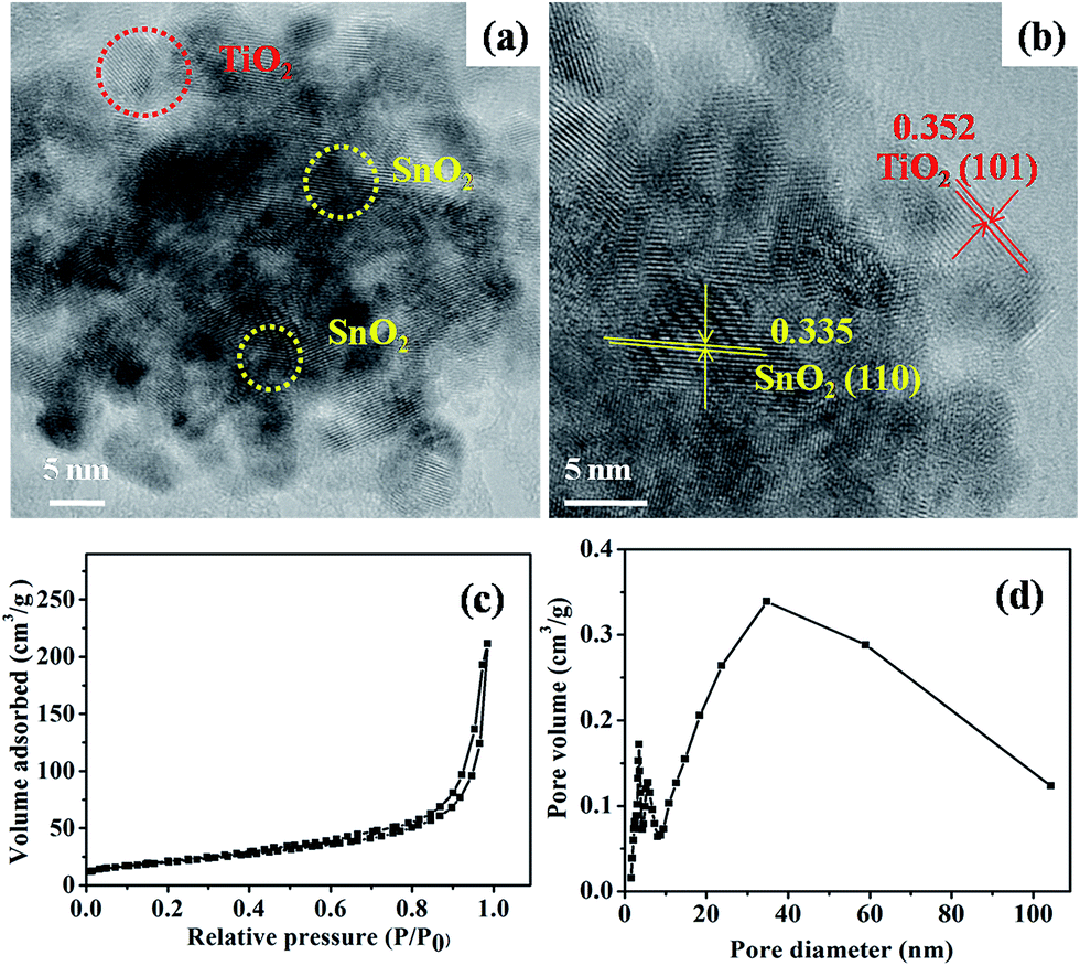 | ||
| Fig. 2 (a and b) HRTEM image, (c) N2 adsorption–desorption isotherm, and (d) the corresponding BJH pore size distribution of SnO2@TiO2-550 product. | ||
XPS measurements were carried out to further investigate the surface composition and chemical state of SnO2@TiO2-550. The typical high-resolution XPS spectral of Ti 2p, Sn 3d and O 1s were presented in Fig. 3. XPS spectra of Sn 3d region were given in Fig. 3b. The peak position corresponding to Sn (3d5/2, 3/2) doublet was observed at about 486.6 and 494.8 eV, which was assigned to Sn4+ species. The Ti 2p XPS spectra depicted in Fig. 3c showed two peaks at about 458.0 and 463.9 eV, which can be assigned to Ti4+ 2p3/2 and Ti4+ 2p1/2, respectively.33 The O 1s XPS spectral (Fig. 3d) were wide and asymmetric, demonstrating that there were at least two kinds of O chemical states. As shown in Fig. 3d, O 1s XPS peak can be divided into two peaks, which may be attributed to Sn–O (531.8 eV)34 and Ti–O (533.2 eV).35
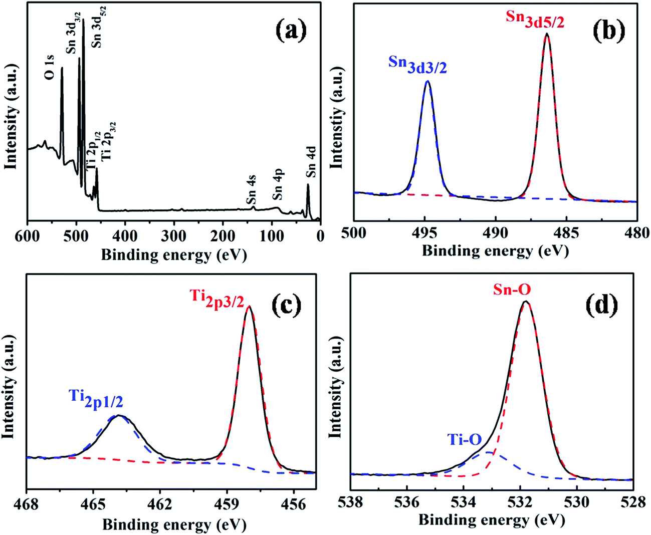 | ||
| Fig. 3 XPS spectra of SnO2@TiO2-550 product. | ||
To insight into the role of carbon, the concentration-dependent experiments were performed, while keeping other conditions constant. The morphology evolution of the products was investigated by TEM technique. At the absence of glucose, the SnO2 product was uniform flower-like morphology, which was composed of a lot of very small nanoparticles (Fig. 4a). Furthermore, plenty spaces of among the nanoparticles could be observed. However, when the glucose/SnSO4 mole ratio increased to 5, the void of among the nanoparticles was indistinct, suggesting the carbon filling (Fig. 4b). While more glucose was added in the reaction system (the glucose/SnSO4 mole ratio increasing from 5 to 10 and 15), the surface of the nanospheres was smoother and thicker, in which the coating carbon layer was 4 nm and 10 nm, respectively. That may be attributed to the pyrolysis of the much glucose during the hydrothermal process. By comparison, we chose the 10 of glucose/SnSO4 mole ratio as the best rate, since the SnO2–C spheres were more dispersed than other products.
 | ||
| Fig. 4 TEM images of as-synthesized SnO2–C products with different glucose/SnSO4 mole ratio: (a) without glucose, (b) 5, (c) 10, and (d) 15. Scale bars: 50 nm. | ||
As reported previously, the calcination temperature had a profound effect on the structure and morphology of the nanomaterials.2,13,36 A series of SnO2@TiO2 nanocomposites were obtained after annealing at the range of temperature from 350 to 750 °C, as shown in Fig. 5. It can be seen in Fig. 5a that once calcined at 350 °C, the small particles were generated and adhered to the spheres compared with the precursor (Fig. 1a). What's more, as increasing the calcination temperature, the amount of the small TiO2 particles became less and less. Upon calcined at 750 °C, the spheres further aggregated and the surface was smoother.
 | ||
| Fig. 5 TEM images of: (a) SnO2@TiO2-350, (b) SnO2@TiO2-450, (c) SnO2@TiO2-650, and (d) SnO2@TiO2-750. Scale bars: 50 nm. | ||
The crystal phase of SnO2@TiO2 nanocomposites was examined by XRD technique. Seen from Fig. 6, the diffraction speaks of all samples were perfectly indexed to the pure tetragonal phase of SnO2 (JCPDS card no. 41-1445). No anatase peaks appeared in the precursor, which may be ascribed to the poor crystallinity of TiO2. Moreover, the intensity of the peaks corresponding to the anatase phase of TiO2 (JCPDS card no. 21-1272) increased with increasing of calcination temperature from 350 to 650 °C, meaning the enhancement of crystallinity in comparsion to the uncalcined sample. However, when the calcination temperature was 750 °C, the crystal phase began to transform from anatase to rutile (JCPDS card no. 21-1276), as marked by asterisk. The phenomenon was common, as reported in previous work.36 Consequently, it could be deduced that the SnO2 tetragonal phase and TiO2 anatase phase coexisted in these nanocomposites, in good agreement with the HRTEM results.
 | ||
| Fig. 6 XRD patterns of various SnO2@TiO2 nanocomposites. | ||
3.2 Photocatalytic properties
As we known, the performance of materials was greatly affected by their intrinsic physicochemical properties including the surface area, crystal phase, particle size, crystallinity, as well as the morphology.37 Considering that, the photocatalytic performances of a series of SnO2@TiO2 samples synthesized at different calcination temperatures were evaluated by the photodegradation of rhodamine B (RhB). Fig. 7 shows the degradation profiles of RhB under UV light over various photocatalysts. The results demonstrated that all SnO2@TiO2 heterostructures calcined possessed excellent properties in photodegradation reactions compared with the uncalcined sample (precursor) owing to the improvement of crystallinity. It was obvious that SnO2@TiO2-550 and SnO2@TiO2-350 catalysts both showed superior photocatalytic performance, the 99.57% and 99.29% of RhB could be degradated within 15 min, respectively. For comparsion, the photocatalytic performance of Degussa P25 was also tested under same conditions, the 99.87% of RhB could be degradated within 15 min, only slightly better than those of SnO2@TiO2 composites, as shown in Fig. 8. The photocatalytic degradation rate could be sequenced as SnO2@TiO2-550 > SnO2@TiO2-350 > SnO2@TiO2-450 > precursor > SnO2@TiO2-650 > SnO2@TiO2-750. The possible reasons will be discussed in subsequent part in detail. In addition, the inset digital images showed the color changes of the resultant liquid from purple to completely transparent colorless with the extension of time, which directly tracked the reaction process. | ||
| Fig. 7 Time-dependent UV-vis spectral changes of RhB aqueous solution in the presence of SnO2@TiO2 nanocomposites: (a) precursor, (b) SnO2@TiO2-350, (c) SnO2@TiO2-450, (d) SnO2@TiO2-550, (e) SnO2@TiO2-650, and (f) SnO2@TiO2-750. | ||
 | ||
| Fig. 8 The comparison of photocatalytic activities among various SnO2@TiO2 catalysts obtained from different calcination temperatures (a), the cycling stability of SnO2@TiO2-550, and TEM (c) images of SnO2@TiO2-550 after cycling 5 times. | ||
More research has down on the difference of photodegradation performance among these products. As described in the experimental part, the remnants RhB concentration (%) after being exposed to light for a specific time was estimated by the following equation:
| %RhB concentration = (1 − Ct/C) × 100% |
 | ||
| Fig. 9 TG curve of the precursor of SnO2@TiO2 nanocomposite (a), and the corresponding digital photos of various products calcined at different temperature (b). | ||
The effect of the carbon from glucose was as the media for the construction of unique core–shell nanostructured SnO2@TiO2 nanocomposites. In addition, the carbon of the SnO2@TiO2-350 and SnO2@TiO2-450 was not completely been removed. Hence, the existence of carbon may improve the separation efficiency of photogenerated electron–hole pairs. As a result, the photocatalytic activity of SnO2@TiO2-350 could be improved. That's why SnO2@TiO2-350 performs better than SnO2@TiO2-450, even though the crystalline improved from 350 to 450 °C. The anatase TiO2 of SnO2@TiO2-550 product has much higher crystallinity than the SnO2@TiO2-350, resulting in better performance in spite of no carbon in the sample. More importantly, the SnO2@TiO2-550 photocatalyst exhibited a good cycling stability, the degradation rates were scarcely changed after 5 cycles, as displayed in Fig. 8b. As was observed in Fig. 8c, the morphology of the product still remained well after 5 cycles. Additionally, the low performance of the SnO2@TiO2-650 and SnO2@TiO2-750 product was related to crystal transformation at 750 °C and the reduction of the specific surface area, which were only 55.4 and 40.0 m2 g−1, respectively.
In order to explore the function of the carbon in the preparation process of the SnO2@TiO2-550 photocatalyst, the photocatalytic properties of pure SnO2 nanoparticles and SnO2@TiO2–N were also measured under same conditions (Fig. 10). As shown in Fig. 10a, the degradation of RhB by the SnO2@TiO2–N was about 40 min, which was much longer than the carbon-mediated SnO2@TiO2-550 of 15 min. And the pure SnO2 nanoparticle was less significant than the other catalysts, and only 68.1% of RhB was degraded after being illuminated for 20 min (Fig. 10c). On one hand, pure SnO2 product has a low excitation efficiency of e−/h+ pairs due to the wide band gap (∼3.6 eV) of SnO2, which leads to a limited absorption of photons. On the other hand, pure SnO2 product has a poor quantum efficiency because of the recombination of photo-generated e−/h+ pairs. For the SnO2@TiO2–N, only 78.3% of RhB was degraded after being illuminated for 20 min. Compared with the SnO2@TiO2–N, the SnO2@TiO2-550 product exhibits higher photocatalytic activity, with a degradation rate of 99.6% for 15 min. As a result, we suspect that the carbon layer may play three roles in the formation of SnO2@TiO2 nanostructures as follows: (I) as morphology controller, (II) improvement the electrical conductivity, (III) as growth-promoting agent so as to construct the heterostructures of SnO2 (tetragonal phase) and TiO2 (anatase phase). The TiO2 Nps with narrow band gap (∼3.2 eV) enhanced the photon absorption and the SnO2 spheres provided a continuous path for the faster transfer of the carrier.
 | ||
| Fig. 10 Time-dependent UV-vis spectral changes of RhB aqueous solution in the presence of SnO2@TiO2 without carbon-mediated (a), the comparison of photocatalytic activities among various catalysts (c); TEM images of SnO2@TiO2–N (b), and SnO2 nanoparticle (d). | ||
Based on the above results, the possible reasons for the superior photocatalytic performance of SnO2@TiO2-550 nanostructures could be explained in detail as follows: (I) the larger specific surface areas (∼105.9 m2 g−1) offer more active sites and absorb more substrates, leading to a higher photocatalytic performance. Moreover, the porous nanostructure and large surface area of the SnO2@TiO2 enhance the mass transfer of RhB and the independence of TiO2 Nps impedes the recombination of photo-generated electrons and holes at the same time.38 (II) SnO2@TiO2 nanocomposites improve the photon utilization efficiency due to their heterostructures. In this band-gap configuration, when photo-generated e−/h+ pairs are generated in SnO2 nanospheres and TiO2 nanoparticles, the electrons on the TiO2 nanoparticles surface transfer swiftly to the conduction band of SnO2 via interfaces; similarly, the holes on the SnO2 surface migrate to TiO2 owing to the different valence band edge (Scheme 2).39 (III) As an unique advantage of our synthetic method, the carbon as the mediate could provide support of the unique structure and conduct the electron, thus enhancing the photocatalytic performance.40
 | ||
| Scheme 2 Schematic diagram for the photodegradation towards RhB over SnO2@TiO2 nanocomposites under UV irradiation. | ||
4. Conclusions
In summary, we present a carbon-mediated approach for preparing the SnO2@TiO2 nanostructures by using SnO2–C nanosphere as the core and TiO2 as the shell, and a subsequent calcination process. After annealing, the TiO2 thin shell was uniformly coated onto the surface of the SnO2 nanoparticles. The property and structure of SnO2@TiO2 nanocomposites were greatly affected by the calcination temperature and the carbon contents. In comparison to the SnO2@TiO2 synthesized without carbon, the SnO2@TiO2 composite exhibited better photocatalytic performance for the decomposition of RhB under UV light irradiation. In particular, carbon-mediated SnO2@TiO2-550 composite processed some excellent properties including a relatively large specific surface area for supplying abundant active sites and unique heterojunctions for charge carriers separation. Consequently, the notable enhancement of photocatalytic performance of the SnO2@TiO2 heterostructures can be attributed to the synergistic effects. We hope that the method of carbon mediation and construction of the heterostructures can be utilized for more potential applications such as battery electrodes, dye sensitization and so on.Acknowledgements
We are grateful to Prof. Shuai Yuan from Shanghai University for kind help. The work is supported by the National Natural Science Foundation of China (11275121, 21471096, 61174011, 21371116), and Program for Innovative Research Team in University (IRT13078).References
- H. Wang, Y. Qu, H. Chen, Z. D. Lin and K. Dai, Highly selective n-butanol gas sensor based on mesoporous SnO2, Sens. Actuators, B, 2014, 201, 153–159 CrossRef CAS PubMed.
- H. J. Zhang, Q. Q. He, X. D. Zhu, D. Y. Pan, X. Y. Deng and Z. Jiao, Surfactant-free solution phase synthesis of monodisperse SnO2 hierarchical nanostructures and gas sensing properties, CrystEngComm, 2012, 14, 3169–3176 RSC.
- A. Bhaskar, M. Deepa and T. N. Rao, Size controlled SnO2 hollow spheres via a template free approach as anodes for lithium ion batteries, Nanoscale, 2014, 6, 10762–10771 RSC.
- K. Kisu, M. Iijima, E. Iwama, M. Saito, Y. Orikasa, W. Naoi and K. Naoi, The origin of anomalous large reversible capacity for SnO2 conversion reaction, J. Mater. Chem. A, 2014, 2, 13058–13068 CAS.
- W. Q. Wu, Y. F. Xu, H. S. Rao, H. L. Feng, C. Y. Su and D. B. Kuang, Constructing 3D branched nanowire coated macroporous metal oxide electrodes with homogeneous or heterogeneous compositions for efficient solar cells, Angew. Chem., Int. Ed., 2014, 126, 4916–4921 CrossRef PubMed.
- J. C. Huo, Y. J. Hu, H. Jiang, W. J. Huang and C. Z. Li, SnO2 nanorod@TiO2 hybrid material for dye sensitized solar cells, J. Mater. Chem. A, 2014, 2, 8266–8272 CAS.
- H. K. Wang, S. Kalytchuk, H. H. Yang, L. F. He, C. Y. Hu, W. Y. Teoh and A. L. Rogach, Hierarchical growth of SnO2 nanostructured films on FTO substrates: structural defects induced by Sn(II) self-doping and their effects on optical and photoelectrochemical properties, Nanoscale, 2014, 6, 6084–6091 RSC.
- J. H. Kim, D. Bhattacharjya and J. S. Yu, Synthesis of hollow TiO2@N-doped carbon with enhanced electrochemical capacitance by anin situ hydrothermal process using hexamethylenetetramine, J. Mater. Chem. A, 2014, 2, 11472–11479 CAS.
- L. Wang, L. Chen, B. Yan, C. G. Wang, F. Zhu, X. F. Jiang, Y. M. Chao and G. Yang, In situ preparation of SnO2@polyaniline nanocomposites and their synergetic structure for high performance supercapacitor, J. Mater. Chem. A, 2014, 2, 8334–8341 CAS.
- X. Zhang, Y. Liu and Z. H. Kang, 3D branched ZnO nanowire arrays decorated with plasmonic Au nanoparticles for high-performance photoelectrochemical water splitting, ACS Appl. Mater. Interfaces, 2014, 6, 4480–4489 CAS.
- J. T. Park, C. S. Lee and J. H. Kim, One-pot synthesis of hierarchical mesoporous SnO2 spheres using a graft copolymer: enhanced photovoltaic and photocatalytic performance, RSC Adv., 2014, 4, 31452–31461 RSC.
- H. S. Zhou, H. J. Zhang, Y. Wang, Y. Miao, L. B. Gu and Z. Jiao, Self-assembly and template-free synthesis of ZnO hierarchical nanostructures and their photocatalytic properties, J. Colloid Interface Sci., 2015, 448, 367–373 CrossRef CAS PubMed.
- H. J. Zhang, G. D. Du, W. Q. Lu, L. L. Cheng, X. D. Zhu and Z. Jiao, Porous TiO2 hollow nanospheres: synthesis, characterization and enhanced photocatalytic properties, CrystEngComm, 2012, 14, 3793–3801 RSC.
- B. Wang, X. Y. Lu, L. K. Yu, J. Xuan, M. K. H. Leung and H. F. Guo, Facile synthesis of TiO2 hollow spheres composed of high percentage of reactive facets for enhanced photocatalytic activity, CrystEngComm, 2014, 16, 10046–10055 RSC.
- M. M. Khan, S. A. Ansari, D. Pradhan, M. O. Ansari, D. H. Han, J. Lee and M. H. Cho, Band gap engineered TiO2 nanoparticles for visible light induced photoelectrochemical and photocatalytic studies, J. Mater. Chem. A, 2014, 2, 637–644 CAS.
- C. L. Wang, J. Y. Liao, Y. B. Zhao and A. Manthiram, Template-free TiO2 hollow submicrospheres embedded with SnO2 nanobeans as a versatile scattering layer for dye-sensitized solar cells, Chem. Commun., 2015, 51, 2848–2850 RSC.
- J. Zhu, M. S. Li, M. H. Lu and J. J. Zhu, Effect of structural properties on catalytic performance in citral selective hydrogenation over carbon–titania composite supported Pd catalyst, Catal. Sci. Technol., 2013, 3, 737–744 CAS.
- Z. Z. Yang, N. Zhang, Y. Cao, M. C. Gong, M. Zhao and Y. Q. Chen, Effect of yttria in Pt/TiO2 on sulfur resistance diesel oxidation catalysts: enhancement of low-temperature activity and stability, Catal. Sci. Technol., 2014, 4, 3032–3043 CAS.
- J. J. Yuan, X. K. Zhang, H. D. Li, K. Wang, S. Y. Gao, Z. Yin, H. J. Yu, X. R. Zhu, Z. Z. Xiong and Y. M. Xie, TiO2/SnO2 double-shelled hollow spheres-highly efficient photocatalyst for the degradation of rhodamine B, Catal. Commun., 2015, 60, 129–133 CrossRef CAS PubMed.
- Y. C. Huang, S. Y. Chang, L. Huang and C. F. Lin, Growing metal trees on tubular semiconductor land: TiO2/(Zn,Sn)Pd heterostructures with high SERS and photocatalytic activity, J. Mater. Chem. A, 2014, 2, 8456–8464 CAS.
- F. Kayaci, S. Vempati, C. O. Akgun, I. Donmez, N. Biyikliab and T. Uyar, Selective isolation of the electron or hole inphotocatalysis: ZnO–TiO2 and TiO2–ZnO core–shell structured heterojunction nanofibers via electrospinning and atomic layer deposition, Nanoscale, 2014, 6, 5735–5745 RSC.
- L. Ciccotti, L. A. S. Vale, T. L. R. Hewerac and R. S. Freire, Fe3O4@TiO2 preparation and catalytic activity in heterogeneous photocatalytic and ozonation processes, Catal. Sci. Technol., 2015, 5, 1143–1152 CAS.
- B. H. Zhou, S. L. Yang, W. Wu, L. L. Sun, M. Lei, J. Pan and X. Xiong, Self-assemble SnO2@TiO2 porous nanowire-nanosheet heterostructures for enhanced photocatalytic property, CrystEngComm, 2014, 16, 10863–10869 RSC.
- X. D. Li, W. Li, M. Li, P. Cui, D. H. Chen, T. Gengenbach, L. H. Chu, H. Y. Liu and G. S. Song, Glucose-assisted synthesis of the hierarchical TiO2 nanowire@MoS2 nanosheet nanocomposite and its synergistic lithium storage performance, J. Mater. Chem. A, 2015, 3, 2762–2769 CAS.
- W. Li, Y. Bai, F. J. Li, C. Liu, K. Y. Chan, X. Feng and X. H. Lu, Core–shell TiO2/C nanofibers as supports for electrocatalytic and synergistic photoelectrocatalytic oxidation of methanol, J. Mater. Chem., 2012, 22, 4025–4031 RSC.
- F. Cui, L. X. Xu, T. Y. Cui, T. J. Yao, J. Yu, X. Zhang and K. N. Sun, Facile synthesis of ultrasmall TiO2 nanocrystals/porous carbon composites in large quantity and their photocatalytic performance under visible light, RSC Adv., 2014, 4, 33408–33415 RSC.
- P. Zhang, C. L. Shao, Z. Y. Zhang, M. Y. Zhang, J. B. Mu, Z. C. Guo and Y. C. Liu, TiO2 @carbon core/shell nanofibers: controllable preparation and enhanced visible photocatalytic properties, Nanoscale, 2011, 3, 2943–2949 RSC.
- S. Chen, Y. L. Xin, Y. Y. Zhou, F. Zhang, Y. R. Ma, H. H. Zhou and L. M. Qi, Branched CNT@SnO2 nanorods@carbon hierarchical heterostructures for lithium ion batteries with high reversibility and rate capability, J. Mater. Chem. A, 2014, 2, 15582–15589 CAS.
- R. Z. Hu, W. Sun, H. Liu, M. Q. Zeng and M. Zhu, The fast filling of nano-SnO2 in CNTs by vacuum absorption: a new approach to realize cyclic durable anodes for lithium ion batteries, Nanoscale, 2013, 5, 11971–11979 RSC.
- B. R. Camacho, H. H. R. Santoyo, J. M. M. Flores and O. A. Martínez, Platinum deposited on TiO2-C and SnO2-C composites for methanol oxidation and oxygen reduction, Electrochim. Acta, 2014, 120, 344–349 CrossRef PubMed.
- Y. Y. Zhou, C. Jo, J. Lee, C. W. Lee, G. J. Qao and S. H. Yoon, Development of novel mesoporous C-TiO2-SnO2 nanocomposites and their application to anode materials in lithium ion secondary batteries, Microporous Mesoporous Mater., 2012, 151, 172–179 CrossRef CAS PubMed.
- J. Z. Chen, L. Yang, Z. X. Zhang, S. H. Fang and S. Hirano, Mesoporous TiO2-Sn@C core–shell microspheres for Li-ion batteries, Chem. Commun., 2013, 49, 2792–2794 RSC.
- J. F. Qian, P. Liu, Y. Xiao, Y. Jiang, Y. L. Cao, X. P. Ai and H. X. Yang, TiO2-coated multilayered SnO2 hollow microspheres for dye-sensitized solar cells, Adv. Mater., 2009, 21, 3663–3667 CrossRef CAS PubMed.
- M. J. Chen, J. P. Yang, Y. Liu, W. Li, J. W. Fan, X. Q. Ran, W. Teng, Y. Sun, W. X. Zhang, G. M. Li, S. X. Dou and D. Y. Zhao, TiO2 interpenetrating networks decorated with SnO2 nanocrystals: enhanced activity of selective catalytic reduction of NO with NH3, J. Mater. Chem. A, 2015, 3, 1405–1409 CAS.
- P. Zhang, C. L. Shao, Z. Y. Zhang, M. Y. Zhang, J. B. Mu, Z. C. Guo, Y. Y. Sun and Y. C. Liu, Core/shell nanofibers of TiO2@carbon embedded by Ag nanoparticles with enhanced visible photocatalytic activity, J. Mater. Chem., 2011, 21, 17746–17753 RSC.
- D. H. Wang, H. J. Zhang, J. X. Guo, H. J. Xu, X. D. Zhu and Z. Jiao, Template-free fabrication of rattle-type TiO2 hollow microspheres with superior photocatalytic performance, RSC Adv., 2014, 4, 37311–37319 RSC.
- D. baiyila, X. H. Wang, X. Li, B. Sharileaodu, X. T. Li, L. Xu, Z. R. Liu, L. M. Duan and J. H. Liu, Electrospun TiO2 nanofibers integrating space-separated magnetic nanoparticles and heterostructures for recoverable and efficient photocatalyst, J. Mater. Chem. A, 2014, 2, 12304–12310 CAS.
- H. J. Zhang, P. P. Xu, G. D. Du, Z. W. Chen, D. Y. Pan, K. Oh and Z. Jiao, A facile one step synthesis of TiO2/graphene composites for photodegradation of methyl orange, Nano Res., 2011, 4, 274–283 CrossRef CAS PubMed.
- Y. Zhao, J. Liu, L. Y. Shi, S. Yuan, J. H. Fang, Z. Y. Wang and M. H. Zhang, Solvothermal preparation of Sn4+ doped anatase TiO2 nanocrystals from peroxo-metal-complex and their photocatalytic activity, Appl. Catal., B, 2011, 103, 436–443 CrossRef CAS PubMed.
- H. Hu, H. Y. Cheng, G. J. Li, J. P. Liu and Y. Yu, Design of SnO2/C hybrid triple layer nanospheres as li-ion battery anodes with high stability and rate capability, J. Mater. Chem. A, 2015, 3, 2748–2755 CAS.
| This journal is © The Royal Society of Chemistry 2015 |