DOI:
10.1039/C5RA08902F
(Paper)
RSC Adv., 2015,
5, 73051-73057
A fluorescence nanosensor for lipase activity: enzyme-regulated quantum dots growth in situ†
Received
13th May 2015
, Accepted 21st August 2015
First published on 21st August 2015
Abstract
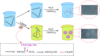
Lipase is a neglected enzyme in the field of quantum dots-based enzyme assays. We have developed a novel analytical assay to detect the lipase activity based on the enzyme-regulated quantum dots growth in situ. Catalytic hydrolytic cleavage of the ester bond in methyl thioglycolate (METG) will produce mercaptoacetate, and Na2S interacts with Cd(NO3)2 to give CdS nanocrystals stabilized by mercaptoacetate. The fluorescence intensity changes, which depends on the absence or presence of lipase, could be used to sense the lipase activity. The detection limit (3σ) as low as 1.2 × 10⁻² mg mL−1 with a linear range from 0.05 to 1.6 mg mL−1 was achieved. A preliminary enzyme activity screening was carried out for four commercial purchased lipase samples, and the results are consistent with ones obtained from titration assay. Moreover, we applied this method to detecting lipase activity in fermentation broth of Bacillus subtilis without any pretreatment. The reported methodology shows potential applications for the development of a simple and inexpensive assay for the hydrolysis enzymes.
1 Introduction
Lipase (E.C.3.1.1.3) has been found to be able to catalyze various hydrolytic and synthetic reactions concerning ester bonds, using nature and synthetic substrates in aqueous and non-aqueous phases and has been widely applied as versatile biocatalyst for ester synthesis, hydrolysis, and interesterification, which makes lipases a good choice for potential applications in the clinic medicine and biochemical engineerings.1–5 As the value of lipases continues to grow, development of the high-sensitivity detection methods that can determine their activity and kinetic parameters is of great importance in the development of novel pharmaceuticals and medical diagnostic devices. It is also important in the biochemical engineering to develop lipases screening tools that can be used to search for the best performance with respect to specific property. The traditional procedures to assay lipase activity, such as titrimetry,6 turbidimetry,7 are tedious and time consuming, and some of instrumental methods, including HPLC,8 GC,9 electrochemical methods,10,11 require either expert operators or expensive instruments and reagents.
Nanoparticles can be used as signal transducers for biosensors because of their remarkable versatility in biological and medical applications,12–14 which ‘will not only yield improved biological sensing but also provide a step change in our ability to probe the biomolecular realm’.15 The nanoparticle-based biosensors have now made it possible to measure minute changes in enzyme activity with high accuracy and precision. However, the nanoparticle-based lipase activity assay is still a virgin ground which has rarely been considered for enzyme assays, because the lipase has not been thoroughly characterized with respect to its biochemical identity and enzymatic catalyzing mechanism.1 A focus on it should not only broaden the spectrum of nanoparticles-based enzyme assays but also stimulate the development of the assays for new targets.
One of the most important advantages of the fluorescent assay is that they allow in vivo imaging.16,17 To date, the majorities of fluorescent enzymatic probes are based on organic fluorophore dyes and require complicated organic synthetic procedure which suffered from laborious synthetic procedures, low sensitivity or complicated detection facilities, thus it severely restricted the extensive applications for sensitive and rapid lipase detection.18,19
Quantum dots (QDs) are a relatively new class of fluorophores with unique photophysical properties that can help address some of the deficiencies encountered by organic fluorophores in bioassays.20,21 Pavlov and his coworkers recently reported a series of detection of enzyme activity based on CdS QDs growth in situ, such as acetylcholinesterase (AChE),22 phosphatase (ALP),23 glucose oxidase (GOx).24
Since 2010, our group has devoted ourself to biosensors for lipase assays based on gold nanoparticles (GNPs),25,26 whose surface plasma resonance (SPR) has been used to develop the colorimetric sensors based on Tween 20 functioned GNPs or one-pot nanoparticle growth. Inspired by Pavlov, we here constructed a novel label-free, sensitive fluorescence nanosensor for detection of lipase activity based on enzyme-controlled CdS QDs growth in situ. The presence of the lipase is the point, and the lipase was just added to the solution containing METG. When the mixture solution was incubated and then added the Na2S and Cd(NO3)2, the CdS QDs were synthesized several minutes later. The QDs solution has significant fluorescence change which could be recorded by fluorescence spectrometer. The relationship between the lipase concentration and the fluorescence intensity makes this approach highly suitable for fabricating a sensitive, simple and cheap sensor for lipases. Furthermore, one of the most important merit is that a one-pot construction process is indeed realized and multiple-step modifications of the QDs when assembling a sensor are omitted. This simple method might be preferable to sophisticated nanoparticle-based enzyme assays, which require complex attachment or linking procedures.
2 Experimental
2.1 Reagents
Cadmium nitrate (Cd(NO3)2), sodium sulfide (Na2S) were purchased from Sigma-Aldrich (Shanghai, China). Mercaptoacetic acid (TGA) and Methyl thioglycolate (METG) were purchased from J&K Chemical Reagent Co., Ltd (Beijing, China). Lipase Novozyme 435 (immobilized on acrylic resin) was purchased from Novozymes. Lipases from Candida rugosa (CRL), Porcine pancreas (PPL) and Rhizopus niveus (RNL), were all ordered from Sigma. Food grade lipase (SBE-01Li) was from Xia Sheng Industrial Group (Beijing, China). Lipase from Bacillus subtilis was fermented by the National Engineering Research Center for Biotechnology in Nanjing Tech University. Enzymatic activity assay based on formation of fluorescent quantum dots were performed in black 96-well microplate (WHB-bio Inc., Shanghai, China), at room temperature (RT). The water used during experiment was ultrapure water (18.2 MΩ cm−1). One unit of lipase hydrolyzes 1.0 μmol of METG per min at pH 8.0 and 35 °C.
2.2 Apparatus
Transmission electron microscopy analysis was performed by using a JEOL JEM-1010 at accelerating voltage of 100 kV (Japan). The samples for TEM characterization were prepared by placing a drop of solution on a 200 mesh carbon-coated copper grid and left to dry in air. Zeta potential was carried out on PALS zeta potential analyzer ver.3.54, Brookhaven Instruments Corp. Ultrapure water was prepared by the Millipore Milli-Q system (Sartorius, Germany). The absorbance and fluorescence spectra were recorded with PerkinElmer Lambda 25 (Thermo Scientific, USA), SpectraMax multi-mode microplate reader M3 (Silicon Valley, USA), respectively.
2.3 Sensing lipase activity
Under the optimized conditions, different concentration of lipases (from 0 to 3.0 mg mL−1) were added to the solution containing 1.5 mM METG in 50 mM Tris–HCl buffer (pH 8.0), and the ultimate volume is 1000 μL. The resulting solution was incubated at 35 °C for 10 min. Then 184 μL of the solution and 10 μL of 25 mM Na2S and 6 μL of 100 mM Cd(NO3)2 were added to each well. Five minutes later, the fluorescence emission spectra were recorded in the wavelength range of 400–620 nm (λex = 350 nm). All measurements were carried out in triplicates; the error bars represent the standard deviation of three independent measurements. The fluorescence intensity of the maximum emission peak was used for the quantitative analysis of lipase, and the fluorescence emission spectrum demonstrates a well-shaped peak at 510 nm which arises from excitonic emission of CdS QDs.27
2.4 Practical application of the strategy
Four commercially purchased lipase samples (RNL, SBE-01Li, PPL and CRL) were used for verifying the practicality of this method. 2.0 mg of different commercial lipase was added into Eppendorf tube, which contained 1.5 mM METG in 50 mM Tris–HCl buffer (pH 8.0), and the ultimate volume is 1000 μL. The resulting solution was incubated at 35 °C for 10 min. We also evaluated whether this method would respond to lipase in living bacterial cells using inactivated and activated fermentation broth of Bacillus subtilis. The enzyme activity was determined by using the protocol for sensing lipase activity.
3 Results and discussion
3.1 Sensing strategy
Scheme 1 shows the sensing strategy for detection of lipase activity. The system includes Cd(NO3)2 and Na2S, methyl thioglycolate (METG) and the lipase, where METG was employed to act as a substrate for lipase. In the presence of lipase, the hydrolytic cleavage of the carboxyl ester bond in METG could produce mercaptoacetate, whose stabilization of in situ growing CdS crystals leads to the generation of fluorescent CdS QDs in the aqueous media and was followed by fluorescence spectroscopy. Whereas, just weak fluorescence signal was observed in the absence of lipase, because the substrates METG could not stabilize the CdS QDs. So the enzymatic hydrolysis-triggered CdS QDs growth in situ was exploited as a platform for fluorescent sensing.
 |
| Scheme 1 Detection of lipase activity by the enzymatic modulation of fluorescent CdS QDs growth in situ. | |
This strategy was performed in a 96 microtiter plate, and it is perfectly suitable for clinical and pharmaceutical application. We can identify lipase with the naked eye by using a commercially available UV lamp (λ = 365 nm), which has distinctive green fluorescence (Fig. 1, inset).
 |
| Fig. 1 UV-Vis absorption (black line) and emission (red line) spectra of CdS QDs produced by the enzymatic hydrolysis of METG. CdS QDs were formed in the presence of lipase (2 mg mL−1), Na2S (1.25 mM) and Cd(NO3)2 (3 mM) in Tris–HCl buffer (50 mM, pH 8.0). Inset: QDs produced in the absence of lipase (image on the left) and in the presence of lipase (image on the right) irradiated by ultraviolet lamp (λ = 365 nm). | |
A number of control experiments were carried out to confirm the suggested mechanism. The main interest was to see how the lipase influenced the CdS QDs growth. Fig. 1 shows UV-Vis absorption and emission spectra of the CdS QDs in presence of the lipase. From the UV-Vis spectrum, we observed a shoulder peak at about 355 nm, which is explained by the excitonic transition between the electron 1S state and the hole 1S state in semiconductor QDs with a diameter about 2.5 nm, according to the work of Peng et al.28 On the other hand, the emission spectrum demonstrates a well-shaped peak at 510 nm which arises from excitonic emission of CdS QDs, indicating a homogeneous distribution of sizes among CdS QDs.29 Transmission Electron Microscopy (TEM) confirms the existence of stable CdS QDs in the reaction mixture (Fig. 2B), and the diameter of formed spherical CdS QDs appeared to be 3 ± 1.75 nm (Fig. S1, ESI†), the main diameter is about 3 nm. When lipase is absent, only weak fluorescence could be observed, and the TEM image revealed that the morphology of the produced nanocrystals was like “nanowire” (Fig. 2A).
 |
| Fig. 2 TEM image of CdS nanoparticles in the (A) absence of lipase; (B) presence of lipase. | |
As shown in Fig. 3, the sample solutions with or without lipase were investigated by fluorescence spectroscopy. In the presence of lipase a significant fluorescence signal was detected and just weak fluorescence signals were observed in absence of lipase (Fig. 3, curve (a and b)). The inactivated lipase, which was deactivated in high temperature (100 °C), was used in those control experiments to ensure the role of the lipase in CdS QDs growth in situ. The fluorescence intensity of the inactivated lipase sample solution (Fig. 3, curve (c)) was the same weak as that without lipase. To demonstrate that the fluorescence signal was caused by CdS QDs, but not by the intrinsic fluorescence of lipase, a control experiment was added (Fig. 3, curve (d)), and the results showed that the active lipase have no fluorescence emission in the visible light wavelength range, the only way to yield fluorescence in our system is the formation of CdS QDs, suggesting that the enzymatic hydrolysis of METG by lipase indeed controlled the growth of the CdS QDs.
 |
| Fig. 3 Emission spectra of the solutions: (a) lipase + METG + Na2S + Cd(NO3)2; (b) METG + Na2S + Cd(NO3)2; (c) inactivated lipase + METG + Na2S + Cd(NO3)2 and (d) lipase. | |
The control experiments were also performed with substituting compounds which have hydrophilic group but no carboxyl group, such as cysteamine and β-mercaptoethanol, to confirm the effect of the carboxyl group on the formation of CdS QDs. The dramatic decrease of the fluorescence intensity (Fig. 4) showed that the presence of amide and hydroxyl group was against the formation of fluorescent CdS QDs, which is consistent with the reported results.30–32
 |
| Fig. 4 Emission spectra of the CdS QDs formed in the presence of the Na2S (1.25 mM), Cd(NO3)2 (3 mM), and TGA (1.5 mM) (a) or different thiols: β-mercaptoethanol (b); cysteamine (c), and without any thiols (d). | |
The zeta potential (ζ), which correlates with the surface charge and the local environment of particles, was measured (Fig. S2, ESI†). The values of the assay solutions were determined to be −14.25 mV and −4.52 mV depending on with and without lipase, demonstrating a decrease in negative charge on the surface of CdS nanoparticles. This implied that the negatively charged residues made by the METG hydrolysis could benefit the formation of CdS QDs.
According to the Kotov's theory,33 we can assumed that the formation of nanowire is based on a crystalline nanowire spontaneously self-assembles from individual nanoparticles. The nanowires formed not through point-initiated vectorial growth but rather by the recrystallization of multiple nanoparticles in a linear aggregate that fused gradually into one crystal. The force capable of producing chain of nanocrystals is believed to be the strong long range attraction. When the lipase is present, the CdS nanocrystals were stabilized by mercaptoacetate anion which came from the hydrolytic cleavage of the carboxyl ester bond in METG, nanowires did not form. However when the lipase is absent, the overall decrease of the stabilizer concentration leads to the decrease of the overall negative charge of the nanoparticles. This reduces the mutual electrostatic repulsion of nanoparticles countering electrostatic attraction and the nanoparticles could fuse together gradually into a nanowire.
3.2 Optimization of the assay conditions
To define the optimal lipase hydrolysis incubation time, kinetic studies of the fluorescence intensity in the presence of various amounts of lipase were carried out (Fig. 5). It was found that the changes of fluorescence intensity F − F0 (where F, F0 are the fluorescence intensity of CdS QDs at 510 nm in the presence and absence of lipase, respectively) increased continuously from a time interval of 0 to 10 min. After 10 min, the F − F0 values showed no significant changes. Therefore, 10 min was selected as the optimal reaction time. Besides, the rate of increase of F − F0 is proportional to the amount of lipase used in the assay system. This indicates that the parameter F − F0 can be used to determine the lipase concentration/activity.
 |
| Fig. 5 Kinetic plots of time-dependent fluorescence intensity values versus those with different concentrations of lipase. | |
Buffer solution can not only affect the fluorescence intensity of QDs, but also affect the interaction between lipase and METG. For this reason, it is important to select an appropriate buffer for assay of lipase activity. From Fig. S3 (ESI†) it can be seen that the fluorescence intensity is optimal, so Tris–HCl was used as buffer solution.
The lipase activity was significantly affected by temperature and pH. Investigations were carried out to optimize the temperature and pH. The results are shown in (Fig. S4A and S4B, ESI†). They imply that lipase has an activity maximum at 35 °C and pH 8.0, which were chosen as the optimal temperature and pH values.
The METG concentration can increase or decrease lipase activity, depending on the nature of cooperativity between the enzyme and its substrate. Na2S is the sulfur source of CdS QDs, its concentration could also affect the size and concentration of CdS QDs, and then the fluorescence intensity. So the effect of the substrate METG concentration was investigated in the range of 0.375–2.25 mM (Fig. S4C, ESI†). 1.5 mM METG was selected as the optimal concentration in the system. The influence of the concentration of Na2S was also explored from 0.2 to 1.5 mM Na2S (Fig. S4D, ESI†). To obtain a high signal, 1.25 mM Na2S was selected.
3.3 Sensitivity of assay
Under the optimized conditions, the fluorescence intensity of the CdS QDs solution was investigated as a function of lipase concentration using fluorescence spectroscopy. For each concentration of lipase, the measurement was repeated three times. As depicted in Fig. 6A, the fluorescence excitation wavelength was 350 nm and the fluorescence intensity of CdS QDs gradually increased at 510 nm with the increasing concentration of lipase from 0 to 3.0 mg mL−1. According to the calibration curve (Fig. 6B), the fluorescence intensity of the samples has a good linear relationship with lipase in the concentration range from 0.05 mg mL−1 to 1.60 mg mL−1 (R2 = 0.9938). The detection limit (3σ) of this assay is as low as 1.2 × 10⁻² mg mL−1. These results were better than our previous report based on gold nanoparticle colorimetric method.25
 |
| Fig. 6 (A) Emission spectra of CdS QDs in the system containing Cd(NO3)2 (3 mM), Na2S (1.25 mM), METG (1.5 mM), and various concentrations of lipase: (a) 0 mg mL−1; (b) 0.05 mg mL−1; (c) 0.2 mg mL−1; (d) 0.4 mg mL−1; (e) 0.6 mg mL−1; (f) 0.8 mg mL−1; (g) 1.0 mg mL−1; (h) 1.2 mg mL−1; (i) 1.4 mg mL−1; (j) 1.6 mg mL−1; (k) 1.8 mg mL−1; (l) 2.0 mg mL−1; (m) 2.5 mg mL−1; (n) 3.0 mg mL−1; (B) calibration curve of lipase at λ = 510 nm (n = 3). | |
3.4 Real sample assays
We conducted a preliminary screening using the developed strategy to test the activity of four commercial purchased lipase samples (RNL, SBE-01Li, PPL and CRL). The fluorescence intensity in the presence of a fixed concentration (2 mg mL−1) of lipase samples was monitored within a fixed time interval (10 min). As shown in Fig. 7A, the fluorescence intensity changes were attributed to the different lipase activities. We concluded that the lipase activity decreases in the following order: CRL > PPL > SBE-01Li > RNL. This order is consistent with the results detected with pH-stat method (Table S1, ESI†). It demonstrates that this assay protocol can be used as an alternative to conventional high throughput screening methods for lipase activity.
 |
| Fig. 7 (A) Plot of the values of fluorescence intensity upon the addition of a fixed concentration (2 mg mL−1) of commercial lipase samples, respectively, and within an interval of 10 min; (B) the fluorescence intensity of the formed CdS QDs upon adding (a) activated and (b) inactivated fermentation broth of Bacillus subtilis. | |
The assay was also applied to fermentation broth of Bacillus subtilis, which is an important lipase-producing microbe.34,35 The supernatant of the crude sample without any pretreatment was incubated with METG in Tris–HCl buffer solution for 10 min, and then with Na2S and Cd(NO3)2. A pronounced fluorescence intensity was observed from Fig. 7B, but the inactivated sample solution, which was deactivated by boiling (100 °C), was no fluorescence signals. This indicates that this protocol provides a useful probing approach for real samples that usually have significant background noise.
4 Conclusions
It was discovered for the first time that lipase can catalyze the hydrolysis of carboxyl-containing substrates METG. The negatively charged hydrolytic product could benefit the formation of fluorescent CdS QDs, which can be employed to design novel fluorescent lipase assays. This proposed lipase assay is cost-effective, convenient and does not require any complicated procedures for the synthesis of fluorescent probes or enzymatic substrates. Even if CdS QDs cannot be used for in vivo detection of biomolecules due to its toxicity,36,37 QDs-based assays still have an advantage over many colorimetric and even FRET assays. The challenge is to replace CdS QDs while maintaining excellent properties. The research work is carrying on finding the alternatives which surpass CdS QDs in terms of optical quality and biofunctionalization.
Acknowledgements
This work was supported by the Natural Science Foundation of Jiangsu Province, China (No. BK2012822), the National Natural Science Foundation of China for Young Scholars (No. 61205195), the Specialized Research Fund for Doctoral College (No. 20123223120011), the Foundation for Distinguished Young Scholars of Jiangsu Province (No. 55135011), the Foundation for Distinguished Professor of Jiangsu Province (No. 54935006), and the Scientific Research Startup Foundation of Nanjing Tech University (No. 3983500115).
Notes and references
- F. Hasan, A. A. Shah and A. Hameed, Biotechnol. Adv., 2009, 27, 782–798 CrossRef CAS PubMed.
- M. H. Sörensen, J. B. S. Ng, L. Bergström and P. C. A. Alberius, J. Colloid Interface Sci., 2010, 343, 359–365 CrossRef PubMed.
- X. Lu, Z. Luo, S. Yu and X. Fu, J. Agric. Food Chem., 2012, 60, 9273–9279 CrossRef CAS PubMed.
- G. Yang, J. Wu, G. Xu and L. Yang, Colloids Surf., B, 2010, 78, 351–356 CrossRef CAS PubMed.
- D. Zhao, C. Peng and J. Zhou, Phys. Chem. Chem. Phys., 2015, 17, 840–850 RSC.
- F. Ferrato, F. Carriere, L. Sarda and R. Verger, Methods Enzymol., 1997, 286, 327–347 CAS.
- F. Plou, M. Ferrer, O. Nuero, M. Calvo, M. Alcalde, F. Reyes and A. Ballesteros, Biotechnol. Tech., 1998, 12, 183–186 CrossRef CAS.
- V. Maurich, M. Zacchigna and A. Pitotti, J. Chromatogr. B: Biomed. Sci. Appl., 1991, 566, 453–459 CrossRef CAS.
- A. Louwrier, G. J. Drtina and A. M. Klibanov, Biotechnol. Bioeng., 1996, 50, 1–5 CrossRef CAS.
- I. Ignatjev, G. Valincius, I. Švedaitė, E. Gaidamauskas, M. Kažemėkaite, V. Razumas and A. Svendsen, Anal. Biochem., 2005, 344, 275–277 CrossRef CAS PubMed.
- G. Valincius, I. Ignatjev, G. Niaura, M. Kažemėkaitė, Z. Talaikytė, V. Razumas and A. Svendsen, Anal. Chem., 2005, 77, 2632–2636 CrossRef CAS PubMed.
- X. Luo, A. Morrin, A. J. Killard and M. R. Smyth, Electroanalysis, 2006, 18, 319–326 CrossRef CAS PubMed.
- K. Saha, S. S. Agasti, C. Kim, X. Li and V. M. Rotello, Chem. Rev., 2012, 112, 2739–2779 CrossRef CAS PubMed.
- E. Hutter and D. Maysinger, Trends Pharmacol. Sci., 2013, 34, 497–507 CrossRef CAS PubMed.
- P. D. Howes, R. Chandrawati and M. M. Stevens, Science, 2014, 346, 1247390–12473910 CrossRef PubMed.
- I. L. Medintz, H. T. Uyeda, E. R. Goldman and H. Mattoussi, Nat. Mater., 2005, 4, 435–446 CrossRef CAS PubMed.
- K. D. Wegner and N. Hildebrandt, Chem. Soc. Rev., 2015, 44, 4792–4834 RSC.
- J. F. Jette and E. Ziomek, Anal. Biochem., 1994, 219, 256–260 CrossRef CAS PubMed.
- D. Resina, A. Serrano, F. Valero and P. Ferrer, J. Biotechnol., 2004, 109, 103–113 CrossRef CAS PubMed.
- V. Pavlov, Part. Part. Syst. Charact., 2014, 31, 36–45 CrossRef CAS PubMed.
- W. R. Algar, K. Susumu, J. B. Delehanty and I. L. Medintz, Anal. Chem., 2011, 83, 8826–8837 CrossRef CAS PubMed.
- L. Saa, A. Virel, J. Sanchez-Lopez and V. Pavlov, Chem.–Eur. J., 2010, 16, 6187–6192 CrossRef CAS PubMed.
- N. Malashikhina, G. Garai-Ibabe and V. Pavlov, Anal. Chem., 2013, 85, 6866–6870 CrossRef CAS PubMed.
- L. Saa and V. Pavlov, Small, 2012, 8, 3449–3455 CrossRef CAS PubMed.
- W. Zhang, Y. Tang, J. Liu, L. Jiang, W. Huang, F.-W. Huo and D. Tian, J. Agric. Food Chem., 2015, 63, 39–42 CrossRef CAS PubMed.
- Y. Tang, W. Zhang, J. Liu, L. Zhang, W. Huang, F. Huo and D. Tian, Nanoscale, 2015, 7, 6039–6044 RSC.
- L. Saa, J. M. Mato and V. Pavlov, Anal. Chem., 2012, 84, 8961–8965 CAS.
- W. W. Yu, L. Qu, W. Guo and X. Peng, Chem. Mater., 2003, 15, 2854–2860 CrossRef CAS.
- P. Mandal, S. S. Talwar, S. S. Major and R. S. Srinivasa, J. Chem. Phys., 2008, 128, 114703–114707 CrossRef CAS PubMed.
- G. Garai-Ibabe, M. Möller and V. Pavlov, Anal. Chem., 2012, 84, 8033–8037 CrossRef CAS PubMed.
- C. Landes, C. Burda, M. Braun and M. A. El-Sayed, J. Phys. Chem. B, 2001, 105, 2981–2986 CrossRef CAS.
- J. R. Cowdery-Corvan, D. G. Whitten and G. L. McLendon, Chem. Phys., 1993, 176, 377–386 CrossRef CAS.
- Z. Tang, N. A. Kotov and M. Giersig, Science, 2002, 297, 237–240 CrossRef CAS PubMed.
- J. Ma, Z. Zhang, B. Wang, X. Kong, Y. Wang, S. Cao and Y. Feng, Protein Expression Purif., 2006, 45, 22–29 CrossRef CAS PubMed.
- R. Gupta, N. Gupta and P. Rathi, Appl. Microbiol. Biotechnol., 2004, 64, 763–781 CrossRef CAS PubMed.
- R. Hardman, Environ. Health Perspect., 2006, 114, 165–172 CrossRef.
- C. E. Bradburne, J. B. Delehanty, K. Boeneman Gemmill, B. C. Mei, H. Mattoussi, K. Susumu, J. B. Blanco-Canosa, P. E. Dawson and I. L. Medintz, Bioconjugate Chem., 2013, 24, 1570–1583 CrossRef CAS PubMed.
Footnotes |
† Electronic supplementary information (ESI) available: See DOI: 10.1039/c5ra08902f |
‡ These authors have contributed equally. |
|
This journal is © The Royal Society of Chemistry 2015 |
Click here to see how this site uses Cookies. View our privacy policy here.