
Robust polylactide nanofibrous membranes by gelation/crystallization from solution
Qingxian Liu,
Ruihua Lv,
Bing Na* and
Yunhui Ju
Fundamental Science on Radioactive Geology and Exploration Technology Laboratory, Faculty of Chemistry, Biology and Materials Science, East China Institute of Technology, Nanchang, 330013, People's Republic of China. E-mail: bna@ecit.edu.cn; bingnash@163.com; Fax: +86 791 83897982
First published on 25th June 2015
Abstract
Most porous membranes are fabricated from petroleum-based polymers without biodegradation, which could pose environmental problems in the disposal stage after use. In this case biodegradable polylactide (PLA) is adopted to produce porous membranes by gelation/crystallization from solution. Abundant nanofibers prevail in the resulting porous membranes, which becomes remarkable with the decrease in the gelation/crystallization temperatures and PLA concentration in solutions. The nanofibers in the porous membranes are well interconnected with each other which is responsible for superior mechanical properties. Notably, very high rejection towards bovine serum albumin is realized by the nanofibrous membranes.
1. Introduction
Porous membranes made from polymers are widely used as separation materials in many applications, ranging from water purification to biomedical treatment.1–6 The separation is mainly based on size exclusion, and only substances with sizes larger than the pore size of the membranes can be rejected. Therefore, morphology tailoring is very critical for the separation application of porous membranes.The phase inversion technique is very popular to fabricate porous membranes from a variety of polymers. It relies on the phase separation of polymer solutions induced by nonsolvents or thermal gradients. The morphology of membranes via nonsolvent induced phase separation is asymmetric, consisting of a very thin separated layer on the surface and large finger-like pores in the bulk.7–11 As a comparison, phase separation induced by thermal gradients yields membranes with nearly symmetric morphology, where pores are penetrated from the surface to bulk.12–15 For crystalline polymers crystallization usually occurs during thermally induced phase separation, which to a large extent dictates the morphology of membranes. Poor mechanical properties, with tensile strength in the order of several MPa, is the main drawback of porous membranes obtained by thermally induced phase separation.13–15
To date, most of porous membranes are produced from petroleum-based polymers without biodegradation, for instance poly(vinylidene fluoride) and its hybrid materials. It could pose environmental problems in the disposal stage after use. For this reason, biodegradable polymers are good candidates to fabricate porous membranes with replacement of petroleum-based polymers. Polylactide (PLA), derived from renewable resources, is the mostly investigated biodegradable polyester due to its superior properties.16–18 Porous PLA membranes have been obtained by phase inversion technique.19–22 Most of researches concentrated in the application of porous PLA membranes as the scaffolds for tissue engineering, whereas their separation performance was less reported.
In this study, robust PLA nanofibrous membranes were produced by gelation/crystallization of the solutions at low temperatures, i.e. thermally induced phase separation. Very high rejection towards bovine serum albumin was realized by the nanofibrous membranes from the solutions with a PLA concentration of 100 mg ml−1 after gelation/crystallization at −18 °C. Moreover, superior mechanical properties were exhibited by these nanofibrous PLA membranes.
2. Experimental section
2.1. Materials
Poly(L-lactide), abbreviated as PLA, was supplied by Changchun Sinobiomaterials Co., Ltd, China, and had a viscosity-average molecular weight of 193 kg mol−1. N,N-Dimethylformamide (DMF) and ethanol were analytical reagents and used as received. Bovine serum albumin (BSA) was purchased from Shanghai BlueGene Biotech Co. Ltd, China, and its purity was better than 99%.2.2. Membrane preparation
A certain amount of PLA was directly dissolved in DMF at 80 °C to form transparent solutions; the PLA concentration in the solutions was 50, 100 and 150 mg ml−1, respectively. After removal of air bubbles the solutions were cast onto a glass plate with controllable thickness, followed by covered by the other glass plate to form a fully sealed sandwich. Immediately, the sandwich was placed for 4 h in a chamber with a preset temperature of 25, 4, and −18 °C, respectively. Through this treatment transparent solutions were transformed into opaque gels. Membranes were obtained by immersion of the gels in a large amount of ethanol overnight and subsequent evaporation of the solvents under slight pressure at room temperature.2.3. Characterizations
The morphology was probed by a Nova NanoSEM 450 scanning electron microscope (SEM). The cross-section of membranes was obtained by cryo-fracture in liquid nitrogen. Prior to observations a thin gold layer was sputtered on the surface of specimens. Crystallization behavior of the solutions was recorded by a TA Q2000 differential scanning calorimetry (DSC) instrument under a flowing nitrogen atmosphere at a cooling rate of 10 °C min−1. Samples were tightly sealed in aluminum pans for liquids measurement to avoid possible solvent evaporation. Thermal properties of membranes were obtained by the DSC instrument at a heating rate of 10 °C min−1. The crystalline structure was revealed by a Bruker D8 ADVANCE X-ray diffractometer (XRD) at room temperature; and the wavelength of the X-ray was 0.154 nm. Mechanical measurements were conducted by an universal testing machine at 5 mm min−1 at room temperature; and five specimens were tested for each sample. Porosity was deduced by the weight of the membranes before and after saturated uptake of an inert liquid (silicon oil); the reported porosity was averaged over three measurements.2.4. Performance evaluation of membranes
Water flux and protein rejection were measured by a stainless cell with an effective area of 23.75 cm2 at a constant feed under an operation pressure of 0.2 MPa. After running for 30 min the permeat was collected; and water flux was deduced from the volume of the permeat divided by the area of measured membranes and the operation period. The protein rejection was evaluated by filtration of BSA aqueous solution with a concentration of 0.5 mg ml−1. The concentration of BSA aqueous solutions before and after filtration was determined by a UV757CRT ultraviolet-visible spectrometer (Shanghai Scientific Instruments Co., Ltd, China). The protein rejection was calculated from the absorbance around 280 nm of the BSA aqueous solutions before and after filtration. Three specimens were measured and their average result was reported for both water flux and BSA rejection.3. Results and discussion
PLA can be easily dissolved in chloroform or dichloromethane at room temperature to generate a transparent solution. However, the solution is stable and no gelation/crystallization occurs even at low temperatures such as −18 °C. On the contrary, DMF is not a good solvent for PLA due to much difference in the solubility parameters. The complete dissolution of PLA in DMF can only be achieved at high temperatures, e.g. 80 °C. Upon cooling PLA tends to crystallize from the solutions, responsible for the appearance of an exothermic peak in the DSC traces. As an example, Fig. 1 gives DSC cooling trace of the solutions with a PLA concentration of 100 mg ml−1. An exothermic peak around 23 °C arises from crystallization of PLA in the solutions.23 Of note, in the measured temperature range DMF remains liquid state and no phase transition is expected.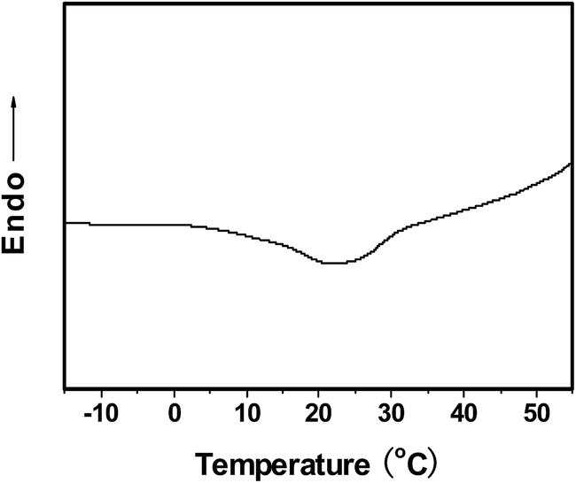 | ||
| Fig. 1 DSC cooling trace of the solutions with a PLA concentration of 100 mg ml−1. | ||
At the same time, crystallization of PLA in the solutions results in the formation of opaque gels. Fig. 2a shows optical photographs of the solutions with a PLA concentration of 100 mg ml−1 before and after being stored in vials at various temperatures. As compared to the solutions, gels cannot flow under gravity because of physical cross-links from PLA crystallites.24 Moreover, gels appear opaque due to significant light scattering by PLA crystallites, whereas the solutions remain transparent. The gelation temperatures seem to have little influence on the visual appearance of gels. To fabricate gels with flat configuration, the solutions were tightly sandwiched between a pair of glass plates, followed by gelation/crystallization at low temperatures. Fig. 2b gives an example of flat gels from the solutions with a PLA concentration of 100 mg ml−1 by gelation/crystallization at −18 °C.
 | ||
| Fig. 2 Optical photographs revealing gelation of the solutions with a PLA concentration of 100 mg ml−1. (a) Sol–gel transition with respect to temperatures, (b) flat gels generated at −18 °C. | ||
After removal of DMF flat gels were transformed into PLA membranes, as demonstrated by the optical photograph in Fig. 3a as an inset. The PLA membranes are crystalline, irrespective of gelation/crystallization temperatures. It is confirmed by the XRD profiles and DSC heating traces in Fig. 3. All membranes exhibit significant diffraction peaks from of (200)/(110) and (203) crystal planes of PLA crystallites.25,26 Moreover, the deduced crystallinity Xc from the XRD profiles is nearly same for all membranes within the experimental errors. The XRD results suggest that crystal form and crystallization degree of PLA has little to do with gelation/crystallization temperatures. On the other hand, melting behaviors of PLA crystals in the membranes depend on gelation/crystallization temperatures. According to DSC heating traces, PLA crystals have one endothermic peak in the membranes from the solutions after gelation/crystallization at −18 and 4 °C, respectively. In contrast, two endothermic peaks are exhibited by the membranes from the solutions after gelation/crystallization at 25 °C. It could arise from melting of two kinds of crystals with different extent of perfection.
 | ||
| Fig. 3 (a) XRD profiles and (b) DSC heating traces of membranes by gelation/crystallization of the solutions with a PLA concentration of 100 mg ml−1 at the indicated temperatures. The deduced crystallinity is labeled in the legend, and the inset is the typical optical photograph of membranes. | ||
Fig. 4 depicts SEM micrographs of membranes obtained from the solutions with a PLA concentration of 100 mg ml−1 by gelation/crystallization at various temperatures. Numerous nanofiber are generated in the membranes after gelation/crystallization at −18 °C. Moreover, nanofibers penetrate throughout the membranes, from the surface to bulk. The nanofibers tend to become large with the increasing of gelation/crystallization temperatures. What is worse, microscale objects, in addition to nanofibers, are observed in the membranes after gelation/crystallization at 25 °C. Nanofibrous morphology is related to lamellar branching of PLA crystals during gelation/crystallization, as indicated by the white circle in Fig. 4a. Lamellar branching becomes significant at low crystallization temperatures due to high nucleation density. This is the reason why nanofibers are overwhelming in the membranes by gelation/crystallization at −18 °C. In contrast, at higher temperatures nucleation and lamellar branching are inhibited to some extent, responsible for larger nanofibers and even microscale objects. On the other hand, remarkable pores prevail among nanofibers and penetrate throughout the membranes due to removal of DMF. Pore size is increased with gelation/crystallization temperatures, which is correlated with the size of nanofibers. Large nanofibers are liable for loose packing to yield large pores, and vice versa.
 | ||
| Fig. 4 SEM micrographs revealing (a–c) surface and (a′–c′) cross-section of membranes by gelation/crystallization of the solutions with a PLA concentration of 100 mg ml−1 at various temperatures: (a and a′) −18 °C, (b and b′) 4 °C and (c and c′) 25 °C. Scale bar, 10 μm. The insets are global view of cross-sections (scale bar, 100 μm). | ||
The morphology of membranes is also affected by PLA concentration in the solutions, as demonstrated by SEM micrographs in Fig. 5. The membranes were produced by gelation/crystallization of the solutions at −18 °C. Nanofibers prevail in all membranes from surface to bulk, irrespective of PLA concentration in the solutions. The size of nanofibers is generally increased with the increasing of PLA concentration in the solutions. It could arise from the overgrowth of nanofibers in the solutions with high PLA concentrations during gelation/crystallization. At the same time, pore size in the membranes seems not to follow the same tendency as the size of nanofibers. As an exception, relatively large pores are generated in the membranes from the solutions with a PLA concentration of 50 mg ml−1. It should be correlated with loose packing of nanofibers induced by high DMF content in the gels. The above results confirm that fibrous PLA membranes are indeed produced and their morphology depends on gelation/crystallization temperatures as well as PLA concentration in the solutions.
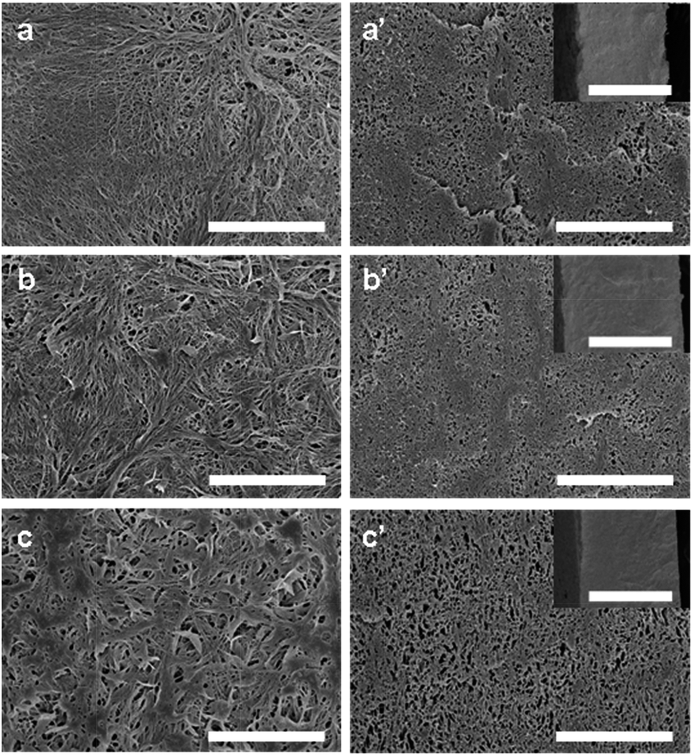 | ||
| Fig. 5 SEM micrographs revealing (a–c) surface and (a′–c′) cross-section of membranes by gelation/crystallization of the solutions with various PLA concentration at −18 °C: (a and a′) 50 mg ml−1, (b and b′) 100 mg ml−1 and (c and c′) 150 mg ml−1. Scale bar, 10 μm. The insets are global view of cross-sections (scale bar, 100 μm). | ||
To obtain further information about pores, porosity of the membranes was measured by weight method. The corresponding results are collected in Fig. 6. For the membranes obtained from the solutions with a PLA concentration of 100 mg ml−1, porosity is increased with gelation/crystallization temperatures (Fig. 6a). On the other hand, porosity in the membranes by gelation/crystallization at −18 °C does not vary monotonously with respect to PLA concentration in the solutions. Lowest porosity is exhibited by the membranes from the solutions with a PLA concentration of 100 mg ml−1. The variation in the porosity of membranes is consistent with the morphological observations in Fig. 4 and 5. That is, large pores correspond to high porosity, and vice versa.
 | ||
| Fig. 6 Variation in the porosity of membranes with respect to (a) gelation/crystallization temperatures and (b) PLA concentration in the solutions, respectively. | ||
The mechanical properties of membranes also depend on gelation/crystallization temperatures as well as PLA concentration in the solutions. As shown in Fig. 7, mechanical properties reach maximum in the membranes from the solutions with a PLA concentration of 100 mg ml−1 by gelation/crystallization at −18 °C. The yield strength and strain at break of these membranes are 16.1 MPa and 0.17, respectively. The mechanical properties are much superior to those of previous membranes by thermally-induced phase separation.13–15 It suggests that good interconnection among nanofibers is achieved in these membranes. In contrast, brittle fracture with lower strength is exhibited by the membranes obtained from other gelation/crystallization temperatures or PLA concentration in the solutions. Generally, mechanical properties are dominated by the pore size and porosity of the membranes. Large pores and high porosity tend to induce immature fracture of membranes during stretching because of severe stress concentration.
 | ||
| Fig. 7 Variation in the yield strength and strain at break of membranes with respect to (a) gelation/crystallization temperatures and (b) PLA concentration in the solutions, respectively. | ||
Fig. 8 depicts water flux and BSA rejection of membranes with respect to gelation/crystallization temperatures and PLA concentration in the solutions. Water flux and BSA rejection follow opposite tendency for all membranes, which is related to pore size and porosity. Large pores and high porosity benefit the increase in water flux but reduce BSA rejection, and vice versa. It is the fact in this case while pore size and porosity of the membranes are taken into account. Impressively, very high BSA rejection up to about 95% is realized by the membranes obtained from the solutions with a PLA concentration of 100 mg ml−1 after gelation/crystallization at −18 °C. It suggests that these porous membranes could be used for protein separation with further considering their superior mechanical properties. What is more, the biodegradability of PLA makes it easy to dispose these porous membranes after use. It is obviously advantageous over porous membranes from petroleum-based polymers without biodegradation.
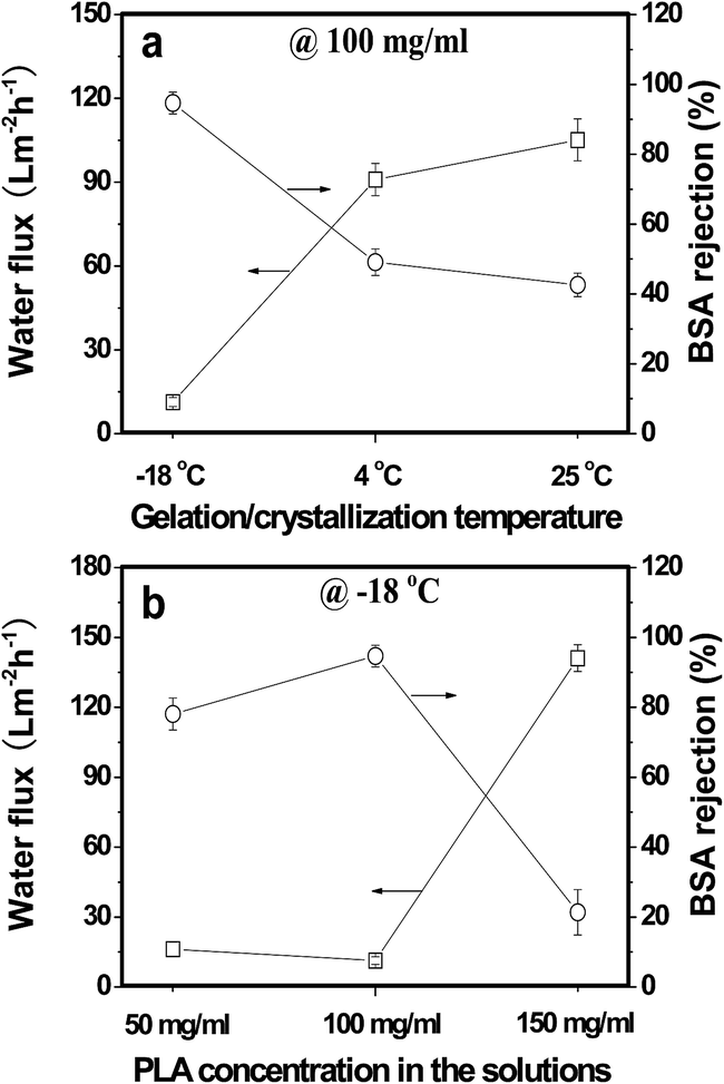 | ||
| Fig. 8 Variation in the water flux and BSA rejection of membranes with respect to (a) gelation/crystallization temperatures and (b) PLA concentration in the solutions, respectively. | ||
4. Conclusion
Porous PLA membranes consisting of nanofibers are produced by gelation/crystallization from the solutions at low temperatures. Formation of nanofibers in the porous membranes is related to lamellar branching of PLA crystals, depending on gelation/crystallization temperatures and PLA concentration in the solutions. It to a large extent accounts for the variation in the morphology of the membranes with respect to fabrication conditions. Good interconnection among nanofibers is achieved in the porous membranes, responsible for superior mechanical properties. Moreover, nanofibrous membranes show very high rejection towards bovine serum albumin because of extremely small pore size. The biodegradability of PLA makes it easy to dispose nanofibrous membranes after use without involvement of environmental problems.Acknowledgements
This work is financially supported by the Major Program of Natural Science Foundation of Jiangxi, China (no. 20133ACB21006), the National Natural Science Foundation of China (no. 21364001) and the Program for Young Scientists of Jiangxi Province (no. 20112BCB23023).References
- D. Rana and T. Matsuura, Surface Modifications for Antifouling Membranes, Chem. Rev., 2010, 110, 2448–2471 CrossRef CAS PubMed.
- M. G. Buonomenna, Membrane processes for a sustainable industrial growth, RSC Adv., 2013, 3, 5694–5740 RSC.
- B. S. Lalia, V. Kochkodan, R. Hashaikeh and N. Hilal, A review on membrane fabrication: Structure, properties and performance relationship, Desalination, 2013, 326, 77–95 CrossRef CAS PubMed.
- Z. Wang, W. Yu and C. Zhou, Preparation of polyethylene microporous membranes with high water permeability from thermally induced multiple phase transitions, Polymer, 2015, 56, 535–544 CrossRef CAS PubMed.
- G. M. Geise, H. S. Lee, D. J. Miller, B. D. Freeman, J. E. Mcgrath and D. R. Paul, Water Purification by Membranes: The Role of Polymer Science, J. Polym. Sci., Part B: Polym. Phys., 2010, 48, 1685–1718 CrossRef CAS PubMed.
- S. Xia and M. Ni, Preparation of poly(vinylidene fluoride) membranes with graphene oxide addition for natural organic matter removal, J. Membr. Sci., 2015, 473, 54–62 CrossRef CAS PubMed.
- P. Wang, J. Ma, Z. Wang, F. Shi and Q. Liu, Enhanced Separation Performance of PVDF/PVP-g-MMT Nanocomposite Ultrafiltration Membrane Based on the NVP-Grafted Polymerization Modification of Montmorillonite (MMT), Langmuir, 2012, 28, 4776–4786 CrossRef CAS PubMed.
- S. Liang, Y. Kang, A. Tiraferri, E. P. Giannelis, X. Huang and M. Elimelech, Highly Hydrophilic Polyvinylidene Fluoride (PVDF) Ultrafiltration Membranes via Postfabrication Grafting of Surface-Tailored Silica Nanoparticles, ACS Appl. Mater. Interfaces, 2013, 5, 6694–6703 CAS.
- H. Wu, B. Tang and P. Wu, Novel Hollow Mesoporous Silica Spheres/Polymer Hybrid Membrane for Ultrafiltration, J. Phys. Chem. C, 2012, 116, 2246–2252 CAS.
- X. Chang, Z. Wang, S. Quan, Y. Xu, Z. Jiang and L. Shao, Exploring the synergetic effects of graphene oxide (GO) and polyvinylpyrrodione (PVP) on poly(vinylylidenefluoride) (PVDF) ultrafiltration membrane performance, Appl. Surf. Sci., 2014, 316, 537–548 CrossRef CAS PubMed.
- S. H. Zhi, J. Xu, R. Deng, L. S. Wan and Z. K. Xu, Poly(vinylidene fluoride) ultrafiltration membranes containing hybrid silica nanoparticles: Preparation, characterization and performance, Polymer, 2014, 55, 1333–1340 CrossRef CAS PubMed.
- L. Wu and J. Sun, Structure and properties of PVDF membrane with PES-C addition via thermally induced phase separation process, Appl. Surf. Sci., 2014, 322, 101–110 CrossRef CAS PubMed.
- H. P. Xu, W. Z. Lang, X. Yan, X. Zhang and Y. J. Guo, Preparation and characterizations of poly(vinylidene fluoride)/oxidized multi-wall carbon nanotube membranes with bi-continuous structure by thermally induced phase separation method, J. Membr. Sci., 2014, 467, 142–152 CrossRef CAS PubMed.
- Z. Cui, N. T. Hassankiadeh, S. Y. Lee, J. M. Lee, K. T. Woo, A. Sanguineti, V. Arcella, Y. M. Lee and E. Drioli, Poly(vinylidene fluoride) membrane preparation with an environmental diluent via thermally induced phase separation, J. Membr. Sci., 2013, 444, 223–236 CrossRef CAS PubMed.
- M. Liu, Y. M. Wei, Z. L. Xu, R. Q. Guo and L. B. Zhao, Preparation and characterization of polyethersulfone microporous membrane via thermally induced phase separation with low critical solution temperature system, J. Membr. Sci., 2013, 437, 169–178 CrossRef CAS PubMed.
- Y. Ikada and H. Tsuji, Biodegradable polyesters for medical and ecological applications, Macromol. Rapid Commun., 2000, 21, 117–132 CrossRef CAS.
- H. Tian, Z. Tang, X. Zhuang, X. Chen and X. Jing, Biodegradable synthetic polymers: Preparation, functionalization and biomedical application, Prog. Polym. Sci., 2012, 37, 237–280 CrossRef CAS PubMed.
- R. Tian, P. Zhang, R. Lv, B. Na, Q. Liu and Y. Ju, Formation of highly porous structure in the electrospun polylactide fibers by swelling–crystallization in poor solvents, RSC Adv., 2015, 5, 37539–37544 RSC.
- C. Tu, Q. Cai, J. Yang, Y. Wan, J. Bei and S. Wang, The Fabrication and Characterization of Poly(lactic acid) Scaffolds for Tissue Engineering by Improved Solid–Liquid Phase Separation, Polym. Adv. Technol., 2003, 14, 565–573 CrossRef CAS PubMed.
- Z. Ma, C. Gao, Y. Gong and J. Shen, Cartilage tissue engineering PLLA scaffold with surface immobilized collagen and basic fibroblast growth factor, Biomaterials, 2005, 26, 1253–1259 CrossRef CAS PubMed.
- A. Gao, F. Liu and L. Xue, Preparation and evaluation of heparin-immobilized poly(lactic acid) (PLA) membrane for hemodialysis, J. Membr. Sci., 2014, 452, 390–399 CrossRef CAS PubMed.
- P. van de Witte, H. Esselbrucce, P. J. Dijkstra, J. W. A. van den Berg and J. A. Feijen, Morphological Study of Membranes Obtained from the Systems Polylactide–Dioxane–Methanol, Polylactide–Dioxane–Water, and Polylactide–N-Methyl Pyrrolidone–Water, J. Polym. Sci., Part B: Polym. Phys., 1996, 34, 2569–2578 CrossRef CAS.
- P. van de Witte, A. Boorsma, H. Esselbrugge, P. J. Dijkstra, J. W. A. van den Berg and J. Feijen, Differential Scanning Calorimetry Study of Phase Transitions in Poly(lactide)–Chloroform–Methanol Systems, Macromolecules, 1996, 29, 212–219 CrossRef CAS.
- J. Zhu, B. Na, R. Lv and C. Li, Enhanced stereocomplex formation of high-molecular-weight polylactides by gelation in an ionic liquid, Polym. Int., 2014, 63, 1101–1104 CrossRef CAS PubMed.
- B. Na, N. Tian, R. Lv, Z. Li, W. Xu and Q. Fu, Evidence of sequential ordering during cold crystallization of poly(L-lactide), Polymer, 2010, 51, 563–567 CrossRef CAS PubMed.
- J. Zhang, Y. Duan, H. Sato, H. Tsuji, I. Noda, S. Yan and Y. Ozaki, Crystal Modifications and Thermal Behavior of Poly(L-lactic acid) Revealed by Infrared Spectroscopy, Macromolecules, 2005, 38, 8012–8021 CrossRef CAS.
| This journal is © The Royal Society of Chemistry 2015 |