One step hydrothermal synthesis of Mn3O4/graphene composites with great electrochemical properties for lithium-ion batteries

YuRong Ren*a, JiaWei Wanga, XiaoBing Huangb, Bo Yanga and JianNing Ding*a
aSchool of Materials Science and Engineering, Jiangsu Collaborative Innovation Center for Photovoltaic Science and Engineering, Jiangsu Province Cultivation base for State Key Laboratory of Photovoltaic Science and Technology, Changzhou University, Changzhou, 213164, Jiangsu, China. E-mail: ryrchem@cczu.edu.cn; ryrchem@163.com; Tel: +86 18761162096, +86 18810891227
bCollege of Chemistry and Chemical Engineering, Hunan University of Arts and Science, Changde 415000, China

Received 5th May 2015 , Accepted 23rd June 2015

First published on 23rd June 2015


Abstract

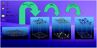
The fabrication and electrochemical performance of Mn3O4/graphene composites are discussed in this work. The main reaction procedures consist of two parts: one is the formation of Mn3O4 particles and the other is the reduction of graphite oxide to graphene. The chemicals, MnCl2·4H2O and NaBH4 are employed as a manganese source and a reduction reagent, respectively. During the formation of Mn3O4 particles, NH3 is added to the reaction system, directly, which simplifies the hydrolysis of amide, and the surfactant, polyvinylpyrrolidone (PVP), is used to ensure great dispersion and size-controlled formation of Mn3O4 particles. The resulting materials are characterized by XRD, SEM, HRTEM, FT-IR, Raman and XPS. Mn3O4 particles dispersing on the surface of graphene have an average diameter of ca. 30 nm. The materials deliver a stable reversible capacity of ca. 500 mA h g−1 at a current density of 60 mA g−1 even after 100 cycles. The reversible capacity of the samples coated with graphene is much better than that of pure materials.


1. Introduction

Energy, as a significant material basis contributing to the development of human society, has various employments in our daily lives. But how is it recycled and stored? Energy storage devices, such supercapacitors,1 redox flow batteries,2 sodium–sulfur cells and lithium-ion batteries,3 are the solution to this matter. But another question that needs to be solved is how to improve the cycling stability and enhance the capacity of energy storage devices. It is found that, as an important part of these energy storage devices, electrode materials are momentous factors that influence their electrochemical performances. In some previous research, various materials were utilized to improve the properties of these devices. Transition metal oxides,4,5 carbon materials and conducting polymers6–9 have all contributed to the progress of these devices, in particular to the lithium-ion battery.

Transition metal oxide materials, like Fe3O4,10 ZnO,11 CuO (12) and Mn3O4,13 were used as anode materials for lithium-ion battery research in some previous works. Among them, Mn3O4 materials known to have a normal spinel structure with tetragonal distortion elongated along the c-axis because of the Jahn–Teller effect on the Mn3+ ion, is one of the most attractive materials employed as an anode electrode for lithium-ion batteries due to the easy availability of manganese, its low cost, environmentally benign nature and a high theoretical specific capacity (approximately 936 mA h g−1). However, the shortcomings of high electrical resistance, poor electrochemical reversibility and low electrical conductivity (∼10−7 to 10−8 S cm−1), etc.14,15 limit its practical application in anode materials for lithium-ion batteries. To overcome these disadvantages, many different methods were taken advantage of in prior studies. For example, adding surfactants,16 doping Mn3O4 with Co,17,18 synthesizing nanosized Mn3O4 anode materials,19 coating Mn3O4 with carbon materials, etc., are all the effective approaches to polish up the electrochemical properties of Mn3O4 materials. In our study, the method of coating Mn3O4 with carbon materials, was operated as an example.

Carbon materials, such as dense and long carbon nanotube arrays,20 activated mesocarbon microbeads,21 uniform carbon layers,22 ball-milled graphite,23 graphene,24,25 etc., combined with Mn3O4 nanoparticles improved the capacity and stability of lithium ion batteries. Especially, graphene, because of its great electrochemical properties (the theoretical specific capacity is ca. 744 mA h g−1), excellent flexibility and large specific surface area, is described as the best selection for the conductive matrix and the support of sediment nanoparticles for anode materials.15 Mn3O4/graphene nanocomposites can be synthesized by various methods, and present great electrochemical performances in many studies. For instant, Liu et al.26 synthesized Mn3O4/graphene nanocomposites by a microwave-assisted hydrothermal method, Park et al.27 prepared the samples by a one-step sonochemical method and Nam et al.28 synthesized Mn3O4/graphene nanocomposite samples by an in situ transformation method. In our experiment, a one-pot hydrothermal synthesis of Mn3O4/graphene nanocomposites was carried out. The main procedures of the reaction consist of two parts: one is the formation of Mn3O4 nanoparticles and the other is the reduction of graphite oxide to graphene. The graphite oxide powders used in this study were prepared by a modified Hummer’s method. In addition, the reactions neither produced any toxic by-products nor required much reaction-energy. Meanwhile, the one-step accomplishment for the oxidation of Mn2+ and the reduction of graphite oxide to graphene economized the reaction-time, as well as simplified the reaction-procedures.

The performances of two samples, Mn3O4 and Mn3O4/graphene, were measured by some characterizations and electrochemical tests. It is found that the specific capacity and cycling stability of the lithium-ion battery are greatly improved by coating the anode materials with carbon materials. To the best of our knowledge, combing transition metal oxide with carbon materials is indeed the most feasible method to achieve the improvement of performances in practice.

2. Experiment details

2.1 Chemicals

Commercial graphite powders (C.P, 98%, Sinopharm chemical reagent Co., Ltd); sulfuric acid (H2SO4, A.R., 95%–98%, Beijing Chemical Works); sodium nitrate (NaNO3, A.R., 99%, Beijing Yili Fing Chemical Co., Ltd.); potassium permanganate (KMnO4, A.R., 99.5%, Beijing Chemical Works); hydrogen peroxide (30%, H2O2, A.R., >30%, Beijing Chemical Works); hydrochloric acid (HCl, A.R., 36%, Beijing Chemical Works); manganese chloride (MnCl2·4H20, A.R., ≥99.0%, Beijing Chemical Works); ammonia solution (NH3·H2O, A.R., 25%, Beijing Chemical Works); polyvinylpyrrolidone (PVP, the molecular weight = 40[thin space (1/6-em)]000 (avg.), Beijing Kebio Bio-Technique Co., Ltd.); sodium borohydride (NaBH4, A.R., ≥98.0%, Tianjin Fuchen Chemical Reagents Factory).

2.2 Sample preparation

2.2.1 Preparation of graphite oxide (GO). The graphite oxide used in this experiment was prepared by a modified Hummer’s method.29,30 1.5 g of NaNO3 powder was added into the 69 ml of H2SO4 solution in a three-necked flask with stirring in the ice-water-bath. When the powder dissolved completely, 3 g of graphite powder and 9 g of KMnO4 were added into the mixture with stirring for 10 min. Then, the ice-water bath was removed and the temperature was kept at 35 °C for 2 h using a water-bath. 137 ml of the deionized water was injected into the resulting solution at 95 °C. The temperature was maintained at 95 °C for 15 min. The resulting solution was diluted to 420 ml with warm-water and 10 ml of H2O2 was added into the solution. Then, the solution became yellow. The products were filtered and washed once with 330 ml of hydrochloric acid solution and three times with distilled water. Finally, the samples were dried for 24 h under an atmosphere at 45 °C.
2.2.2 Preparation of graphene and Mn3O4/graphene (MGC). Firstly, 40 mg of GO powder was added into 30 ml of the deionized water with stirring for 10 min and ultrasonic irradiation for 2 h. Next, 2 mmol of MnCl2·4H2O and 1.2 g of PVP were dissolved in 10 ml of the deionized water with stirring for 3 h. Then, 0.8 ml of NH3 was inject into the resulting solution with a pipette with stirring for 1 h. Afterwards, 20 mg of NaBH4 was added into the previous solution with stirring for 10 min. Finally, the resulting mixture was aged in an autoclave at 140 °C for 8 h. After cooling to room temperature, the samples were filtered and washed three times with distilled water and absolute ethyl alcohol, respectively. The Mn3O4/graphene nanoparticles were precipitated by drying at 80 °C for 4 h. For comparison, a sample of pure graphene was also prepared using a similar method (Scheme 1).
image file: c5ra08250a-s1.tif
Scheme 1 The schematic for the fabrication of Mn3O4 on graphene oxide and the reduction of graphene oxide to graphene.
2.2.3 Preparation of Mn3O4. The preparation of Mn3O4 nanoparticles was similar to the preparation of Mn3O4/graphene nanoparticles, except the first step. GO powder was needless in this preparation.

2.3 Characterization of the samples

The structure and phases of the samples were characterized by X-ray diffraction (XRD, Bruker D8 advance with Cu Kα, λ = 1.5418 Å, 40 kV, 40 mA). The morphology of the samples was investigated by transmission electron microscopy (TEM, Hitachi H7650B, 120 kV), high resolution transmission electron microscopy (HRTEM, TECNAI G2 20, 200 kV) and scanning electron microscopy (SEM, JEOL JSM-6360LA, 120 kV). The morphology of pristine graphene was investigated by transmission electron microscopy (TEM, JEOL JEM-2100). The surface chemical environments of the samples were analyzed by X-ray photoelectron spectroscopy (XPS, 250XI X-ray photoelectron spectrometer with an Al/Mg-Kα/Al X-ray source). The content of graphene was measured by thermogravimetric analysis (TGA, NETZSCH TG 209F1 Libra) from room temperature to 850 °C at a heating rate of 10 °C min−1 under O2. Raman spectra were measured on a RM2000 Raman Spectrometer (Renishaw, British). FT-IR spectra were tested by a Nicolet 560 Fourier transform infrared spectrophotometer.

2.4 Electrochemical analysis

The working electrode was prepared by mixing 80 wt% of the samples as the active material, 10 wt% Super P as a conductive additive, and 10 wt% sodium carboxymethyl cellulose (CMC) as a binder of the total electrode mass. The three components were mixed with deionized water as the solvent to produce slurry. It was uniformly loaded on a Cu foil with a doctor blade as a current collector and compressed to prepare a film-type electrode. The sample was cut into circular electrodes and dried for 12 h under a vacuum at 105 °C. The cells were assembled in an Ar-filled glovebox (ZKX2, Nanjing University Instrument Factory) with lithium foil as the anode, and a solution of 1.0 M LiPF6 dissolved in 1[thin space (1/6-em)]:[thin space (1/6-em)]1 (v/v) EC/DEC as the electrolyte. All electrochemical measurements were carried out on a CT2001A Land battery testing system in the potential range from 0.01 V to 3 V (vs. Li+/Li). The AC impedance data were recorded in the frequency range 10−2 Hz to 105 Hz using CHI760E electrochemical station (Shanghai Chenhua).

3. Results and discussion

3.1 Characterizations

3.1.1 Structure and morphology analysis. To study the crystalline nature of the prepared samples, the XRD pattern was recorded in the 2θ range 20–100° and HRTEM was brought to bear.

The crystalline structures of the Mn3O4/graphene composite materials were examined by XRD analysis, as in Fig. 1. The main characteristic peaks of the XRD curve (Fig. 1d) have great agreement with the JCPDS card (no. 80-0382), which is meant to be the tetragonal structure of Mn3O4. In accordance with the formula: 1/d2 = (h2 + k2)/a2 + l2/c2, where d is the interplanar spacing of the samples, (h k l) is the symbol of indicates of the crystal face, ‘a’ and ‘c’ are the lattice parameters of the tetragonal structure of Mn3O4, the actual value of the lattice parameters can be figured out (average size: a = 5.742 Å, c = 9.448 Å) and the peak at ca. 24° is indexed to the graphitic planes (002) of the graphene nanosheets.25 Observing the XRD patterns of GO and rGO (Fig. 1d), the characteristic stacking peak shifted from ca. 10°to ca. 24°, which indicates that GO has been converted into graphene with the help of a reductant and hydrothermal treatment. Moreover, from the HRTEM images of the Mn3O4/graphene nanocomposites (Fig. 1e), it can be observed that Mn3O4 nanoparticles are composed of nanocrystal lines with a lattice spacing of about 0.464 nm. Contrasting with data obtained from the XRD analysis, it is the (101) plane of the hausmannite structure. Meanwhile, the polycrystalline nature of the Mn3O4 nanoparticles can be seen from the SAED patterns (Fig. 1f).


image file: c5ra08250a-f1.tif
Fig. 1 (a)–(c), (e) HRTEM images, (d) XRD spectra of GO, rGO and Mn3O4/graphene, (f) SAED pattern of Mn3O4/graphene and (g) TEM images of pristine graphene.

The TEM images of pristine graphene show that the graphene we prepared has a wrinkled morphology and a multilayer structure (Fig. 1g). After the introduction of Mn3O4 particles, the electrochemically active surface area of graphene would be enlarged with the anchored Mn3O4 nanoparticles on the surface of the graphene platelets. The Mn3O4 nanoparticles are dispersed on the surface of graphene evenly (Fig. 1a and c). The size of these particles is approximately 30 nm (Fig. 1b), and this value is close to the outcome (about 27 nm) calculated by the Scherrer equation for XRD analysis: D = Kl/b[thin space (1/6-em)]cos[thin space (1/6-em)]q where D is the grain size of the Mn3O4 nanoparticles, K is the shape factor (fixed as 0.9), l is the X-ray wavelength, b is the full width at half maximum intensity (FWHM) in radians and q is the Bragg angle.

The morphology of the samples obtained by FE-SEM investigation is shown in Fig. 2. In Fig. 2a (scale bar = 1 μm) and Fig. 2b (scale bar = 100 nm), the substrate surface is covered uniformly with prism-like Mn3O4 nanomaterials. These prisms are interlocked with each other or aggregated, and the diameter of the cross section for these prism is approximately 100 nm and their length is 700 nm–1100 nm. Apart from these prisms, some sphere-like and cluster-like nanoparticles can be observed, as well due to the overgrowth of samples.


image file: c5ra08250a-f2.tif
Fig. 2 SEM images of (a) and (b) Mn3O4, (c) and (d) Mn3O4/graphene, and (e) the grain distribution of Mn3O4/graphene.

In Fig. 2c (scale bar = 100 nm) and Fig. 2d (scale bar = 100 nm), the Mn3O4 nanoparticles are closely attached and highly dispersed on the surface of graphene. However, perhaps because of the overdose of Mn3O4, some extra Mn3O4 nanoparticles not absorbed by graphene are found in Fig. 2c (marked by a black circle). The gain size of Mn3O4 absorbed on the surface of graphene is mainly distributed in the range of 30 nm to 70 nm (Fig. 2e).

3.1.2 FT-IR study. The IR spectrum of the samples synthesized is delineated in Fig. 3a. Functional groups that the samples contain can be analyzed on the basis of the position of the peaks. The peak at ca. 3428 cm−1 symbolizes the existence of “OH” groups and the peak at ca. 1730 cm−1 illustrates the presence of “C[double bond, length as m-dash]O” groups. Comparing the peaks in the same positions of 3428 cm−1 and 1730 cm−1, respectively, the peaks of Mn3O4/graphene become less intensive than those of graphite oxide. Even the peak at ca. 1730 cm−1 is invisible in the above solid line. It’s supposed that when graphite oxide was reduced to graphene, these functional groups at the surface of the samples were substituted by “C[double bond, length as m-dash]C” groups and Mn3O4 molecules, and this is the reason why the peaks of the “COOH” groups and “–C–O–C–” groups within the range of 1500 cm−1 to 1000 cm−1 have become weaker as well. From the foregoing analysis, although there are still a few oxygen molecules left over, the reduction effect of NaBH4 is satisfactory. The peak at ca. 1634 cm−1, as marked in Fig. 3a, signifies that there are some “C[double bond, length as m-dash]C” groups in the samples. This is a specific structure of graphene, denoting the sp2 hybridization of carbon structures. In addition, two obvious absorption bands, which are related with the coupling mode between the Mn–O stretching modes of the tetrahedral and octahedral sites, can be found at ca. 500 cm−1 in the curve of the Mn3O4/graphene materials.31 Therefore, the FT-IR results provide further evidence of the formation of Mn3O4 nanoparticles and the reduction of graphite oxide to graphene.
image file: c5ra08250a-f3.tif
Fig. 3 (a) FT-IR spectrum of graphite oxide (GO) and Mn3O4/graphene (MGC) and (b) Raman curves of graphite oxide (GO), reduced graphene oxide (rGO) and Mn3O4/graphene (MGC).
3.1.3 Raman test. Fig. 3b exhibits the Raman curves of graphite oxide, graphene and Mn3O4/graphene. The only difference found in the curves is a small peak in Raman shift at ca. 655 cm−1, which reflects the existence of Mn3O4 nanoparticles. The other peaks are the characteristic Raman shifts of graphene. The peak at ca. 1350 cm−1 called the D-peak, represents the lattice defects of carbon atoms and reflects the randomness of graphite layers, and the peak at ca. 1595 cm−1 called the G-peak, delegating sp2 hybridization of carbon structures, suggests the symmetry and the crystallinity of graphene. The more intensive the G-peak is, the more excellent the quality of graphene, but the D-peak is on the contrary. In theory, if the prepared graphene is unilaminar, there should be only one 2D-peak originating from the inelastic scattering of two double-phonons in the Raman curve and the intensity of the 2D-peak ought to be higher than the G-peak. According to Fig. 3b, it is obvious that the graphene we prepared is multilayered, and this result can be confirmed by TEM analysis as well (Fig. 1g).
3.1.4 Composition analysis. The elemental compositions of the sample were examined by XPS analysis. Fig. 4a shows a typical survey XPS spectrum of Mn3O4/graphene nanoparticles. All of the elements consist of five contributions of manganese species (3p, 3s, 2p3/2, 2p1/2), carbon species (1s), nitrogen species (1s), oxide species (1s) and fluorine species. To do a deeper analysis, the XPS species for the C1s region exhibited in Fig. 4b are made up of three dedications at ca. 284.84, 285.94 and 287.93 eV, which correspond to carbon sp2 bonding (C–C), epoxy/hydroxyl groups (C–O), and carbonyl/carboxyl groups (O–C[double bond, length as m-dash]O/C[double bond, length as m-dash]O), respectively. By accessing some literature,17 it was found that the peaks indicating epoxide/hydroxyl groups (C–O), and carbonyl/carboxyl groups (C[double bond, length as m-dash]O) should have become extensive, and the peak associated with “C–C” bond should have been predominant, while the graphite oxide was reduced to graphene completely. However, the intensity of the peak signifying the “C–O” bond, observed in Fig. 4b, is almost as intensive as “C–C” bond. So, the result of the reducing agent we employed is unsatisfactory. The manganese oxidation state is affirmed by the analysis of the Mn (3s, 2p) peak splitting width. As shown in Fig. 4c, The splitting width of Mn3s is ca. 5.9 eV, which in good agreement with other earlier articles32 on Mn3O4 as well as the splitting width of Mn3s (ca. 12.4 eV). So, it is proved that the product of the reaction is indeed Mn3O4/graphene. To study the real mass content of Mn3O4 and graphene in MGC, TGA analysis was conducted. The mass loss beginning from 150 °C to 400 °C indicates the decomposition of graphene into CO2 and H2O.33 To avoid the interference of mass variation of Mn3O4, pristine Mn3O4 was measured under the same circumstances. And the mass-loss of pristine Mn3O4 from 150 °C to 400 °C is attributed to the decomposition of PVP. The mass content of Mn3O4 and graphene are estimated to be 86.53% and 13.47% (Fig. 4e and f), respectively. While reaching above 500 °C, the mass of the samples does not undergo any distinct degradation, which indicates the thermostability of Mn3O4. On the basis of these data and the theoretical specific capacity of each composition, the theoretical specific capacity of the sample can be figured out with the formula. Taking this sample as an example, sum = 744 mA h g−1 × 13.47% + 936 mA h g−1 × 86.53% = 910.14 mA h g−1.
image file: c5ra08250a-f4.tif
Fig. 4 XPS spectra for (a) the survey spectra, (b) the C1s regions, (c) the Mn3s regions, (d) the Mn2p regions, and TGA profile of (e) Mn3O4 and (f) the Mn3O4/graphene nanocomposites.

3.2 Electrochemical measurement

Fig. 5a displays the comparison of the discharge–charge capacity between the Mn3O4 and Mn3O4/graphene electrodes at the current density of 60 mA g−1 for 100 cycles. In the initial cycle, the discharge and charge capacity of the Mn3O4 electrode are ca. 291.1 mA h g−1 and ca. 130.4 mA h g−1, respectively, and those of the Mn3O4/graphene electrode are ca. 897.2 mA h g−1 and ca. 456.4 mA h g−1, respectively. It is interesting to note that the initial discharge capacity of Mn3O4/graphene is much lower than the theoretical value calculated by TGA analysis (910.14 mA h g−1). This phenomenon is ascribed to the incomplete utilization of the active materials. Moreover, for the Mn3O4 and Mn3O4/graphene electrodes, in the voltage range of 1.3 to 0.3 V, both have an inclined curve (Fig. 5b). This is caused by the formation of a very thick solid electrolyte interface (SEI) film on the surface of the electrodes. However, the capacity efficiency of the Mn3O4/graphene electrode (ca. 50%) is much higher than that of the Mn3O4 electrode (ca. 45%). The unsatisfactory initial charge–discharge might be due to the reaction between the electrolyte and the oxygen-containing groups that the samples have.
image file: c5ra08250a-f5.tif
Fig. 5 (a) The comparison of the discharge–charge capacity between the Mn3O4 and Mn3O4/graphene electrodes at a current density of 60 mA g−1 for 100 cycles, (b) galvanostatic discharge–charge profiles of the Mn3O4 and Mn3O4/graphene electrodes at a current density of 200 mA g−1, (c) the rate capacity of the Mn3O4 and Mn3O4/graphene electrodes.

In the first 30 cycles, the capacity of the Mn3O4/graphene and pristine Mn3O4 electrodes are both not stable. The ups and downs of the capacity of the Mn3O4/graphene electrode might result from the scaling of the excess of Mn3O4 particles (marked in Fig. 2c), which are not coated with graphene. With the ongoing cycling, because of the gradually improved stability of the material structure which owns to the supporting function of graphene, the capacity of the Mn3O4/graphene electrode can be maintained at a level of ca. 500 mA h g−1 after 30 cycles, but the capacity of Mn3O4 electrode shows a little attenuation to ca. 140 mA h g−1 after 50 cycles (without the support of graphene, the pristine Mn3O4 electrodes undergo a large volume change in the process of lithium insertion and escape, resulting in disintegration of the crystals and loss of the connection between the electrode materials and the current collector34). All of these illustrate that the cycling performance of the samples is optimized after being coated with graphene.

The rate capacity of the samples was measured at current densities of 60, 250, 500, 750 mA g−1 (Fig. 5c). The capacities of the Mn3O4 electrode are ca. 150, ca. 110, ca. 60, ca. 50 mA h g−1, respectively, and those of the Mn3O4/graphene electrode are ca. 450, ca. 320, ca. 250, ca. 220 mA h g−1, respectively. The capacities of Mn3O4/graphene and pristine Mn3O4 at a current density of 60 mA g−1 are not stable, and this phenomenon is ascribed to the instability of the material structure in the initial several cycles (the scaling of an excess of Mn3O4 particles in MGC or the large volume change of pristine Mn3O4). However, with the ongoing cycling, the capacity of the samples is stable even at the higher current densities because of the gradually improved stability of the material structure. Meanwhile, the capacity of Mn3O4/graphene is always much higher than that of pristine Mn3O4. The graphene coating enhances the capacity of the samples.

Fig. 6 shows the Nyquist plots of the Mn3O4 and Mn3O4/graphene electrodes obtained at a current density of 60 mA g−1 after different cycling numbers.


image file: c5ra08250a-f6.tif
Fig. 6 Nyquist plots of the Mn3O4 and Mn3O4/graphene electrodes.

The Nyquist plots of the Mn3O4 and Mn3O4/graphene electrodes consist of two parts: a semicircle in the high frequency region and a vertically linear spike in the low frequency region, which are associated with the process of the charge transfer in the electron/ion conductive junction and the diffusion of lithium ion through the active material, respectively (Fig. 6a and b). From the figures, it is obvious that with the increasing in cycling number, the electrical resistance of the charge transfer becomes smaller, and the diffusion coefficient increases a little. This may have something to do with the activation of the samples. But in the partial enlargement (Fig. 6b), it is observed that two semicircles show up in the high frequency region, which are due to the formation of a SEI film. The absence of this semicircle in other curves may result as the electrical resistance of the SEI film is not big enough to be observed.

The electrochemical parameters are obtained by the fitting of an equivalent circuit (Fig. 6c), and the raw data and fitting data of two samples obtained after 10 cycles are exhibited in Fig. 6d and e, respectively.

The common equivalent circuit of the lithium ion battery is shown in Fig. 7.35,36 In this article, the Constant Phase Element (CPE1) is employed to replace the capacitor.

 
ZCPE1 = [T × (j × w)⁁P]−1 (1)
 
ZW1 = R × ctnh[(j × T × w)⁁P]/(j × T × w)⁁P (2)


image file: c5ra08250a-f7.tif
Fig. 7 Equivalent circuit used for analysis of impedance spectra of the lithium ion insertion/desertion in the intercalation electrode.

Observing the data shown in Table 1, the electrochemical resistance of the porous diaphragm, electrolyte, etc. (Rs) and charge transfer (R1) of the Mn3O4 electrode are both higher than that of the Mn3O4/graphene electrode. The open circuit terminus (W1) is defined by three values: W1R, W1T and W1P. W1T = L⁁2/D, where L is the effective diffusion thickness and D is the effective diffusion coefficient of the particle. According to the slope of the vertically linear spike in the low frequency region, the two samples have similar effective diffusion coefficients, but the gap between the values of W1T is big. In association with the formula, the effective diffusion thickness of the Mn3O4/graphene electrode is much bigger than that of the Mn3O4 electrode, and this might be one reason why the capacity of the Mn3O4/graphene electrode is much higher than that of the Mn3O4 electrode. The high electrical resistance of the Mn3O4 electrode influences the transfer velocity of lithium ion, which leads to its poor rate capacity as well. So the Mn3O4/graphene electrode obtains much greater electrochemical properties than the Mn3O4 electrode.

Table 1 Electrochemical parameters of the samples
Sample Mn3O4 MGC
Rs (Ω) 2.38 2.074
CPE1T−1 × cm−2 × s⁁P) 0.00011035 0.00021644
CPE1P 0.69288 0.51166
R1 (Ω) 223.1 96.88
W1R (Ω) 27.5 4572
W1T (s) 0.0010632 71.89
W1P 0.28607 0.58608


3.3 The formation mechanism of the samples37–39

In this synthesis, Mn3O4 nanoparticles were grown on graphene nanosheets by a one-pot hydrothermal method to prepare Mn3O4/graphene nanocomposite materials.

For this procedure, firstly, graphite oxide prepared by a modified Hummer’s method provided many oxygen-containing functional groups, which are on the surface of graphite oxide for the attachment of Mn2+ ions via electrostatic interactions. In the reaction, these Mn2+ ions were oxidized with oxygen contained in a functional group on the surface of graphite oxide.

With stirring under room-temperature, free Mn2+ ions were released from the stable precursor. Then, ammine complexes of Mn(NH3)n2+ (n = 1–4) were formed when NH3 (aq.) was added into the solution to react with the Mn2+ ions:

 
Mn2+ + nNH3 ⇄ Mn(NH3)n2+ (3)

The intermediate products, Mn(NH3)n2+ (n = 1–4), were unstable in the reaction conditions (due to the addition of NH3 (aq.), the solution is alkalescent), so the following reaction occurred:

 
Mn(NH3)n2+ ⇄ Mn2+ + nNH3 (4)

Finally, oxidation of Mn2+ to Mn3+ takes place as

 
6Mn2+ + O2 + 12OH ⇄ 2Mn3O4 + 6H2O (5)

The reagent, NaBH4, was employed in the reaction of graphite oxide, while PVP reagent was added to ensure great dispersion and size-controlled formation of the samples, which would avoid the excessive size of Mn3O4.

4. Conclusion

In summary, the Mn3O4/graphene nanocomposites are fabricated by a one-pot hydrothermal method with MnCl2·4H2O as a manganese donor, NaBH4 for the successful reduction of graphene oxide to graphene, and the chemical, PVP, which indeed plays a good role as a control agent that ensures the great dispersion and size-controlled formation of Mn3O4 on the surface of graphene. The electrochemical performances of samples get a great improvement after being coated with graphene.

For further optimization, doping with other metal elements is a valuable approach to attempt. And the initial charge–discharge efficiency of the samples leaves much to be desired, being coated with some other polymer may be effective in improving the performances of the materials.

Conflict of interest

The authors declare that there is no conflict of interest.

Acknowledgements

This study was supported by the National Natural Science Foundation of China (51304077 and 51374175), the Science and Technology Department of Science and Technology of Project in Jiangsu Province (BY2014037-31), the Privileged Development Program of Jiangsu High Education on New energy material science and engineering, the Opening Project of State Key Laboratory of Alternate Electrical Power System with Renewable Energy Sources (Grant No. LAPS15001), the Priority Academic Program Development of Jiangsu Higher Education Institutions, Qing Lan Project of Education Department of Jiangsu Province and the Material Corrosion and Protection Key Laboratory of Sichuan province (2014CL15).

References

  1. J. M. Miller, New Ultracapacitor Applications, Institution of Engineering & Technology, Stevenage, 2011 Search PubMed.
  2. R. Badrinarayanan, J. Y. Zhao, K. J. Tseng and S. K. Maria, J. Power Sources, 2014, 270, 576–586 CrossRef CAS PubMed.
  3. T. R. Crompton, Battery Reference Book, Science Direct, Newnes, 2000 Search PubMed.
  4. S. Komaba, T. Tsuchikawa, A. Ogata, N. Yabuuchi, D. Nakagawa and M. Tomita, Electrochim. Acta, 2012, 59, 455–463 CrossRef CAS PubMed.
  5. W. F. Zhang, F. Liu, Q. Q. Li, Q. L. Shou, J. P. Cheng, L. Zhang, B. J. Nelson and X. B. Zhang, Phys. Chem. Chem. Phys., 2012, 14, 16331–16337 RSC.
  6. S. Sahoo, G. Karthikeyan, G. C. Nayak and C. K. Das, Synth. Met., 2011, 161, 1713–1719 CrossRef CAS PubMed.
  7. D. C. Zhang, X. Zhang, Y. Chen, P. Yu, C. H. Wang and Y. W. Ma, J. Power Sources, 2011, 196, 5990–5996 CrossRef CAS PubMed.
  8. C. H. Xu, J. Sun and L. Gao, J. Mater. Chem., 2011, 21, 11253–11258 RSC.
  9. P. Si, S. J. Ding, X. W. Lou and D. H. Kim, RSC Adv., 2011, 1, 1271–1278 RSC.
  10. A. P. Hu, X. H. Chen, Q. L. Tang and B. Zeng, Ceram. Int., 2014, 40, 14713–14725 CrossRef CAS PubMed.
  11. E. R. Ezeigwe, M. T. T. Tan, P. S. Khiew and C. W. Siong, Ceram. Int., 2015, 41, 715–724 CrossRef CAS PubMed.
  12. J. Y. Xiang, J. P. Tu, L. Zhang, Y. Zhou, X. L. Wang and S. J. Shi, J. Power Sources, 2010, 195, 313–319 CrossRef CAS PubMed.
  13. S. H. Guo, M. Zhang, G. N. Zhang, L. Zheng, L. P. Kang and Z. H. Liu, Powder Technol., 2012, 228, 371–376 CrossRef CAS PubMed.
  14. J. Y. Qu, F. Gao, Q. Zhou, Z. Y. Wang, H. Hu, B. B. Li, W. B. Wan, X. Z. Wang and J. S. Qiu, Nanoscale, 2013, 5, 2999–3005 RSC.
  15. L. E. F. F. Torres, S. Roche and J. C. Charlier, Introduction to Graphene-Based Nanomaterials-From Electronic structure to Quantum Transport, Cambridge University Press, New York, 2014 Search PubMed.
  16. Y. F. Lee, K. H. Chang, C. C. Hu and Y. H. Chu, J. Power Sources, 2012, 206, 469–475 CrossRef CAS PubMed.
  17. J. Z. Wang, N. Du, H. Wu, H. Zhang, J. X. Yu and D. R. Yang, J. Power Sources, 2013, 222, 32–37 CrossRef CAS PubMed.
  18. Z. Q. Li, N. N. Liu, X. K. Wang, C. B. Wang, Y. X. Qi and L. W. Yin, J. Mater. Chem., 2012, 22, 16640–16648 RSC.
  19. J. Z. Wang, N. Du, H. Wu, H. Zhang, J. X. Yu and D. R. Yang, J. Power Sources, 2013, 222, 32–37 CrossRef CAS PubMed.
  20. X. W. Cui, F. P. Hu, W. F. Wei and W. X. Chen, Carbon, 2011, 49, 1225–1234 CrossRef CAS PubMed.
  21. H. Q. Wang, Z. S. Li, J. H. Yang, Q. Y. Li and X. X. Zhong, J. Power Sources, 2009, 194, 1218–1221 CrossRef CAS PubMed.
  22. C. B. Wang, L. W. Yin, D. Xiang and Y. X. Qi, ACS Appl. Mater. Interfaces, 2012, 4, 1636–1642 CAS.
  23. J. Y. Cao, Y. M. Wang, Y. Zhou, D. C. Jia, J. H. Ouyang and L. X. Guo, J. Electroanal. Chem., 2012, 682, 23–28 CrossRef CAS PubMed.
  24. S. H. Yang, X. F. Song, P. Zhang and L. Gao, J. Mater. Chem. A, 2013, 1, 14162–14169 CAS.
  25. X. Zhang, X. Z. Sun, Y. Chen, D. C. Zhang and Y. W. Ma, Mater. Lett., 2012, 68, 336–339 CrossRef CAS PubMed.
  26. C. L. Liu, K. H. Chang, C. C. Hu and W. C. Wen, J. Power Sources, 2012, 217, 184–192 CrossRef CAS PubMed.
  27. G. Park, L. Bartolome, K. G. Lee, S. J. Lee, D. H. Kim and T. J. Park, Nanoscale, 2012, 4, 3879–3885 RSC.
  28. I. Nam, N. D. Kim, G. P. Kim, J. S. Park and J. Yi, J. Power Sources, 2013, 244, 56–62 CrossRef CAS PubMed.
  29. W. Hmmners, J. Am. Chem. Soc., 1958, 80, 1339 CrossRef.
  30. L. Shahriary and A. Athawale, International Journal of Renewable Energy and Environmental Engineering, 2014, 02, 58–63 Search PubMed.
  31. Y. Z. Wu, S. Q. Liu, H. Y. Wang, X. W. Wang, X. Zhang and G. H. Jin, Electrochim. Acta, 2013, 90, 210–218 CrossRef CAS PubMed.
  32. J. W. Lee, A. S. Hall, J. D. Kim and T. E. Mallouk, Chem. Mater., 2012, 24, 1158–1164 CrossRef CAS.
  33. B. G. S. Raj, R. N. R. Ramprasad, A. M. Asiri, J. J. Wu and S. Anandan, Electrochim. Acta, 2015, 156, 127–137 CrossRef CAS PubMed.
  34. G. W. Li, B. M. Zhang, F. Yu, A. A. Novakova, M. S. Krivenkov, T. Y. Kiseleva, L. Chang, J. C. Rao, A. O. Polyakov, G. R. Blake, R. A. D. Groot and T. T. M. Palstra, Chem. Mater., 2014, 26, 5821–5829 CrossRef CAS.
  35. A. Rahmoun, M. Loske and A. Rosin, Energy Procedia, 2014, 46, 204–213 CrossRef PubMed.
  36. E. Barsoukov and J. R. Macdonald, Impedance Spectroscopy Theory, Experiment, and Applications, A John Wiley & Sons Inc Publication, Hoboken, 2005 Search PubMed.
  37. L. X. Yang, Y. Liang, H. Chen, Y. F. Meng and W. Jiang, Mater. Res. Bull., 2009, 44, 1753–1759 CrossRef CAS PubMed.
  38. D. P. Dubal, D. S. Dhawale, R. R. Salunkhe, S. M. Pawar, V. J. Fulari and C. D. Lokhande, J. Alloys Compd., 2009, 484, 218–221 CrossRef CAS PubMed.
  39. D. P. Dubal, D. S. Dhawale, R. R. Salunkhe, V. J. Fulari and C. D. Lokhande, J. Alloys Compd., 2010, 497, 166–170 CrossRef CAS PubMed.

This journal is © The Royal Society of Chemistry 2015
Click here to see how this site uses Cookies. View our privacy policy here.