Determination of tryptophan and tyrosine by chemiluminescence based on a luminol–N-bromosuccinimide–ZnS quantum dots system

Shifeng Li*, Min Xing, Haiyan Wang, Li Zhang, Yiman Zhong and Lei Chen
The Key Laboratory of Functional Molecular Solids, Ministry of Education, College of Chemistry and Materials Science, Anhui Normal University, Wuhu 241000, China. E-mail: lsf2008@mail.ahnu.edu.cn; Fax: +86 13605536845

Received 21st April 2015 , Accepted 3rd July 2015

First published on 3rd July 2015


Abstract

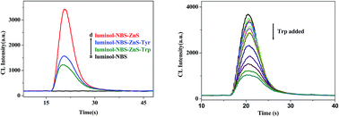
In this work, water-soluble ZnS quantum dots (QDs) modified with 3-mercaptopropionic acid (MPA), L-cysteine (L-Cys) and thioglycolic acid (TGA) were synthesized. The nanostructure and optical properties were characterized by X-ray powder diffraction (XRD), Transmission electron microscopy (TEM), UV-Vis absorption spectrum and fluorescence spectrum. Through the study of the ZnS QDs effects on the chemiluminescence (CL) system of luminol, we found that ZnS QDs could obviously enhance the CL of the luminol–N-bromosuccinimide (NBS) system in an alkaline medium. At the optimal experimental conditions, we researched the impact of 17 kinds of amino acids on this system. It was found that L-tryptophan (Trp) and L-tyrosine (Tyr) had significantly inhibitory effects on luminol–NBS–ZnS QDs CL. Based on the inhibitory effects, a novel CL method with a wider linear range and lower detection limit for determination of Trp and Tyr was developed. The detection limits were 1.5 × 10⁻¹¹ g mL−1 and 2.0 × 10⁻¹⁰ g mL−1, respectively. The possible CL reaction mechanism was also discussed briefly.


Introduction

Tryptophan (2S-2-amino-3-(1H-indol-3-yl)propanoic acid) and tyrosine (4-hydroxyphenylalanine) are two aromatic amino acids which are important precursors for synthetic peptide hormones, antibiotics, dopamine and some drugs.1–3 Meanwhile, Trp and Tyr play crucial roles on synthesizing melanin and melanocytes in the blood. Many means for detecting Trp and Tyr have been reported including electrochemistry,4,5 high performance liquid chromatography,6,7 spectrofluorometry,8 mass spectrometry9 and capillary electrophoresis.10 However, these methods often suffer from complicated pretreatment, poor reproducibility and low sensitivity defect. Therefore, it is necessary to design a rapid and sensitive method for detection of Trp and Tyr.

Flow injection analysis with low power consumption, wide liner range, low detection limit advantage has been widely used in biological immune analysis.11–13 Luminol as an ordinary CL reagent has been extensively studied by researchers, including luminol–ferricyanide,14 luminol–potassium permanganate,15–17 luminol–hydrogen peroxide system18,19 and so on.20,21 Many of these oxidants have color interfere with analysis, therefore, finding more stable and no color oxidants become our primary problem. HBrO as a strong oxidant could be applied in the field of CL. The biggest drawback is the reagent poor stability which limited its application scopes. In order to solve this problem, we choose NBS as oxidant. NBS can be hydrolyzed in aqueous solution for producing HBrO and the chemical properties are stable than HBrO. In addition, this agent can both dissolved in the polar and non-polar solvent, which has a greatly potential application in the organic phase CL. Many researchers have used different methods to enhance luminol–NBS system CL, including Au nanoparticles,22 some drugs.23

Recently, QDs or colloidal semiconductor nanocrystals have stimulated great interest in many fields due to their unique electronic and optical properties.24–28 ZnS QDs is direct broadband semiconductor, its fluorescence wavelength can cover ultraviolet to infrared, high fluorescence efficiency, narrow and continuous excitation spectrum etc. advantages, which make it has a wide application prospect in optoelectronic aspect. For example, as a light emitting diode, quantum dots laser, biological fluorescent probes and optical luminescence catalyst.29–36

In this paper, we studied the presence of ZnS QDs in the CL reaction of luminol–NBS system, it was found that ZnS QDs could catalyze luminol–NBS CL. Through researching the 17 kinds of amino acids effects on this system, Trp and Tyr could significantly inhibit luminol–NBS–ZnS QDs system CL intensity. Based on this inhibition, we have developed a flow injection method for measuring Trp and Tyr. This method has a wider liner range and lower sensitivity advantages for the samples analysis with satisfactory results.

Experimental section

Reagents and chemicals

All the chemicals were of analytical-reagent grade and used without further purification, and prepared in double-distilled water. 3-Aminophthal-hydrazide (luminol) was purchased from Alfa Aesar (Britain). N-Bromosuccinimide (NBS), zinc chloride (ZnCl2) and ethanol were purchased from Sinopharm Chemical Reagent Co. (Shanghai, China). Sodium sulfide (Na2S) was purchased from Wuxi Chemical Co. (Jiangsu, China). MPA and TGA were purchased from Aladdin Chemistry Co. (Shanghai, China). Amino acids were purchased from Huixing Chemical Reagent Co. (Shanghai, China).

Apparatus

All the CL measurements were carried out on the ultra weak luminescence analyzer (IFFM-E, Remex Electronic Institute Limited Co., Xi'an China). The CL signals were monitored by the PMT adjacent to the flow CL cell. When the CL system was used for investigating the effects of amino acids on the CL system, the sample solution and ZnS QDs were injected simultaneously and mixed with each other before further reaction with luminol–NBS solutions. Amino acids were injected after the optimized experimental concentrations.

Procedure

The FI–CL system procedure is shown in Scheme 1. The solutions of luminol, NBS and carrier water were delivered into the flow cell by peristaltic pumps at 3.0 mL min−1 flow rate, respectively. The ZnS QDs colloid solution was injected by a valve injector with 100 μL sample loop. The light output from the CL reaction was detected and amplified by the PMT and luminometer. The signal was imported to the computer for date acquisition. The determination of certain analytes of interest was based on changes in CL intensity ΔI = IsIo, where Is and Io were the CL intensity of sample and blank solutions, respectively, which was used for quantitative analysis of amino acid molecules.
image file: c5ra07233f-s1.tif
Scheme 1 Schematic diagram of the FI–CL system.

Synthesis of ZnS QDs capped with MPA (TGA or L-Cys)

As it was cited in the literature,37–39 0.3408 g of zinc chloride (ZnCl2) and 0.21 mL MPA (0.21 mL TGA or 0.2908 g L-Cys) as a stabilizing regent were dissolved in 265 mL of deionized water then stirred for 10 min to get a homogeneous mixture. The pH of the resulting solution was adjusted to 10.2 by adding 1 mol L−1 NaOH solution then transferred to the water bath with 70 °C for preheating 10 min, after that dropwise added a certain concentration of sodium sulphide solution for reacting 2 h. Then the resultant MPA capped ZnS QDs was centrifuged and washed with absolute ethanol three times. Finally, ZnS QDs was dried in 60 °C for 6 h. The sample obtained in preparation for the subsequent experiments.

Results and discussion

Characterization of ZnS QDs

As shown in Fig. 1A, the size of the as-prepared MPA/ZnS QDs was characterized by TEM and statistically analyzed as 5 ± 1 nm in diameter. TEM images of TGA or L-Cys modified ZnS QDs were also investigated (see ESI material Fig. S1). A histogram of approximate distribution of MPA/ZnS QDs as shown in Fig. 1B based on TEM observations. Fig. 1C was 70 °C ZnS X-ray power diffraction pattern, the peak location is consistent with cubic ZnS standard card 05-0556. Fig. 1D was different modifier modified ZnS QDs UV-Vis absorption and fluorescence spectra, the ultraviolet absorption peak appear at 285 nm, the emission peak of fluorescence was appeared at 438 nm.
image file: c5ra07233f-f1.tif
Fig. 1 (A) TEM of the MPA/ZnS QDs at 70 °C, (B) the size distribution histogram, (C) XRD image of MPA/ZnS QDs and (D) UV-Vis absorption spectrum and fluorescence spectrum ZnS QDs (a–c: TGA/ZnS, L-Cys/ZnS, MPA/ZnS, respectively).

Kinetic curves of CL reaction

In our experiments, we had synthesized and characterized three different modifiers capped ZnS QDs. All of the size were about 5 nm, which meant that the effect of particle size on catalytic properties could basically be eliminated. As it shown in Fig. 2 insert, on injection of ZnS QDs into the luminol–NBS system (curve a), CL intensity was substantially increased. Compared with two other modifiers (TGA/L-Cys, curve b/c), the enhance effect of MPA modified ZnS QDs was the best (curve d).
image file: c5ra07233f-f2.tif
Fig. 2 Kinetic curves of CL reaction (insert a–d): luminol–NBS, luminol–NBS–TGA/ZnS, luminol–NBS–L-Cys/ZnS, luminol–NBS–MPA/ZnS. (Luminol: 3.5 × 10⁻⁷ mol L−1, NaOH: 0.02mol L−1, NBS: 2 × 10⁻⁵ mol L−1, ZnS: 2 × 10⁻⁵ mol L−1) and (a–d): luminol–NBS, luminol–NBS–MPA/ZnS–Trp, luminol–NBS–MPA/ZnS–Tyr, luminol–NBS–MPA/ZnS. (Luminol: 3.5 × 10⁻⁷ mol L−1 NaOH: 0.025 mol L−1, NBS: 2 × 10⁻⁵ mol L−1, ZnS: 4 × 10⁻⁵ mol L−1, Trp and Tyr: 1 × 10⁻⁶ g mL−1).

So we selected MPA modified ZnS QDs as the reaction catalyst. Under the optimal condition, through the sensing research of 17 amino acids, the results showed that Trp and Tyr significantly inhibit the CL intensity (Fig. 2).

Optimization of the experimental conditions

Effect of NaOH concentration

Considering luminol in the alkaline medium has a strong CL signal, we chose the same concentration of NaOH, Na2CO3, and NaHCO3 to add into the luminol solution. NaOH was selected as a reactive medium because of the strongest signal intensity. We chose 0.025 mol L−1 NaOH (Fig. 3a) as the reaction medium for consequent research.
image file: c5ra07233f-f3.tif
Fig. 3 Effects of reaction on the CL intensity: (a) NaOH concentration, luminol, 2.5 × 10⁻⁷ mol L−1; NBS, 2.0 × 10⁻⁵ mol L−1; MPA/ZnS, 5 × 10⁻⁵ mol L−1 (b) luminol concentration (0.025 mol L−1 NaOH); NBS, 2.0 × 10⁻⁵ mol L−1; MPA/ZnS, 5 × 10⁻⁵ mol L−1 (c) NBS concentration, luminol, 3.5 × 10⁻⁷ mol L−1 (0.025 mol L−1 NaOH); MPA/ZnS, 5 × 10⁻⁵ mol L−1 (d) MPA/ZnS concentration, luminol, 3.5 × 10⁻⁷ mol L−1 (0.025 mol L−1 NaOH), NBS, 2.0 × 10⁻⁵ mol L−1.

Effect of luminol concentration

The effect of luminol concentration on the CL intensity was examined from 7.5 × 10⁻⁸ to 1 × 10⁻⁶ mol L−1. The results showed that the CL intensity increased with luminol added (Fig. 3b). Considering too large concentration was unstable and difficultly test, 3.5 × 10⁻⁷ mol L−1 was chosen as the optimum concentration of luminol for further research.

Effect of NBS concentration

The effect of the NBS concentration ranging from 8.0 × 10⁻⁶ to 9 × 10⁻⁵ mol L−1 on the CL signal was investigated. It was obvious found 2.0 × 10⁻⁵ mol L−1 NBS solution had a maximum CL intensity (Fig. 3c). Therefore, 2.0 × 10⁻⁵ mol L−1 NBS was chosen for the subsequent experiment.

Effect of ZnS QDs concentration

The effect of ZnS QDs concentration on the changes in relative CL intensity was studied at different concentrations from 6.0 × 10⁻⁶ to 7 × 10⁻⁵ mol L−1. The maximum CL intensity appeared at the concentration of 4 × 10⁻⁵ mol L−1, so 4 × 10⁻⁵ mol L−1 ZnS (Fig. 3d) was selected for further research.

Mechanism discussion

The CL spectra of the reactions is obtained by using the modified F-4500 spectrofluorimeter, whose light entrance slot was shut. We mixed each substance and found the peak appeared at 425 nm, as shown in Fig. 4a, indicating the same maximal peak as some literature reported.18–20 Thus, we explored that the main emitters of luminol–NBS–ZnS QDs CL system were related to the excited state of 3-aminophthalate molecule with characteristic maximum emissions of around 425 nm.40,41 The enhanced CL signals are thus ascribed to the possible catalysis from ZnS QDs.20
image file: c5ra07233f-f4.tif
Fig. 4 A CL spectra of CL system: (a) luminol, (b) luminol–NBS, (c) (b)-ZnS; (b) UV-Vis absorption spectra of CL system: (a) Trp, (b) ZnS, (c) luminol, (d) luminol–NBS, (e) (d)-ZnS, (f) (e)-Trp.

We also used UV-Vis absorption spectrum to explore the possible mechanism, shown as Fig. 4b. We could clearly see the absorption peak of luminol–NBS system appear at 302 nm and 352 nm. Merging with ZnS, the CL system absorption peak location was no change. The light absorption of the mixed system was approximately equal to the sum of the light absorption of the two individual systems. Therefore, we concluded that ZnS QDs might be used as the catalyst for this reaction.

Using the flow injection analyzer, we could not found signal when the lower concentration of luminol and NBS mixed. When ZnS QDs added into the system, it could be observed a significant CL signal. A lot of reactive oxygen and hydroxyl radical could be produced. Moreover, ZnS QDs and reactive oxygen could catalyze luminol and NBS reaction intermediates reaction, which might greatly improve the speed of light-emitting and signal strength. Therefore, we explored this CL system mechanism as following in Scheme 2.


image file: c5ra07233f-s2.tif
Scheme 2 Possible mechanism for the luminol–NBS–ZnS system.

Analytic application

Under the optimum conditions, we researched the behavior of amino acids with luminol–NBS–ZnS QDs CL. It was found that Trp and Tyr both had stronger inhibition for the above CL, as shown in Table 1. The CL inhibitory ratios of amino acids on this system were shown in Fig. 5. Based on this inhibition effects, a novel CL method for detection of Trp and Tyr was developed. Compared with other conventional method, this mean has a greatly lower detection limit and wide linear range advantage for samples analysis (Fig. 6). We obtained that Trp with a linear range from 4.0 × 10⁻¹¹ to 2.0 × 10⁻⁶ g mL−1, Tyr with a linear range from 6.0 × 10⁻¹⁰ to 1.0 × 10⁻⁶ g mL−1. The detection limits were 1.5 × 10⁻¹¹ g mL−1, 2.0 × 10⁻¹⁰ g mL−1 of Trp and Tyr, respectively. The relative standard deviation (R.S.D., n = 11) for Trp in water samples at 4.0 × 10⁻⁷ g mL−1 was 0.92% (Fig. 7). Moreover, recovery tests of standard addition in Trp samples showed the recoveries between 98.6% and 103.8%, which were at confidence interval of 100 ± 5% (Table 2). The results showed that this flow injection CL was a sensitive and accurate method for the Trp and Tyr detection.
Table 1 Effects of amino acids on luminol–NBS–ZnS CL systema
Amino acids ΔI
a Concentration of amino acids: 1 × 10⁻⁶ g mL−1, ΔI: net CL intensity. CL conditions: luminol, 3.5 × 10⁻⁷ mol L−1 (0.02 mol L−1 NaOH); NBS, 2.0 × 10⁻⁵ mol L−1; ZnS QDs, 4.0 × 10⁻⁵ mol L−1; flow rate: 3.0 mL min−1, negative high voltage: 450 V. I0 obtained by luminol–NBS–ZnS QDs CL system CL single intensity was 2160.
L-Tryptophan −1380
L-Tyrosine −1180
L-Cysteine −130
L-Valine +62
L-Hitsidine +95
L-Aspartic acid +105
L-Threonine +105
L-Proline +125
L-phenylalanine +125
L-Cystine +145
L-Asparagine +215
L-Glycine +255
L-Isoleucine +270
L-Methionine +285
L-Leucine +290
L-Arginine +320
L-Serine +335


image file: c5ra07233f-f5.tif
Fig. 5 The chemiluminescence inhibitory ratios of amino acids on this system.

image file: c5ra07233f-f6.tif
Fig. 6 (a) Analysis characteristic curve of Trp (illustration is linear relationship) and (b) analysis characteristic curve of Tyr (illustration is linear relationship).

image file: c5ra07233f-f7.tif
Fig. 7 Parallel determination (n = 11) for Trp at 4.0 × 10⁻⁷ g mL−1.
Table 2 The recoveries of the determination of Trp in sample
Sample Initially present (g mL−1) Added (g mL−1) Found (g mL−1) Recovery (%)
1 4.72 × 10⁻⁷ 2.0 × 10⁻⁷ 6.97 × 10⁻⁷ 103.7
3.0 × 10⁻⁷ 8.01 × 10⁻⁷ 103.8
2 6.46 × 10⁻⁷ 1.0 × 10⁻⁷ 7.48 × 10⁻⁷ 100.3
3.0 × 10⁻⁷ 9.41 × 10⁻⁷ 99.5
3 6.98 × 10⁻⁷ 1.0 × 10⁻⁷ 8.05 × 10⁻⁷ 100.9
2.0 × 10⁻⁷ 8.85 × 10⁻⁷ 98.6


Conclusion

In this paper, we successfully synthesized MPA functionalized ZnS QDs which could catalyze the luminol-NBS CL system. Through consulting relevant articles, UV-Vis and CL spectra experiments, we explored the possible reaction mechanism. With 17 kinds of common amino acids detection and analysis, we found that Trp and Tyr could significantly inhibited the CL intensity. Based on this phenomenon, we could design a sensitive and rapid detection of Trp and Tyr sensor.

Acknowledgements

This work is supported by the special funding support from the National Natural Science Foundation of China (21001005).

References

  1. J. S. Meyer, K. M. Welch, V. D. Deshmukh, F. I. Perez, R. H. Jacob, D. B. Haufrect, N. T. Mathew and R. M. Morrell, J. Am. Geriatr. Soc., 1977, 25, 289 CrossRef CAS PubMed.
  2. A. J. Gelenberg, C. J. Gisbon and J. D. Wojcik, Psychopharmacol. Bull., 1982, 18, 7 CAS.
  3. Y. Zhu, Y. Yang, G. Zhou, M. Li, C. Jiang, C. Zhang and S. Chen, Food Chem., 2010, 118, 159 CrossRef CAS.
  4. Q. Wang, A. Vasilescu, P. Subramanian, A. Vezeanu, V. Andrei, Y. Coffinier, M. Li, R. Boukherroub and S. Szunerits, Electrochem. Commun., 2013, 35, 84 CrossRef CAS.
  5. M. Y. Wan, W. F. Li, Y. M. Long and Y. F. Tu, Anal. Methods, 2012, 4, 2860 RSC.
  6. M. Sa, L. Ying, A. G. Tang, L. D. Xiao and Y. P. Ren, Clin. Chim. Acta, 2012, 413, 973 CrossRef CAS PubMed.
  7. J. Yang, S. Wang, J. Liu and A. Raghani, J. Chromatogr. A, 2007, 1156, 174 CrossRef CAS PubMed.
  8. K. Lotte, R. Plessow and A. Brockhinke, Photochem. Photobiol. Sci., 2004, 3, 348 CAS.
  9. T. Miron and M. Wilchek, Anal. Biochem., 2013, 440, 12 CrossRef CAS PubMed.
  10. B. Arvidsson, N. Johannesson, A. Citterio, P. G. Righetti and J. Bergquist, J. Chromatogr. A, 2007, 1159, 154 CrossRef CAS PubMed.
  11. Y. Chi, J. Duan, S. Lin and G. Chen, Anal. Chem., 2006, 78, 1568 CrossRef CAS PubMed.
  12. D. Du, J. Wang, J. N. Smith, C. Timchalk and Y. Lin, Anal. Chem., 2009, 81, 9314 CrossRef CAS PubMed.
  13. Z. P. Wang, J. Li, B. Liu, J. Q. Hu, X. Yao and J. H. Li, J. Phys. Chem. B, 2005, 109, 23304 CrossRef CAS PubMed.
  14. C. Guo, H. J. Zeng, X. J. Ding, D. He, J. J. Li, R. Yang and L. B. Qu, J. Lumin., 2013, 134, 888 CrossRef CAS.
  15. X. Chen, C. Wang, X. Tan and J. Wang, Anal. Chim. Acta, 2011, 689, 92 CrossRef CAS PubMed.
  16. B. Du, T. Wang, S. Han, X. Cao, T. Qu, F. Zhao and H. Yao, Spectrochim. Acta, Part A, 2015, 136, 149 CrossRef CAS PubMed.
  17. Z. Y. Yan, A. Xiao, H. Lu, Z. Liu and J. Q. Chen, New Res. Carbon Mater., 2014, 29, 216 CrossRef.
  18. X. Zhong, Y. Q. Chai and R. Yuan, Talanta, 2014, 128, 9 CrossRef CAS PubMed.
  19. M. Deng, S. J. Xu and F. N. Chen, Anal. Methods, 2014, 6, 3117 RSC.
  20. S. F. Li, X. Z. Li, Y. Q. Zhang, F. Huang, F. F. Wang and X. W. Wei, Microchim. Acta, 2009, 167, 103 CrossRef CAS.
  21. Z. H. Song and S. Hou, J. Pharm. Biomed. Anal., 2002, 28, 683 CrossRef CAS PubMed.
  22. J. X. Du, J. Quan and Y. D. Wang, Talanta, 2012, 90, 117 CrossRef CAS PubMed.
  23. Z. P. Wang, Z. J. Zhang, Z. F. Fu, W. F. Luo and X. Zhang, Talanta, 2004, 62, 611 CrossRef CAS PubMed.
  24. N. Wang, R. J. Zou, H. H. Chen, H. H. Chen, Y. G. Sun, J. H. Wu, Q. W. Tian, J. M. Yang, Z. G. Chen and J. G. Hu, CrystEngComm, 2011, 13, 1518 RSC.
  25. A. Khataee, A. Hasanzadeh, M. Iranifam and S. W. Joo, Sens. Actuators, B, 2015, 215, 272 CrossRef CAS.
  26. I. Costas-Mora, V. Romero, I. Lavilla and C. Bendicho, Anal. Chim. Acta, 2013, 788, 114 CrossRef CAS PubMed.
  27. P. T. Shao, Q. H. Zhang, Y. G. Li and H. Z. Wang, J. Mater. Chem., 2011, 21, 151 RSC.
  28. M. Hosseini, M. R. Ganjali, Z. Vaezi, F. Faridbod, B. Arabsorkhi and M. H. Sheikhha, Spectrochim. Acta, Part A, 2014, 121, 116 CrossRef CAS PubMed.
  29. S. K. Panda, S. G. Hickey, H. V. Demir and A. Eychmüller, Angew. Chem., Int. Ed., 2011, 50, 4432 CrossRef CAS PubMed.
  30. J. L. Zhang and B. X. Li, Spectrochim. Acta, Part A, 2014, 125, 228 CrossRef CAS PubMed.
  31. D. I. Son, H. H. Kim, D. K. Hwang, S. Kwon and W. K. Choi, J. Mater. Chem. C, 2014, 2, 510 RSC.
  32. P. Wu, T. Zhao, S. L. Wang and X. D. Hou, Nanoscale, 2014, 6, 43 RSC.
  33. A. Mandal, A. Dandapat and G. de, Analyst, 2012, 137, 765 RSC.
  34. J. Kang, X. Li, J. Geng, L. Han, J. Tang, Y. Jin and Y. Zhang, Food Chem., 2012, 134, 2383 CrossRef CAS PubMed.
  35. X. Y. Cao, F. Shen, M. W. Zhang, J. X. Bie, X. Liu, Y. L. Luo, J. J. Guo and C. Y. Sun, RSC Adv., 2014, 4, 16597 RSC.
  36. P. Wu and X. P. Yan, Chem. Soc. Rev., 2013, 42, 5489 RSC.
  37. W. Vogel, P. H. Borse, N. Deshmukh and S. K. Kulkarni, Langmuir, 2000, 16, 2032 CrossRef CAS.
  38. B. Gilbert, F. Huang, Z. Lin, C. Goodell, H. Zhang and J. F. Banfield, Nano Lett., 2006, 6, 605 CrossRef CAS PubMed.
  39. L. Shen, X. Cui, H. Qi and C. Zhang, J. Phys. Chem. C, 2007, 111, 8172 CAS.
  40. S. F. Li, X. M. Zhang, W. X. Du, Y. H. Ni and X. W. Wei, J. Phys. Chem. C, 2009, 113, 1046 CAS.
  41. G. Merenyi, J. Lind and T. E. Eriksen, J. Biolumin. Chemilumin., 1990, 5, 53 CrossRef CAS PubMed.

Footnote

Electronic supplementary information (ESI) available. See DOI: 10.1039/c5ra07233f

This journal is © The Royal Society of Chemistry 2015
Click here to see how this site uses Cookies. View our privacy policy here.