Using nano-QSAR to determine the most responsible factor(s) in gold nanoparticle exocytosis

Arafeh Bigdelia, Mohammad Reza Hormozi-Nezhad*ab and Hadi Parastara
aDepartment of Chemistry, Sharif University of Technology, Tehran, Iran. E-mail: hormozi@sharif.edu; Tel: +98 21 6616 5337
bInstitute for Nanoscience and Nanotechnology (INST), Sharif University of Technology, Tehran, Iran

Received 7th April 2015 , Accepted 11th June 2015

First published on 12th June 2015


Abstract

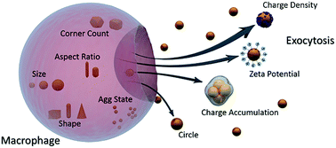
There are, to date, few general answers to fundamental questions related to the interactions of nanoparticles (NPs) with living cells. Studies reported in the literature have delivered only limited principles about the nano–bio interface and thus the biological behavior of NPs is yet far from being completely understood. Combining computational tools with experimental approaches in this regard helps to precisely probe the nano–bio interface and allows the development of predictive and descriptive relationships between the structure and the activity of nanomaterials. In the present contribution, a nano-quantitative structure-activity relationship (nano-QSAR) model has been statistically established using the Partial Least Squares Regression (PLSR) model. Also, variable importance on PLS projections (VIP) has been used to find the most responsible factors in NP exocytosis. Physicochemical properties of a set of different sized gold NPs with different surface coatings were strongly correlated to their exocytosis in macrophages. The results suggest that among the pool of physicochemical properties defined as nano-descriptors, charge density and surface charge seem to be the paramount factors leading to higher exocytosis values. Furthermore, charge accumulation and circularity of NPs are in the next level of priority among other nano-descriptors. The regression based nano-QSAR model reported here is satisfactory in both statistical quality and interpretability. The results could serve as a quantitative framework for better understanding the mechanisms that govern the interactions at the nano–bio interface.


1. Introduction

The interaction of NPs with living cells is dictated by many factors, including size, shape, chemical composition, crystallinity, hydrophobicity, porosity, surface charge, aggregation state, and surface coating, plus characteristics of the suspending media.1–8 In addition to the type of surface chemical functions, their relative arrangement also plays a key role in nanoparticle–cell interactions.9 Amongst these numerous factors affecting NP behavior at the “nano–bio” interface, the decision about their priority is far from being completely understood. Which factor is most responsible? How could one decide which factor to tune among the pool of parameters in order to adjust a specific cellular response? With the increasing number of medicinal and therapeutic applications of NPs, these questions gain even more attention. Answers to these questions could provide remarkable clues for preparing safe and efficacious NPs as diagnostic and drug delivery tools. Moreover, there is still a lot to find out about what exactly happens at the nano–bio interface. Knowing the exact contribution of each parameter in NPs’ behavior could help to deeply discover the interactions and to design NPs with enhanced efficiency. Meanwhile, it would be noteworthy if it was possible to quantitatively adjust and distinguish between the most important variables involved.

Various case studies have taken into account the role of different factors on NPs’ cellular uptake, cytotoxicity and exo/endocytosis. Chan et al.10 investigated the impact of NP size on active and passive tumor targeting efficiency. Rotello et al.11 quantified the exocytosis behavior of NPs with different surface functionalities. Crespy et al.12 showed how shape can influence the uptake of anisotropic polymeric NPs. Kanaras et al.13 reported that the penetration of gold NPs through skin is influenced by the charge, morphology and function of the NPs. In another study, Chan et al.14 investigated the role of size and surface chemistry in mediating serum protein adsorption to gold NPs and their subsequent uptake by macrophages.

All these studies together with other similar ones reported in the literature15–19 have delivered only limited principles about how various parameters affect the NPs’ behavior at the nano–bio interface. In other words, they have argued that the biological response to a NP is generally a complex function of multiple parameters and is still not fully understood. Thus, there is a need for a comprehensive tool that could gather this individual information and provide a broader framework for understanding the interactions beyond the nano–bio interface. This knowledge is also important from the perspective of developing quantitative relationships that are able to predict the biological response profiles of NPs from their physicochemical properties.

Recently, Park et al.20 have investigated the effect of native surface chemistries of gold nanoparticles (GNPs) and their subsequent opsonization by serum proteins on their exocytosis pattern in macrophages. They have reported the exocytosis rates of a set of different sized and different charged GNPs. It was demonstrated that between size and surface charge, the latter seems to play a more crucial role in determining the exocytosis pattern which is confirmed by other similar related works. Again, this was another case study that considered only two factors simultaneously, size and surface charge. As an alternative, what if it was possible to take this a step further and take a more comprehensive look? Is surface charge still the most dominant factor if a wider pool of variables are considered? To answer this question, a quantitative structure-activity relationship framework may be helpful.

In this study, we have proposed a nano-quantitative structure-activity relationship (nano-QSAR) model to investigate the effects of various parameters on GNP exocytosis in macrophages. The statistical model quantitatively detects the most prominent factors affecting the exocytosis of GNPs among a pool of physicochemical properties of NPs (called nano-descriptors). A set of morphological nano-descriptors has therefore been extracted from their corresponding TEM images by applying image processing methods (based on our previous work).21 In addition, a set of experimental parameters, together with a combinatorial set of nano-descriptors (a combination of experimental and image extracted descriptors) have also been provided. Partial Least Squares Regression (PLSR) has been carried out to analyze the data both for predictive and descriptive purposes. Actually, from a predictive point of view, the regression model finds the best possible correlation between the physicochemical properties of NPs and their cellular response profiles (here exocytosis). Consequently, the established regression model can be applied to predict the exocytosis for unknown GNPs. On the other hand, with a descriptive perspective, variable importance on PLS projections (VIP) has been used to indicate the most dominant factors that are responsible for determining the exocytosis of GNPs.

2. Methods

2.1. Nano-QSAR approach

There are a lot of parameters to control when investigating the biological behavior of NPs interacting with living cells. Many of these parameters are strongly inter-correlated. Besides, it may be difficult to vary individual properties while keeping others constant. To overcome these constraints, attempts have been made to develop predictive models which relate the physicochemical properties of NPs to their biological response(s).22–30 However, this is a great challenge and requires the use of both powerful computational tools and experimental methods. Applying QSAR models to comprehensive datasets (collected from reliable experimental parameters and several physicochemical properties of NPs) could manifest the real parameters underlying a considered interaction and therefore provide substantial details about the nano–bio interface. The more data provided, the greater the authenticity and accuracy of the constructed model.

2.2. Descriptor generation

To gather the required data set for the nano-QSAR approach, three subsets of variables were provided: (a) TEM extracted nano-descriptors including size, surface area, aspect ratio, corner count, curvature, aggregation state, and shape; (b) experimental parameters including zeta potential, hydrodynamic diameter, and maximum wavelength both before and after protein coating; and (c) combinatorial nano-descriptors including charge density, adjusted aspect ratio, charge accumulation, spectral size, spectral surface area, spectral aspect ratio and spectral aggregation. The first subset was prepared by performing image processing on the TEM images shown in Fig. 1. The details on extracting these twelve image nano-descriptors can be found elsewhere.21 The second subset was inserted from the characterization information of surface-functionalized GNPs before and after serum coating.20 In order to extract more information on the morphology and surface of the NPs, an idea was to combine image nano-descriptors with experimental ones. Consequently, a set of ten new meaningful combinatorial nano-descriptors was provided as a combination of some previously defined image extracted and experimental descriptors. The aim of defining the third subset was to provide maximum informative data for the corresponding GNPs. The complete set of nano-descriptors is shown in Table 1. The last column comprises of the numerical values of the exocytosis of different sized and coated GNPs extracted from the reference article.20 In addition, Table 2 demonstrates the calculation of the new defined combinatorial descriptors: charge density accounts for the amount of charge per unit surface area which can be calculated based on the zeta potential before or after protein coating (ChDensB and ChDensA); the adjusted aspect ratio was defined to distinguish between NPs with similar aspect ratios but different lengths which can be calculated from multiplying the aspect ratio with either the size extracted from TEM or the hydrodynamic size before protein coating (AdjAR1, AdjAR2); and charge accumulation takes into account the amount of available charge surrounding aggregated NPs. It is obvious that the amount of free charge is different between the situations in which the particles are either dispersed in the media or have formed an aggregate. Again, this combinatorial variable can be calculated from multiplying the aggregation state value by the zeta potential of the NPs before or after protein binding (ChAccumB, ChAccumA). The last combined nano-descriptors have been defined based on the strong dependency of NPs’ plasmon shift to their size, surface area, aspect ratio and amount of aggregation (SpecSize, SpecSA, SpecAR, SpecAgg).
image file: c5ra06198a-f1.tif
Fig. 1 TEM visualization (top) and size distribution histograms (bottom) of all GNPs in the dataset with different sizes and different coatings.20 The number and letter after the GNP identifier designate the size and type of coating (anionic, cationic, zwitterionic and PEGylated surfaces), respectively. Scale bar is 50 nm. Reprinted with permission from ref. 20. Copyright 2014 ACS.
Table 1 Whole set of nano-descriptors and the two last columns consist of the observed and predicted exocytosis values
NP ID Nano-descriptors
Image extracteda Experimentalb
Size Surface area Curvature Aspect ratio Corner count Circle Rod Dog bone Triangle Square Hexagon Agg state HD_B ZP_B Peak_B HD_A ZP_A Peak_A
a TEM extracted image nano-descriptors calculated by image analysis on TEM images in Fig. 1. Please see ref. 21 for more information on the image analysis process.b Gold NPs characteristics extracted from Fig. 1c in ref. 20.c Combinatorial descriptors calculated based on the formula presented in the last column of Table 2.
G10A 18.800 267.200 0.793 1.056 7.133 0.655 0.168 0.151 0.754 0.578 0.777 0.121 14.410 −29.280 520.000 36.030 −28.390 525.000
G10C 16.700 227.600 1.280 1.061 7.176 0.712 0.138 0.143 0.533 0.663 0.798 0.065 13.480 26.330 520.000 30.640 −20.090 525.000
G10Z 14.000 165.800 1.519 1.073 7.455 0.794 0.132 0.139 0.545 0.670 0.832 0.041 16.260 −0.920 520.000 36.120 −21.270 525.000
G10P 19.600 274.100 0.687 1.044 7.571 0.669 0.118 0.120 0.450 0.579 0.714 0.052 25.420 −27.540 525.000 37.220 −29.610 530.000
G20A 31.800 816.800 0.831 1.008 6.833 0.653 0.102 0.110 0.545 0.649 0.798 0.230 26.860 −38.400 525.000 48.220 −40.010 530.000
G20C 36.800 999.700 0.981 1.113 7.444 0.752 0.173 0.166 0.442 0.566 0.671 0.238 29.020 47.120 525.000 40.790 −23.580 530.000
G20Z 28.800 679.600 0.924 1.007 6.909 0.700 0.110 0.119 0.529 0.629 0.792 0.161 27.230 0.120 525.000 47.510 −15.560 530.000
G20P 37.300 1027.100 0.840 1.072 7.333 0.751 0.193 0.182 0.464 0.572 0.731 0.221 35.930 −13.550 530.000 44.090 −34.740 535.000
G40A 46.100 1925.200 0.560 0.843 6.200 0.655 0.049 0.050 0.437 0.522 0.674 0.137 45.490 −35.500 535.000 66.280 −36.450 540.000
G40C 51.100 1956.600 1.105 1.035 6.625 0.679 0.185 0.180 0.407 0.582 0.638 0.211 47.790 40.030 535.000 62.310 −22.070 540.000
G40Z 50.800 2131.800 0.549 0.923 6.375 0.641 0.051 0.052 0.482 0.582 0.741 0.141 48.840 0.130 535.000 65.380 −25.890 540.000
G40P 46.700 1917.400 0.613 0.882 6.600 0.724 0.086 0.089 0.402 0.485 0.647 0.180 55.150 −15.180 540.000 66.020 −30.980 545.000

NP ID Nano-descriptors Observed exocytosis Predicted exocytosis
Combinatorialc
Charge density_B Charge density_A AdjAR1 AdjAR2 ChargeAccum_B ChargeAccum_A SpecSize SpecSA SpecAR SpecAgg
G10A −0.110 −0.106 19.864 15.219 −3.552 −3.444 9780.160 138[thin space (1/6-em)]944.000 549.182 63.087 46.7 51.6
G10C 0.116 −0.088 17.698 14.300 1.721 −1.313 8674.952 118[thin space (1/6-em)]345.760 551.642 33.992 80 71.1
G10Z −0.006 −0.128 14.981 17.440 −0.037 −0.863 7263.100 86[thin space (1/6-em)]225.360 557.736 21.110 36.7 55.5
G10P −0.100 −0.108 20.497 26.546 −1.436 −1.544 10[thin space (1/6-em)]304.385 143[thin space (1/6-em)]925.075 548.263 27.368 20 27.2
G20A −0.047 −0.049 32.058 27.071 −8.849 −9.220 16[thin space (1/6-em)]698.990 428[thin space (1/6-em)]793.750 529.132 120.983 58.6 43.2
G20C 0.047 −0.024 40.923 32.296 11.207 −5.608 19[thin space (1/6-em)]305.353 524[thin space (1/6-em)]825.175 584.262 124.861 82.7 66.9
G20Z 0.000 −0.023 29.024 27.420 0.019 −2.512 15[thin space (1/6-em)]132.075 356[thin space (1/6-em)]808.900 528.665 84.762 65.5 65.2
G20P −0.013 −0.034 39.953 38.521 −2.993 −7.673 19[thin space (1/6-em)]751.245 544[thin space (1/6-em)]368.300 568.213 117.068 31 46.2
G40A −0.018 −0.019 38.845 38.362 −4.861 −4.991 24[thin space (1/6-em)]643.866 1[thin space (1/6-em)]029[thin space (1/6-em)]982.000 451.168 73.255 37.5 26.1
G40C 0.020 −0.011 52.901 49.483 8.448 −4.657 27[thin space (1/6-em)]334.113 1[thin space (1/6-em)]046[thin space (1/6-em)]791.700 553.950 112.902 67.8 62.5
G40Z 0.000 −0.012 46.856 45.069 0.018 −3.647 27[thin space (1/6-em)]165.107 1[thin space (1/6-em)]140[thin space (1/6-em)]486.250 493.695 75.369 48.2 58.4
G40P −0.008 −0.016 41.170 48.620 −2.735 −5.582 25[thin space (1/6-em)]217.838 1[thin space (1/6-em)]035[thin space (1/6-em)]396.000 476.057 97.300 21.4 22.2


Table 2 New combinatorial nano-descriptors
ID Definition Abbreviation Calculation
1 Charge density B ChDensB ZP_B/surface area
2 Charge density A ChDensA ZP_A/surface area
3 Adjusted aspect ratio 1 AdjAR1 Aspect ratio × size
4 Adjusted aspect ratio 2 AdjAR2 Aspect ratio × HD_B
5 Charge accumulation B ChAccumB Agg state × ZP_B
6 Charge accumulation A ChAccumA Agg state × ZP_A
7 Spectra size SpecSize PeakB × size
8 Spectra surface area SpecSA PeakB × surface area
9 Spectra aspect ratio SpecAR PeakB × aspect ratio
10 Spectra aggregation state SpecAgg PeakB × agg state


2.3. Data analysis

The PLSR model was utilized to analyze the data. PLS as a powerful chemometric tool in data analysis has been widely applied to numerous datasets in order to predict a set of dependent variables (responses) from a set of independent variables (predictors or descriptors). PLS finds the best correlation between these two datasets by extracting a small number of latent variables (LVs). The LVs form a new set of basis vectors that span a new space of the original variables and are aligned in directions in which both the captured variance and the correlation between x and y are maximized. A detailed explanation of PLS can be found elsewhere.31 The PLS model is also able to reveal the most important variables that participated in the construction of the LVs and actually, it measures the variable significance. This is done by assessing the PLS model parameters such as weights, regression coefficients, selectivity ratios (SRs) and the scores of variable importance on projections (VIP).32 In the present contribution, a PLS model with four LVs was built according to the minimum value of the root mean square error of cross-validation (RMSECV) to find the best correlation between the data presented in Table 1 and the exocytosis of GNPs in macrophages reported by Park et al. PLSR was performed using the PLS Toolbox vs. 5.8 (Eigenvector Research, Inc., Wenatchee, WA) and the resulting statistical model was constructed using the calibration set (X-block (12 × 28) and Y-vector (12 × 1)). The model was then validated by different cross-validation methods. The aim was to mathematically correlate the X-block to the Y-block using PLSR. The descriptor pool was preprocessed previous to modeling by autoscaling, a common preprocessing method. This approach is necessary when the data consists of variables with different scaling and is applied to equalize the scale of different descriptors. Therefore the model can then be constructed based on the relative changes in the variables rather than being concerned with their absolute values. Autoscaled data have a mean expression of zero and a standard deviation of one. This is achieved by subtracting the column mean from each descriptor and dividing by the standard deviation of each column (descriptor). For the values of each nano-descriptor, the corresponding autoscaled column can be achieved by following:
 
image file: c5ra06198a-t1.tif(1)
where [X with combining macron] and SD respectively stand for the mean value of each column in the data set and its corresponding standard deviation.

The prediction ability of the PLS regression model was evaluated using the leave-one-out cross-validation (LOO-CV) method. Iteratively, eleven out of twelve NPs were used to generate a PLS model (as the training set) and the left-out NP was tested as an unknown sample (as the test set). This process was repeated until each sample was left out once, and the results were compiled to determine the mean cross-validation regression coefficient (RCV2) and root mean square error (RMSECV) values. In order to assure the prevention of over fitting in the model, the adjusted R-squared (Radj2) value was also calculated. These statistical parameters were calculated as follows:

 
image file: c5ra06198a-t2.tif(2)
 
image file: c5ra06198a-t3.tif(3)
 
image file: c5ra06198a-t4.tif(4)
where yi and ŷi stand for the observed and predicted exocytosis value for each ith sample, respectively. n and p are the total number of samples and the number of parameters in the model, respectively. ȳ is the mean exocytosis value of the GNPs. It must be noticed that the proper number of LVs was chosen based on the maximum explained variance and minimum RMSECV value corresponding to each LV (See Fig. S1).

3. Results and discussion

Regression results are illustrated in Table 3. The statistical significance of the developed model is reflected from the acceptable values of RCal2 (0.971) and RMSEC (3.45) together with other parameters reported in this table. The least possible deviations of the predicted exocytosis endpoints from the corresponding observed/measured ones are further implied from the satisfactory values of RCV2 (0.707) and Radj2 (0.780). Fig. 2 shows the percent of GNPs exiting the macrophages measured by ICP/MS vs. their corresponding predicted values estimated by the PLS regression model. As can be seen, an acceptable correlation33–35 is obtained and the resulting graph shows that the points are close to the line of fit. This again implicated the predictive ability of the developed PLSR model. The PLSR model uses latent variables (linear combinations of initial descriptors) to predict the exocytosis values. The exact amount of the contribution of the descriptors in each predictor (latent variable) can be extracted from the loadings plot (Fig. S2b). The greater the loading value of the descriptor, the more the descriptor contributes in predicting the response (exocytosis). In the present study, 4 latent variables were chosen to build the PLS model. The amounts of the loadings for each descriptor on these latent variables can be seen from their corresponding loading plot. Furthermore, the order of importance of the descriptors towards the exocytosis of the GNPs has been demonstrated using the variable importance on projections. The VIP scores calculated for individual variables are demonstrated in Fig. 3. Descriptors with VIP scores over the cutoff contribute more highly in the regression model. Actually, the descriptors are ranked according to the descending order of the VIP scores. Similarly, the calculated values of the SRs and regression vectors for all the variables can be seen in Fig. S3b and c. Another informative plot in the PLSR model is called the “Biplot” which graphically demonstrates the association between the samples (GNPs) and the model variables (nano-descriptors) (Fig. 4). Showing the scores and loadings in one plot helps to interpret significant variables while looking at the samples’ location.
Table 3 Statistical results of the PLS model
Statistical parameter LVs RCal2 RCV2 RMSEC RMSECV Radj2 Rel. errora
a Relative error in percent is calculated from: image file: c5ra06198a-t5.tif.
PLS model 4 0.971 0.707 3.456 11.129 0.78 20.7%


image file: c5ra06198a-f2.tif
Fig. 2 The predicted versus observed exocytosis values of gold nanoparticles displayed as the percent of GNPs leaving the macrophages.

image file: c5ra06198a-f3.tif
Fig. 3 Variable importance on projections (VIP) scores calculated for all the nano-descriptors in the PLS model. The descriptors with VIP scores higher than the cutoff (VIP = 1) are important.

image file: c5ra06198a-f4.tif
Fig. 4 Biplot (combined score and loading plots) showing the samples (red rectangles) and the nano-descriptors (blue squares) together in one plot.

The interpretation and importance of the descriptors appearing in the regression are discussed below. The results revealed that among the nano-descriptors inserted into the model, the charge density and zeta potential, together with the charge accumulation and circularity have the highest influence on gold NP exocytosis. This conclusion confirms previous studies20,36,37 and suggests that the amount of charge density is a better predictor for the exocytosis of a particular GNP than the amount of its surface charge. Moreover, the positive sign of the regression vector for the most important surface charge related descriptors (ZP_B and ChAccum_B) in Fig. S3b indicates that surface charge has a direct effect on exocytosis of the corresponding NPs, i.e. a positive zeta potential leads to a higher number of GNPs leaving the macrophages (the exocytosis value), which again is consistent with the results published by Park et al.20 In addition, the results in Fig. 2 clearly represent the importance priority of the selected descriptors. It must be noticed that, as it can be seen, all the important descriptors derived from the PLS model attribute to parameters before protein coating. This finding suggests that the properties of the NPs previous to entering the biological media intensely control their behavior, compared to the parameters after protein coating. This assumption can be supplemented by comparing the model based variable importance derived from the VIP scores, SRs and regression vectors. As expected, important variables from all these viewpoints are in common and belong to parameters “before protein coating”. Therefore, as a complement to previous findings38–40 which discuss that the protein corona (formed after the NPs enter into biological media) determines the NPs’ fate and transport, one might conclude from the results displayed here that the initial conditions of the NP previous to entering the cell can actually have the same level of importance. In other words, the properties of NPs prior to moving into the cell can influence the nano–bio interface by dictating the protein corona formed around a specific NP.

Moreover, the low VIP scores for the size and surface area nano-descriptors are quite below the cut off value, indicating that they do not seem to be significant factors in the exocytosis of GNPs within this size range (about 10–70 nm) in comparison to other descriptors. This result was further investigated and confirmed based on the low SR and regression vector values that appeared for these variables (Fig. S3). It should be mentioned here that in contrast to the endocytosis which has been reported to be influenced mainly by the size and shape of GNPs,36,41–43 the results herein reveal that GNP exocytosis is rarely dependent on size.

On the other hand, the relatively high importance value for the aggregation state and its derivatives (charge accumulation and spectral aggregation) manifests the effect of these features which had been poorly mentioned and discussed in previous reports.37,44,45 It can be concluded that the amount of variables’ contribution to a specific biological endpoint might vary when looking into a wider framework. From another viewpoint, a negative but large regression vector value of the circle index favors lower exocytosis of GNPs in macrophages. Compared to variables with positive regression vectors, this variable contributes to the exocytosis in a reverse manner. The greater the circularity, the lesser the exocytosis of GNPs. It is noteworthy to mention that this shape-type descriptor that has appeared among the set of important contributing factors, along with the rest of the newly defined descriptors in this study (columns 3, 5–12 and 19–28 of Table 1) have been introduced and investigated for the first time in such a nano-QSAR approach. It can be noticed from Fig. 3 and S3 that circularity and charge accumulation before protein binding approximately contribute equally (but with opposite directions) to the exocytosis of GNPs, signifying the important effect of the newly defined shape descriptor. Among the other shape descriptors, the square-like feature also displays a pretty high VIP score. The appearance of the circle and square descriptors above the VIP cut off value can be explained from the TEM images of the considered GNPs, displaying rather spherical particles. As a result, these two shape descriptors are expected to disclose higher impacts compared to others.

Although extending the findings of this study to wider sets of NPs and diverse biological endpoints requires taking into account larger data sets, the method developed in this study has revealed the potential benefits of using chemometric approaches, such as nano-QSAR modeling to obtain both predictive and interpretative knowledge for a sample set of GNPs entering macrophages. This knowledge can be utilized to improve the experimental design of safe and effective GNPs for specific purposes and can be further applied to other sets of NPs. Considering all these points, the proposed model provides useful information for screening NP libraries seeking different biological endpoints.

4. Conclusion

The biological behavior of NPs is a complicated function of multiple parameters and requires powerful tools to derive accurate correlations between the nanostructures and their biological responses. To gain an in-depth understanding of the relationship between the physicochemical properties of gold NPs and their exocytosis in macrophages, the PLS regression model as a powerful data analysis tool was proposed. The nano-QSAR PLS model was derived from a pool of nano-descriptors consisting of image extracted features, experimental parameters and combinatorial descriptors. In addition to the predictive ability of the developed PLS regression model, major contributing features for the exocytosis of GNPs were also identified. Inspection of the results suggests that surface charge, charge density, circularity and charge accumulation seem to exhibit the highest impact on the exocytosis of GNPs among other variables. In addition, the results revealed that parameters attributing to the NPs before protein binding have more of an influence on their exocytosis, compared to the ones after protein binding. Thus, controlling the initial conditions of the NPs previous to entering into the cell media is of great importance. The constructed model that quantitatively correlates the exocytosis of gold NPs to their basic physicochemical properties could allow researchers to predict biological response profiles (cellular uptake, cytotoxicity, etc.) of NPs and design potential NPs for particular means.

References

  1. A. Verma and F. Stellacci, Small, 2010, 6, 12 CrossRef CAS PubMed.
  2. A. E. Nel, L. Madler, D. Velegol, T. Xia, E. M. V. Hoek, P. Somasundaran, F. Klaessig, V. Castranova and M. Thompson, Nat. Mater., 2009, 8, 543 CrossRef CAS PubMed.
  3. X. Duan and Y. Li, Small, 2013, 9, 1521 CrossRef CAS PubMed.
  4. M. Zhu, G. Nie, H. Meng, T. Xia, A. Nel and Y. Zhao, Acc. Chem. Res., 2013, 46, 622 CrossRef CAS PubMed.
  5. S. T. Kim, K. Saha, C. Kim and V. M. Rotello, Acc. Chem. Res., 2013, 46, 681 CrossRef CAS PubMed.
  6. L. A. Dykman and N. G. Khlebtsov, Chem. Rev., 2014, 114, 1258 CrossRef CAS PubMed.
  7. D. Bartczak, S. Nitti, T. M. Millar and A. G. Kanaras, Nanoscale, 2012, 4, 4470 RSC.
  8. A. Albanese, P. S. Tang and W. C. W. Chan, Annu. Rev. Biomed. Eng., 2012, 14, 1 CrossRef CAS PubMed.
  9. A. Verma, O. Uzun, Y. Hu, Y. Hu, H. S. Han, N. Watson, S. Chen, D. J. Irvine and F. Stellacci, Nat. Mater., 2008, 7, 588 CrossRef CAS PubMed.
  10. E. A. Sykes, J. Chen, G. Zheng and W. C. W. Chan, ACS Nano, 2014, 8, 5696 CrossRef CAS PubMed.
  11. C. S. Kim, N. D. B. Le, Y. Xing, B. Yan, G. Y. Tonga, C. Kim, R. W. Vachet and V. M. Rotello, Adv. Healthcare Mater., 2014, 3, 1200 CrossRef CAS PubMed.
  12. L. Florez, C. Herrmann, J. M. Cramer, C. P. Hauser, K. Koynov, K. Landfester, D. Crespy and V. Mailänder, Small, 2012, 8, 2222 CrossRef CAS PubMed.
  13. R. Fernandes, N. R. Smyth, O. L. Muskens, S. Nitti, A. Heuer-Jungemann, M. R. Ardern-Jones and A. G. Kanaras, Small, 2014, 11, 713 CrossRef PubMed.
  14. C. D. Walkey, J. B. Olsen, H. Guo, A. Emili and W. C. W. Chan, J. Am. Chem. Soc., 2012, 134, 2139 CrossRef CAS PubMed.
  15. E. Oh, J. B. Delehanty, K. E. Sapsford, K. Susumu, R. Goswami, J. B. Blanco-Canosa, P. E. Dawson, J. Granek, M. Shoff, Q. Zhang, P. L. Goering, A. Huston and I. L. Medintz, ACS Nano, 2011, 5, 6434 CrossRef CAS PubMed.
  16. A. K. Suresh, D. A. Pelletier and M. J. Doktycz, Nanoscale, 2013, 5, 463 RSC.
  17. E. C. Cho, L. Au, Q. Zhang and Y. Xia, Small, 2010, 6, 517 CrossRef CAS PubMed.
  18. K. Huang, H. Ma, J. Liu, S. Huo, A. Kumar, T. Wei, X. Zhang, S. Jin, Y. Gan, P. C. Wang, S. He, X. Zhang and X. J. Liang, ACS Nano, 2012, 6, 4483 CrossRef CAS PubMed.
  19. P. Rivera-Gil, D. Jimenez de Aberasturi, V. Wulf, B. Pelaz, P. del Pino, Y. Zhao, J. M. de la Fuente, I. Ruiz de Larramendi, T. Rojo, X. J. Liang and W. J. Parak, Acc. Chem. Res., 2013, 46, 743 CrossRef CAS PubMed.
  20. N. Oh and J. H. Park, ACS Nano, 2014, 8, 6232 CrossRef CAS PubMed.
  21. A. Bigdeli, M. R. Hormozi-Nezhad, M. Jalali-Heravi, M. R. Abedini and F. Sharif-Bakhtiar, RSC Adv., 2014, 4, 60135 RSC.
  22. D. Fourches, D. Pu, C. Tassa, R. Weissleder, S. Y. Shaw, R. J. Mumper and A. Tropsha, ACS Nano, 2010, 4, 5702 CrossRef PubMed.
  23. V. C. Epa, F. R. Burden, C. Tassa, R. Weissleder, S. Shaw and D. A. Winkler, Nano Lett., 2012, 12, 5808 CrossRef CAS PubMed.
  24. T. Puzyn, D. Leszczynska and J. Leszczynski, Small, 2009, 5, 2494 CrossRef CAS PubMed.
  25. B. Rasulev, A. Gajewicz, T. Puzyn, D. Leszczynska and J. Leszczynski, Towards Efficient Designing of Safe Nanomaterials: Innovative Merge of Computational Approaches and Experimental Techniques, RSC, 2012, ch. 10, pp. 220–256 Search PubMed.
  26. T. Le, V. C. Epa, F. R. Burden and D. A. Winkler, Chem. Rev., 2012, 112, 2889 CrossRef CAS PubMed.
  27. X. R. Xia, N. A. Monteiro-Riviere and J. E. Riviere, Nat. Nanotechnol., 2010, 5, 671 CrossRef CAS PubMed.
  28. X. R. Xia, N. A. Monteiro-Riviere, S. Mathur, X. Song, L. Xiao, S. J. Oldenberg, B. Fadeel and J. E. Riviere, ACS Nano, 2011, 5, 9074 CrossRef CAS PubMed.
  29. X. Hu, S. Cook, P. Wang and H. M. Hwang, Sci. Total Environ., 2009, 407, 3070 CrossRef CAS PubMed.
  30. K. P. Singh and S. Gupta, RSC Adv., 2014, 4, 13215 RSC.
  31. S. Wold, M. Sjostrom and L. Eriksson, Chemom. Intell. Lab. Syst., 2001, 58, 109 CrossRef CAS.
  32. C. M. Andersen and R. Bro, J. Chemom., 2010, 24, 728 CrossRef CAS PubMed.
  33. D. M. Hawkins, C. B. Basak and D. Mills, J. Chem. Inf. Comput. Sci., 2003, 43, 579 CrossRef CAS PubMed.
  34. P. P. Roy and K. Roy, QSAR Comb. Sci., 2008, 27, 302 CAS.
  35. J. H. Kalivas and P. Gemperline, Practical Guide to Chemometrics, Taylor and Francis, 2006, ch. 5, pp. 125–131 Search PubMed.
  36. N. Oh and J. H. Park, Int. J. Nanomed., 2014, 9, 51 Search PubMed.
  37. R. Sakhtianchi, R. F. Minchin, K. B. Lee, A. M. Alkilany, V. Serpooshan and M. Mahmoudi, Adv. Colloid Interface Sci., 2013, 201–202, 18 CrossRef CAS PubMed.
  38. M. Rahman, S. Laurent, N. Tawil, L. Yahia and M. Mahmoudi, Protein-Nanoparticle Interactions, Springer-Verlag, 2013, ch. 2, pp. 21–44 Search PubMed.
  39. I. Lynch, A. Salvati and K. A. Dawson, Nat. Immunol., 2009, 4, 546 CAS.
  40. M. Mahmoudi, S. N. Saeedi-Eslami, M. A. Shokrgozar, K. Azadmanesh, M. Hassanlou, H. R. Kalhor, C. Burtea, B. Rothen-Rutishauser, S. Laurent, S. Sheibani and H. Vali, Nanoscale, 2012, 4, 5461 RSC.
  41. B. D. Chithrani and W. C. W. Chan, Nano Lett., 2007, 7, 1542 CrossRef CAS PubMed.
  42. W. Jiang, B. Y. S. Kim, J. T. Rutka and W. C. W. Chan, Nat. Nanotechnol., 2008, 3, 145 CrossRef CAS PubMed.
  43. W. G. Kreyling, S. Hirn, W. Moller, C. Schleh, A. Wenk, G. Celik, J. Lipka, M. Schaffler, N. Haberl, B. D. Johnston, R. Sperling, G. Schmid, U. Simon, W. J. Parak and M. Semmler-Behnke, ACS Nano, 2014, 8, 222 CrossRef CAS PubMed.
  44. M. Tarantola, A. Pietuch, D. Schneider, J. Rother, E. Sunnick, C. Rosman, S. Pierrat, C. Sönnichsen, J. Wegener and A. Janshoff, Nanotoxicology, 2011, 5, 254 CrossRef CAS PubMed.
  45. J. A. Yang, S. E. Lohse and C. J. Murphy, Small, 2014, 10, 1642 CrossRef CAS PubMed.

Footnote

Electronic supplementary information (ESI) available. See DOI: 10.1039/c5ra06198a

This journal is © The Royal Society of Chemistry 2015
Click here to see how this site uses Cookies. View our privacy policy here.