Free nitrous acid breaks down extracellular polymeric substances in waste activated sludge

Tingting Zhanga, Qilin Wanga, Javaid Khanb and Zhiguo Yuan*a
aAdvanced Water Management Centre (AWMC), The University of Queensland, QLD 4072, Australia. E-mail: zhiguo@awmc.uq.edu.au; Fax: +61 7 3365 4726; Tel: +61 7 3365 4374
bAustralian National Fabrication Facility-Queensland Node (ANFF-Q), Australian Institute for Bioengineering and Nanotechnology (AIBN), The University of Queensland, QLD 4072, Australia

Received 6th April 2015 , Accepted 8th May 2015

First published on 8th May 2015


Abstract

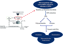
Free nitrous acid (FNA) has been demonstrated to be effective in enhancing the degradability of waste activated sludge (WAS). Considering that extracellular polymeric substances (EPS) are a major component in sludge flocs, the chemical breakdown of EPS components by FNA has been hypothesized to account for the improvement of sludge biodegradability in addition to enhanced cell lysis. EPS extracted from WAS was treated with FNA at 2.0 mg HNO2-N per L (260 mg NO2-N per L and pH 5.5). The molecular weight distribution of EPS showed the breakdown of macromolecules into smaller molecules. The chemical structure analysis of EPS using Fourier transform infrared spectroscopy ascribed the breakdown to FNA-induced deamination of proteins, amino sugars and nucleic acids, implying that the main targets of FNA in EPS are protein-like substances. Particle size distribution analysis on the original WAS with the same FNA treatment revealed that FNA treatment of sludge significantly reduces the flocs sizes, which supported that FNA breaks down EPS in activated sludge flocs.


1. Introduction

Activated sludge processes produce substantial amounts of waste activated sludge (WAS), the treatment and disposal of which incur large costs.1–3 Aerobic and anaerobic sludge digestion is the mainstream technology for sludge reduction and stabilization prior to disposal, but their effectiveness is limited by the poor degradability of WAS. Various methods including mechanical, thermal and chemical treatment have been proposed to enhance the WAS biodegradability prior to sludge digestion.3–7 However, the above approaches require either intensive energy input (e.g. high pressure or high temperature) or large chemical consumption (e.g. chlorine, ozone or alkali), incurring substantial economic costs.

Free nitrous acid (FNA, i.e. HNO2), a renewable and low cost chemical that can be produced on site by nitritation of the anaerobic digestion liquor,8 has been demonstrated to be effective in causing cell lysis9,10 and improving sludge biodegradability.11 For example, aerobic digestion tests showed that the degradation of WAS with FNA pre-treatment at 2.0 mg N per L for 48 h was more than two times higher than that of WAS without FNA pre-treatment.10 Wang et al.12 reported that WAS production in a reactor treating synthetic domestic wastewater was reduced by 28% by treating part of the returned activated sludge with FNA at 2.0 mg N per L for 24 h. Recently, it has been demonstrated that methane production from a full-scale WAS, with FNA pre-treatment at 2.0 mg N per L for 24 h, was improved by approximately 30% at an anaerobic digestion time of 20 days in comparison with that from the WAS without FNA pre-treatment.11 The FNA-based sludge treatment technology was further proposed to be economically attractive,11–13 which poses a significant advantage over most of the current available cost-intensive technologies. However, a detailed understanding of the mechanisms involved in FNA treatment is still in need, which is essential for the optimization of the technology. It has been hypothesized that the improved sludge biodegradability was due to the enhanced cell lysis caused by FNA treatment.9,10,12

However, the sludge consists of not only cells, but also extracellular polymeric substances (EPS), which comprise different types of biopolymers (e.g. proteins and polysaccharides) to form the three-dimensional scaffold structure of the sludge aggregates and facilitate them function effectively. In fact, EPS can account for up to 33–42% of the volatile solids in sludge12,14 and their components and structure have significant impacts on sludge biodegradability.15–18 Many studies have proven that both aerobic and anaerobic biodegradability of WAS was enhanced effectively by removing the EPS from the WAS flocs through lytic bacterial strains, bacteria secreted enzymes or surfactant.19–22 Therefore, the enhanced cell lysis may not be the sole reason for the improved sludge biodegradability, and FNA may also break down EPS by reacting with the components and changing their chemical structures.

This study aims to verify the hypothesis that FNA breaks down EPS in activated sludge flocs, which accounts for the partial release of EPS into soluble phase and the improvement of the sludge biodegradability in addition to the enhanced cell lysis. To verify this hypothesis, EPS extraction was used to investigate the working principles of the FNA-based sludge treatment technology. EPS extracted from WAS was treated with FNA at 2.0 mg HNO2-N per L (260 mg NO2-N per L and pH 5.5), with untreated EPS and EPS treated at pH 5.5 alone as controls. The molecular weight (MW) distribution and the chemical structure of EPS were analyzed and compared by gel permeation chromatography (GPC) and Fourier transform infrared (FTIR), respectively. The same treatments were also directly applied to WAS and then the particle size distribution of sludge flocs was measured and compared.

2. Materials and methods

2.1. Sludge source

WAS used in this experimental study was collected from a local biological nutrient removal wastewater treatment plant treating domestic wastewater at three different time (27/08/2013, 04/05/2014 and 23/05/2014) with 2 L each time. Following collections, it was transported to the lab within 2 hours and stored at 4 °C overnight prior to EPS extraction and sludge treatment as described in Sections 2.2 and 2.3. The main characteristics of WAS were: total solids (TS) 9.6 ± 0.1 g L−1, volatile solids (VS) 7.3 ± 0.1 g L−1, total chemical oxygen demand (TCOD) 9.2 ± 0.0 g L−1, soluble chemical oxygen demand (SCOD) 0.11 ± 0.01 g L−1, and pH = 7 ± 0.2 (with standard errors obtained from triplicate measurements of all samples).

2.2. EPS extraction

The sludge was firstly centrifuged at 2000g, 4 °C for 15 min and the supernatant was decanted to remove the soluble microbial products in the bulk water. The pellet was then dissolved to their original volume with extraction buffer consisting of 2 mM Na3PO4, 4 mM NaH2PO4, 9 mM NaCl and 1 mM KCl at pH 7 with the similar ionic strength to that in the original sludge according to ref. 23 to extract both loosely and tightly bound EPS. The modified cation exchange resin (CER) method (DOWEX 50 × 8, 20–50 mesh in Na+, Sigma-Aldrich) was then used for EPS extraction, which is capable of maintaining the biochemical properties of EPS components in comparison with the chemical extraction methods.24 The CER was added to the extraction beaker with the ratio of 70 g/g VS with the redissolved sludge and the mixture was stirred at 600 rpm, 4 °C for 6 hours. Afterwards, the mixture was settled for 5 min for the separation of the resin from the sludge. Then the mixture was centrifuged at 12[thin space (1/6-em)]000g at 4 °C for 20 min. The supernatant filtered through disposable Millipore filter units (0.45 μm pore size) was the final EPS extract. EPS concentrations throughout the study are expressed in bovine serum albumin (protein), D-glucose (carbohydrates) and calf thymus (DNA) equivalents per gram VS. The yield of EPS extraction was 5% (VS basis), which is comparable with the previous studies.23,25 The FTIR analyses on both original sludge and EPS extract (data not shown) showed that their chemical compositions are highly similar, suggesting that the EPS extract was sufficiently representative for the chemical functional groups even though the extraction yield was not very high. The main compositions were proteins (38.3 ± 1.8 mg g−1 VS) and polysaccharides (19.1 ± 0.5 mg g−1 VS), accounting for up to 66% and 33% of the total EPS extract, respectively. The DNA concentration was 0.50 ± 0.02 mg g−1 VS, which is in consistency with previous studies,23,25 indicating a minimal cell lysis during the extraction.

2.3. FNA treatment on EPS extract and WAS

The tests described below were carried out on all the three sludge samples collected at different time (see Section 2.1).

In each set of tests, three batch reactors each with 100 mL EPS extract were used to investigate the effect of FNA on molecular weight distribution and chemical structures of the components in EPS extract. Two served as controls, with the other as the experimental reactor. The operating conditions are as summarised in Table 1. A nitrite stock solutions (40 g N per L) was added to the reactor to achieve the designed level of 260 mg N per L. Since the pH increased after FNA addition, 1 M HCl was used for pH control via a programmable logic controller. The FNA concentration achieved was estimated to be 2.0 mg HNO2-N per L using the formula image file: c5ra06080j-t1.tif with the Ka value determined as a function of temperature T (°C) by Ka = e−2300/(273+T) (22 °C in this study).23,26 The effectiveness of FNA on anaerobic methane production at different concentrations have been investigated. In combination with the economic analysis, FNA treatment at 2.0 mg HNO2-N per L for 24 hours resulted in the highest methane production with significant economic advantages.11 Therefore, these conditions were selected in this study. After the 24 hour treatment, the EPS extract in each reactor was divided into two halves, with half for molecular weight distribution measurement and the other half freeze-dried for chemical structure analysis.

Table 1 Treatment conditions applied in this study
Treatment FNA (mg N per L) NO2–N (mg N per L) pH Treatment time (h)
a pH was not controlled.b pH was controlled.
Control I 0 0 6.8–7.0a 24
Control II 0 0 5.5 ± 0.2b 24
FNA 2.0 260 5.5 ± 0.2b 24


In parallel to the above reactors, three other batch reactors each with 100 mL WAS, the same as that used for EPS extraction, were used to study the impacts of FNA treatment on particle size distribution of WAS. The WAS in the three reactors was subject to the same treatments conditions as applied to the EPS extract. The WAS collected after 24 hours treatment was analysed for its particle size distribution in less than two hours after the treatment completed.

2.4. Chemical analyses

Concentrations of proteins and DNA in the EPS extracts were analysed with Peirce™ Coomassie (Bradford) Protein Assay kit (Pierce Biotechnology, USA) and DNA Quantification kit (Fluorescence assay using bisBenzimide H 33258, Sigma Aldrich, USA), respectively. Carbohydrates were quantified with the phenol–sulfuric acid method.27 Bovine serum albumin (BSA), D-glucose (Sigma Aldrich) and calf thymus DNA were used to prepare standard curves in the same extraction buffer as EPS extraction. To normalize the concentrations of the three major components in EPS, the measured volume concentrations (mg BSA-, mg D-glucose- and mg calf-thymus equivalents per L) were then divided by the volatile solids concentration (g VS L−1) in the original sludge samples to obtain the mass-based concentrations (mg BSA-, mg D-glucose- and mg calf-thymus equivalents per g VS). All measurements were done in triplicate. The concentrations of NO2, NO3, and NH4+ were determined using a Lachat QuikChem8000 Flow Injection Analyzer (Lachat Instrument, Milwaukee, Wisconsin). TS, VS, TCOD and SCOD was determined according to the standard method.28

2.5. Analyses of molecular weight distribution and chemical structure of EPS

The molecular weight (MW) distribution of EPS was analysed with Agilent Aqueous Gel permeation chromatography 1260 Infinity System connected with refractive index (RI) and ultraviolet (UV) detectors for data acquisition and equipped with Agilent GPC/SEC software for operation and data analysis. Two Agilent Aqua gel PL Aquagel-OH mixed-M, 8 μm columns were kept at 40 °C with Milli-Q water as eluent at a flow rate of 1 mL min−1 during the analysis. UV and RI detectors were used in series to provide complementary information with regards to MW distribution of the mixture consisting of UV non-absorbing (polysaccharides) and absorbing (protein, amino sugars and nucleic acids) constituents. The Pullulan Polysaccharide calibration kit (MW: 180, 667, 6100, 9600, 21[thin space (1/6-em)]100, 47[thin space (1/6-em)]100, 107[thin space (1/6-em)]000, 194[thin space (1/6-em)]000, 344[thin space (1/6-em)]000, 708[thin space (1/6-em)]000 g mol−1) was used to establish the calibration curve.

The chemical structure of the components in EPS extract was analysed by Fourier transform infrared spectroscopy (FTIR) using Nicolet 5700 FTIR spectrometer with the resolution at 4 and Gain 8 with 64 scans in the spectral range of 400–4000 cm−1. The diamond smart accessory with DTGS Tec detector was used for analysing the sample at room temperature. The chamber was purged continuously with the nitrogen gas during the operation.

2.6. Analysis of particle size distribution of WAS flocs

The particle size distribution of sludge with and without FNA treatment was analysed by Mastersizer 2000 particle size analyzer (Malvern Co.) equipped with Hydro 2000MU sample dispersion unit with the measurement range from 0.02 to 2000 μm. Distilled water (2 L) was used as a liquid dispersant. Sludge samples were well mixed and then added to the dispersant until the laser obscuration reached the optimum range and then the particle size distribution was measured with the technique of laser diffraction, which is based on the principle that particles passing through a laser beam will scatter light at an angle that is directly related to their size. The measurement for each sample was repeated for 5 times to ensure that the results were reproducible.

3. Results

3.1. Effect of FNA on molecular weight distribution of EPS

The molecular weight distribution and chemical structure analyses were applied to the EPS extracted from the three batches of sludge. The results were reproducible and herewith only the results from 04/05/2014 were interpreted as a representative.

Fig. 1 shows the molecular weight distribution of EPS with and without FNA treatment. The corresponding molecular weights of the peaks at different retention times are listed in Table 2. Substantial peak shifts were observed from UV 254 nm detector after FNA treatment. The peak at 15.36 min (in Control I) shifted to 15.67 min after FNA treatment, corresponding to the decrease of MW from 57[thin space (1/6-em)]300 Da to 42[thin space (1/6-em)]015 Da. Similarly, the peak at 17.12 min shifted to 17.60 min, representing the decrease of MW from 10[thin space (1/6-em)]048 Da to 7238 Da after FNA treatment. In the case of Control II, the decrease of the MWs in comparison with that in Control I was also observed but to a much less extent than that after FNA treatment, from 57[thin space (1/6-em)]300 Da at 15.36 min to 52[thin space (1/6-em)]149 Da at 15.46 min and from 10[thin space (1/6-em)]048 Da at 17.12 min to 9690 Da at 17.22 min, respectively. Besides peaks shifts, the intensity of the peak at around 17.60 min was significantly intensified after FNA treatment in comparison to that of Control I and Control II, indicating the breakdown of large molecules into smaller ones. In addition to the intensity changes, the shoulder peak at 16.25 min in Control I representing MW of 15[thin space (1/6-em)]949 Da disappeared after FNA treatment, while no significant change was observed in the case of Control II. Also, FNA treatment yielded more new peaks in the range with MW lower than 180 Da. All the evidence collectively suggested the breakdown of proteins, nucleic acids and/or amino sugars.


image file: c5ra06080j-f1.tif
Fig. 1 GPC results of molecular weight distribution of EPS with and without FNA treatment. (UV detector: A 27/08/2013, B 04/05/2014, C 23/05/2014; RI detector: A 27/08/2013, B 04/05/2014, C 23/05/2014).
Table 2 Retention times and corresponding MWs of the peaks with and without FNA treatment
  Peaks Control I Control II FNA treatment
Time (min) MW (Da) Time (min) MW (Da) Time (min) MW (Da)
UV 254 nm detector Peak 1 15.36 57[thin space (1/6-em)]300 15.46 52[thin space (1/6-em)]149 15.67 42[thin space (1/6-em)]015
Peak 2 17.12 10[thin space (1/6-em)]048 17.22 9690 17.60 7238
RI detector Peak 1 15.05 32[thin space (1/6-em)]732 15.15 30[thin space (1/6-em)]856 15.23 29[thin space (1/6-em)]945
Peak 2 17.47 29[thin space (1/6-em)]71 17.52 2889 17.80 2525
Peak 3 Not applicable Not applicable 19.83 245


The RI detector showed a similar trend. Substantial peak shifts were observed from RI detector after FNA treatment. The peak acquired from Control I at 15.05 min shifted to 15.23 min, representing the decrease of MW from 32[thin space (1/6-em)]732 Da to 29[thin space (1/6-em)]945 Da after FNA treatment. The more significant peak shift took place from 17.47 min to 17.80 min, implying the MW decreased from 2971 Da to 2525 Da after FNA treatment. In the case of Control II, slight decrease of MWs in comparison with that in Control I was observed but also to a much less extent compared to that after FNA treatment from 32[thin space (1/6-em)]732 Da at 15.05 min to 30[thin space (1/6-em)]856 Da at 15.15 min and from 2971 Da at 17.47 min to 2889 Da at 17.52 min. The peak at around 17.80 min after FNA treatment was substantially intensified compared to those of Control I and Control II. Besides the peaks shifts, the new peak representing MW of 245 Da emerged only after FNA treatment. These results collectively evidenced that FNA could also break down polysaccharides.

However, it is noticeable that the MW shifts from RI detector are not as distinct as those from UV detector, implying the main target of FNA in the EPS is UV absorbing substances, such as protein, amino sugars and nucleic acids.

3.2. Effect of FNA on chemical structures of EPS

The FTIR results revealed the chemical structure changes caused by FNA treatment as shown in Fig. 2. Four main characteristic functional groups were identified: 1700–1600 cm−1 for amide I region; 1500–1300 cm−1 for carboxylic group; 1200–900 cm−1 for carbohydrates and nucleic acid, and 900–600 cm−1 for fingerprint region. The broad band near 3300 cm−1 corresponds to the stretching of both νO–H and νN–H. The bands around 1640 cm−1 and 1550 cm−1 represent νsC[double bond, length as m-dash]O stretching and δN–H and νsC–N stretching, respectively, indicating the existence of amide I and amide II associated with proteins. The shoulder band around 1129 cm−1 represents δC–OH, δC–O, and νC–O associated with amino acids. The band around 1060 cm−1 corresponds to νC–OH of phosphorylated proteins and polysaccharides. The wide band around 950 cm−1 represents νasO–P–O stretching associated with nucleic acids. Bands at 600–900 cm−1 denote the existence of ring vibration associated with νC–C and νC–OH from aromatic amino acids (e.g. Phe, Trp and Tyr) and nucleotides.29 The band assignment is summarised in Table 3.
image file: c5ra06080j-f2.tif
Fig. 2 FTIR spectra of EPS with and without FNA treatment (A 27/08/2013, B 04/05/2014, C 23/05/2014).
Table 3 FTIR spectra band assignment30–32
Frequencies (cm−1) Band assignment
3300 νO–H and νN–H associated with alcohols and amines
1640 νsC[double bond, length as m-dash]O stretching (amide I) and νC[double bond, length as m-dash]N stretching associated with proteins
1550 δN–H and νsC–N stretching in amide II associated with proteins
1129 δC–OH, δC–O, and νC–O associated with amino acids
1060 νO–H deformation, νC–O stretching, ring vibration νP[double bond, length as m-dash]O, C–O–C and C–O–P in phosphodiesters and polysaccharides
950 νasO–P–O stretching associated with nucleic acids
900–600 Ring vibrations associated with νC–C and νC–OH from aromatic amino acids and nucleotides


The peak at 1550 cm−1 was weakened after FNA treatment, while in the case of Control II, no changes happened to this peak. For the shoulder peak at 1129 cm−1, a slight peak shift was observed in the case of Control II while FNA treatment extended the shoulder band to an obvious peak in comparison to that in Control I. This is likely due to the deaminative depolymerisation of proteins and/or amino sugars by FNA and/or its derivatives.33,34 The band at 950 cm−1 split into two vibration bands near 950 cm−1 and 990 cm−1 only after FNA treatment, implying the presence of FNA-induced mutagenic changes of nucleic acids. It was reported that this mutagenesis was also accredited to the deamination of the amino bases in nucleic acids.32 Slight impairment of the peaks between 600–900 cm−1 after FNA treatment indicated that FNA is capable of destructing the ring structure, which likely improves the biodegradability of the hardly biodegradable organic matters.35 It should be noted that the breakdown of the macromolecules is ascribed to the chemical reactions between them and FNA and/or its derivatives instead of the microbial degradation. This is because that all the EPS samples have been filtered through 0.45 μm pore size disposable filters, which could remove the majority of the microorganisms. In addition, previous studies have demonstrated that FNA at a concentration of parts per million (ppm) level has a very strong biocidal effect on microorganisms.9,36 Therefore, it is believed that, at the FNA concentration used in our study (2.0 mg HNO2-N per L), a residual amount of microorganisms that might have gone through the filters have been inactivated and would not contribute to the breakdown of the macromolecules.

3.3. Effects of FNA on particle size distribution of WAS flocs

Fig. 3 shows the sludge particle size distribution with and without FNA treatment. The d50 values are also shown in the figure. In comparison to the d50 in Control I (102 ± 2 μm), it remained without significant changes in the Control II (99 ± 1 μm) (p > 0.05), whereas after FNA treatment, it was reduced significantly to 69 ± 1 μm (p < 0.01), 32% and 30% lower than that in Control I and Control II. The shift of particle size distribution after FNA treatment indicated the drastic decrease in particle size. These imply that the sludge flocs were disintegrated, supporting that FNA led to the breakdown of EPS in flocs.
image file: c5ra06080j-f3.tif
Fig. 3 Sludge particle size distribution with and without FNA treatment.

4. Discussion

This study reveals for the first time that FNA breaks down EPS. This was evidenced by the changes in molecular weight distribution in the EPS extract and the chemical structures. The chemical structure analysis of EPS extract with and without FNA treatment revealed that the breakdown is due to the oxidative and deaminative depolymerisation of proteins, amino sugars and amino bases in nucleic acids by FNA or its derivatives. FNA has been reported to be able to cause oxidative deamination of NH2 group in adenine or cytosine to ether group36,37 and chemically destroy DNA structure by converting adenine to hypoxanthine (which pairs with C), cytosine to uracil (which pairs with A) and guanine to xanthine (which still pairs with C).32 The deaminative cleavage of model amino sugar glycosides and glycosaminoglycuronans by FNA was also experimentally demonstrated.37 Besides, it was also reported that the reactive nitrogen species (RNSs) derived from nitric oxide, which is one of the derivatives from FNA,38 could chemically depolymerize carbohydrates through deamination.39 The results from our study are consistent with the previous studies. However, it should be noted that our study, for the first time, demonstrated the breakdown of the microbial produced molecules (i.e. EPS) by FNA while the previous studies focused on the chemistry between FNA and the synthetic pure macromolecules.

It has been demonstrated that extracellular proteins are strongly involved in the aggregation of bacteria into flocs.39,40 Therefore, the breakdown of proteins caused by FNA treatment could result in the disintegration of EPS matrix. Furthermore, the breakdown of the macromolecules and ring structures due to FNA-induced deamination and oxidation potentially converted hardly biodegradable substances into more easily biodegradable ones,41 which likely contributed to the enhanced sludge biodegradability.11 A recent study showed that the toxic metal removal from WAS with FNA treatment at 19.2 mg HNO2-N per L (i.e. 20 mg NO2-N per L at pH 2) was significantly enhanced in comparison to that from WAS with acid treatment at pH 2 alone.42 Further analysis verified that the improvement of toxic metal removal mainly resulted from the release of organically bound metals. This observation can be explained by the results reported in our study.

The FNA based sludge treatment technology has been experimentally demonstrated to be an effective and economically attractive method for improving sludge biodegradability.11 This study significantly improved the understanding of the working principles of this technology, which is essential in the optimization of this technology. Additionally, it is also of importance in providing the fundamental knowledge for the development of new possible sludge treatment technologies using FNA in combination with other chemicals.

5. Conclusions

The effect of FNA on EPS from waste activated sludge was investigated through the molecular weight distribution and chemical structure analyses of the components in EPS extract with and without FNA treatment. The main conclusions are:

(1) FNA could break down macromolecules in EPS, resulting in disintegration of activated sludge flocs.

(2) The breakdown is mainly due to the oxidative and deaminative depolymerisation of proteins, amino sugars and amino bases in nucleic acids by FNA and/or its derivatives.

Acknowledgements

The authors acknowledge Australian Research Council for funding support through Discovery Project DP120102832 and Linkage Project LP130100361, and the funding support from the University of Queensland Early Career Research Grant (ECR040) awarded to Dr Qilin Wang. Ms Tingting Zhang acknowledges the scholarship support from China Scholarship Council and also acknowledges Dr Bogdan Donose for his professional help with FTIR analyses. This work was performed in part at the Queensland node of the Australian National Fabrication Facility, a company established under the National Collaborative Research Infrastructure Strategy to provide nano and micro-fabrication facilities for Australia's researchers.

References

  1. T. Shimizu, K. Kudo and Y. Nasu, Biotechnol. Bioeng., 1993, 41, 1082–1091 CrossRef CAS PubMed .
  2. A. Canales, A. Pareilleux, J. Rols, G. Goma and A. Huyard, Water Sci. Technol., 1994, 30, 97–106 CAS .
  3. L. Appels, J. Baeyens, J. Degreve and R. Dewil, Prog. Energy Combust. Sci., 2008, 34, 755–781 CrossRef CAS PubMed .
  4. G. Lissens, A. B. Thomsen, L. De Baere, W. Verstraete and B. K. Ahring, Environ. Sci. Technol., 2004, 38, 3418–3424 CrossRef CAS .
  5. H. Carrere, C. Dumas, A. Battimelli, D. J. Batstone, J. P. Delgenes, J. P. Steyer and I. Ferrer, J. Hazard. Mater., 2010, 183, 1–15 CrossRef CAS PubMed .
  6. P. Foladori, G. Andreottola and G. Ziglio, Sludge reduction technologies in wastewater treatment plants, IWA Pub., London, New York, 2010 Search PubMed .
  7. M. Carballa, C. Duran and A. Hospido, Environ. Sci. Technol., 2011, 45, 10306–10314 CrossRef CAS PubMed .
  8. Y. Y. Law, L. Ye, Q. L. Wang, S. H. Hu, M. Pijuan and Z. G. Yuan, Chem. Eng. J., 2015, 259, 62–69 CrossRef CAS PubMed .
  9. G. M. Jiang, O. Gutierrez and Z. G. Yuan, Water Res., 2011, 45, 3735–3743 CrossRef CAS PubMed .
  10. M. Pijuan, Q. L. Wang, L. Ye and Z. G. Yuan, Bioresour. Technol., 2012, 116, 92–98 CrossRef CAS PubMed .
  11. Q. Wang, L. Ye, G. Jiang, P. D. Jensen, D. J. Batstone and Z. Yuan, Environ. Sci. Technol., 2013, 47, 11897–11904 CrossRef CAS PubMed .
  12. Q. Wang, L. Ye, G. Jiang and Z. Yuan, Water Res., 2013, 47, 3663–3672 CrossRef CAS PubMed .
  13. Q. Wang and Z. Yuan, RSC Adv., 2015, 5, 19128–19134 RSC .
  14. B. Frølund, R. Palmgren, K. Keiding and P. H. Nielsen, Water Res., 1996, 30, 1749–1758 CrossRef .
  15. Y. Magara, S. Nambu and K. Utosawa, Water Res., 1976, 10, 71–77 CrossRef CAS .
  16. B. Q. Liao, D. G. Allen, I. G. Droppo, G. G. Leppard and S. N. Liss, Water Res., 2001, 35, 339–350 CrossRef CAS .
  17. Z. W. Wang, Y. Liu and J. H. Tay, Appl. Microbiol. Biotechnol., 2007, 74, 462–466 CrossRef CAS PubMed .
  18. C. Park, R. F. Helm and J. T. Novak, Water Environ. Res., 2008, 80, 2219–2227 CrossRef CAS .
  19. S. Kavitha, S. Adish Kumar, K. N. Yogalakshmi, S. Kaliappan and J. Rajesh Banu, Bioresour. Technol., 2013, 150, 210–219 CrossRef CAS PubMed .
  20. S. Kavitha, C. Jayashree, S. Adish Kumar, I. T. Yeom and J. Rajesh Banu, Bioresour. Technol., 2014, 168, 159–166 CrossRef CAS PubMed .
  21. S. Kavitha, S. Adish Kumar, S. Kaliappan, I. T. Yeom and J. Rajesh Banu, Bioresour. Technol., 2014, 169, 700–706 CrossRef CAS PubMed .
  22. M. V. Lakshmi, J. Merrylin, S. Kavitha, S. A. Kumar, J. R. Banu and I. T. Yeom, Environ. Sci. Pollut. Res. Int., 2014, 21, 2733–2743 CrossRef CAS PubMed .
  23. H. Liu and H. H. Fang, J. Biotechnol., 2002, 95, 249–256 CrossRef CAS .
  24. Y. Liu and H. H. Fang, Crit. Rev. Environ. Sci. Technol., 2003, 33(3), 237–273 CrossRef CAS PubMed .
  25. P. D'Abzac, F. Bordas, E. Van Hullebusch, P. N. Lens and G. Guibaud, Appl. Microbiol. Biotechnol., 2010, 85, 1589–1599 CrossRef PubMed .
  26. A. C. Anthonisen, R. C. Loehr, T. B. S. Prakasam and E. G. Srinath, J. – Water Pollut. Control Fed., 1976, 48, 835–852 CAS .
  27. G. Kochert, in Handbook of phycological methods: physiological and biochemical methods, Cambridge University Press London, 1978, pp. 95–97 Search PubMed .
  28. A. D. Eaton and M. A. H. Franson, Standard methods for the examination of water and wastewater, American Public Health Association, 2005 Search PubMed .
  29. A. R. Badireddy, S. Chellam, P. L. Gassman, M. H. Engelhard, A. S. Lea and K. M. Rosso, Water Res., 2010, 44, 4505–4516 CrossRef CAS PubMed .
  30. R. L. Taylor and H. E. Conrad, Biochemistry, 1972, 11, 1383–1388 CrossRef CAS .
  31. J. Duan, F. Y. Avci and D. L. Kasper, Proc. Natl. Acad. Sci. U. S. A., 2008, 105, 5183–5188 CrossRef CAS PubMed .
  32. H. Schuster, Biochem. Biophys. Res. Commun., 1960, 2, 320–323 CrossRef .
  33. D. R. Klein, Organic Chemistry, John Wiley, Hoboken, 2nd edn, 2013 Search PubMed .
  34. W. S. Klug and M. R. Cummings, Concepts of genetics, Pearson Education, Inc, 2003 Search PubMed .
  35. H. V. Malling, Environ. Mol. Mutagen., 2004, 44, 372–386 CrossRef CAS PubMed .
  36. S. S. Yoon, R. Coakley, G. W. Lau, S. V. Lymar, B. Gaston, A. C. Karabulut, R. F. Hennigan, S. H. Hwang, G. Buettner, M. J. Schurr, J. E. Mortensen, J. L. Burns, D. Speert, R. C. Boucher and D. J. Hassett, J. Clin. Invest., 2006, 116, 436–446 CAS .
  37. J. E. Shively and H. E. Conrad, Biochemistry, 1970, 9, 33–43 CrossRef CAS .
  38. M. J. Higgins and J. T. Novak, J. Environ. Eng., 1997, 123, 479–485 CrossRef CAS .
  39. F. Martinez, E. Favela-Torres and J. Gomez, Appl. Biochem. Biotechnol., 2000, 87, 177–188 CrossRef CAS .
  40. A. Ramesh, D. J. Lee and S. G. Hong, Appl. Microbiol. Biotechnol., 2006, 73, 219–225 CrossRef CAS PubMed .
  41. B. J. Ni, R. J. Zeng, F. Fang, W. M. Xie, G. P. Sheng and H. Q. Yu, Water Res., 2010, 44, 2292–2302 CrossRef CAS PubMed .
  42. F. Du, S. Freguia, Z. Yuan, J. Keller and I. Pikaar, Environ. Sci. Technol., 2015 DOI:10.1021/es504507m .

This journal is © The Royal Society of Chemistry 2015
Click here to see how this site uses Cookies. View our privacy policy here.