Azine or hydrazone? The dilemma in amidinohydrazones

Ashok Ramakrishnan, Sumit S. Chourasiya and Prasad V. Bharatam*
Department of Medicinal Chemistry, National Institute of Pharmaceutical Education and Research, (NIPER) Sector-67, S.A.S. Nagar-160 062, Mohali, Punjab, India. E-mail: pvbharatam@niper.ac.in; Fax: +91-172-2214692; Tel: +91-172-2292018

Received 29th March 2015 , Accepted 26th May 2015

First published on 26th May 2015


Abstract

Azines belong to an important class of compounds that are found to have numerous applications in medicinal chemistry. Hydrazones are related to azines and are more widely known compounds that carry many biochemical applications. Hydrazones with appropriate substituents can show azine–hydrazone tautomerism. There are many cases in which azines are wrongly considered to be hydrazones. In this article, we report the tautomeric energy differences of azine and hydrazone and provide the structural details of amidinohydrazones, which prefer azine structure rather than that of hydrazone structure, an important example being the anti-hypertensive drug, guanabenz. The importance of appropriate tautomeric representation of guanabenz has been established in terms of its molecular interactions with a known enzyme.

Introduction

Hydrazones are characterized by the presence of an imine (C[double bond, length as m-dash]N) linked to an amino (–NHR) moiety (–C[double bond, length as m-dash]N–NHR).1 Azines (also known as 2,3-diazabutadienes) carry two ‘C–N’ double bonds in conjugation with an ‘N–N’ linker and prefer to exhibit in (E/E) configuration (–C[double bond, length as m-dash]N–N[double bond, length as m-dash]C–).2 Recently, hydrazones and azines have been explored for various chemical and biological applications.1–19 Hydrazones3 and azines4 serve as synthons for the synthesis of many heterocyclic compounds. The formation of stable complexes with most of the transition metal ions has increased the interest in hydrazone5 and azine6 complexes because it was recognized that many of these complexes may serve as models for biologically important species and were reported to act as enzyme inhibitors. Hydrazone derivatives have been suggested for several pharmacological applications, including antimicrobial,7 antimycobacterial,8 antimalarial,9 anticonvulsant,10 antidepressant,11 analgesic and anti-inflammatory,12 anticancer13 and antiplatelet.14 Similarly, azine derivatives are being studied for antibacterial,15 antifungal,15 antifilarial,16 anticancer,17 opiate antagonist18 and molluscacidal19 activities.

Amidinohydrazones (a) (also known as ‘guanylhydrazones’) are a special class of hydrazones containing an amidino group (Fig. 1).20 Amidinohydrazones exhibit several biological activities. For example, guanabenz (Wy-8678) is an α-2 adrenoreceptor agonist, which is being marketed as an anti-hypertensive agent.21 Other molecules containing amidinohydrazone moiety, such as CNI-1493 (inhibits macrophage activation and subsequent proinflammatory cytokine production)22 and CGP-48664 (S-adenosylmethionine decarboxylase, SAMdc, inhibitor acting as an anti-proliferative agent)23 are currently in clinical trials. Methylglyoxalbisguanylhydrazone (MGBG)20,24,25 is an agent with a unique mechanism of action of polyamine biosynthesis inhibition (Fig. 2). Other pharmacological actions reported by amidinohydrazones include thrombin inhibition,26 furin inhibition in many bacterial and viral diseases,27 and neurodegenerative disorders.28 Recently, they have been explored for their use as thermally stable energetic materials.29


image file: c5ra05574a-f1.tif
Fig. 1 General representations of (a) amidinohydrazone and (b) 1,1-diamino-2,3-diazabutadiene (azine) tautomer.

image file: c5ra05574a-f2.tif
Fig. 2 Some important biologically active molecules containing amidinohydrazone moiety in their traditional representation.

A notable phenomenon that takes place in drug molecules is tautomerism.30 Tautomers interconvert with a relatively low activation energy of less than 20 kcal mol−1 for the common keto–enol tautomerism.31 The molecular structure and polarity of the solvent define the tautomeric form. In addition, pH of the environment is also an important factor.32 It is important to establish the preferred tautomeric state of drug molecules because drug action can vary as a function of the preferred tautomeric state. In particular, when the drug action of any therapeutic agent is being explored using molecular modelling methods, any erroneous representation can lead to undesirable conclusions and expectations. Quantum chemical studies can be used to establish the preferred tautomeric state of any chemical species and to explain the reasons for such preference.31,34 The smaller the ΔE or ΔG between the tautomers, the greater is the possibility of equilibrium between two tautomeric states. The preferred structures of the anti-diabetic agent, metformin, and the anti-malarial agent, proguanil, were conclusively established using quantum chemical studies.33k Furthermore, the design of novel guanyl thiourea derivatives (anti-malarial) was considered on the basis of the data obtained on the tautomeric studies using electronic structure analysis.33i,35

Amidinohydrazones are generally represented with structure a. They can also exist in the corresponding tautomeric state b as 1,1-diamino-2,3-diazabutadiene (azine) (Fig. 1). In the past, hydrazone–azine tautomerism has been studied and reported (i) in a 14-membered macrocyclic ligand by Bell and co-workers,36 (ii) in 4-(1-alkylbenzimidazol-2-ylazo)-2-pyrazolin-5-ones by Morkovnik and co-workers37 and (iii) in aldazines by Silva and co-workers.38 The crystal structures of glyoxal bis guanylhydrazone (GBG)39 and its derivatives, EMGBG,40 MPGBG41 and PhGBG,42 were indeed established as azines rather than as hydrazones. Similarly, the crystal structures of 2-(1-phenylethylideneamino)guanidine43 and guanabenz were established as azines.21b The existence of the two tautomeric forms, hydrazone and azine, of amidinohydrazones has been experimentally proven by Zoltan and co-workers.44 However, in published reports of both medicinal and organic chemistry, the title compounds are represented as hydrazones. In particular, molecular docking studies have been performed using the hydrazone structure.28,45 This leads to a fundamental question as to which representation is more suitable (a or b) for amidinohydrazones, which is explicitly addressed in this article. Quantum chemical analysis on the azine–hydrazone tautomerism in amidinohydrazones has been carried out, and the conditions that influence the tautomeric energy have been explored. Finally, the importance of representing the drug molecule, guanabenz, in the azine form has been evaluated using a molecular docking analysis.

Methods of calculation

The quantum chemical calculations were carried out using GAUSSIAN09 package.46 Full geometry optimizations were carried out by ab initio MO calculations47 using MP248 and G2MP249 methods and DFT calculations50 using B3LYP,51 M0652 and CBS-Q53 methods. The basis set used was 6-311++G(d,p). An implicit solvent study was performed using the B3LYP/6-311++G(d,p) and IEFPCM solvent model.54 Explicit solvent studies were also carried out in the gas phase using the same method for up to four water molecules. Frequencies were computed analytically for all the optimized species for the characterization of each stationary state as that of a minimum or transition. The Gibbs free energy (ΔG) difference between the two tautomers has been considered in all the observations discussed below. The partial atomic charges were estimated using NBO analysis.55 The electron localization function (ELF)56 calculations were performed on the most stable tautomers using Multiwfn 3.3.6 software57 to estimate the total electron density localization at the nitrogen centres. The molecular electrostatic potentials (MESP)58 were obtained on the B3LYP/6-311++G(d,p) optimized geometries of hydrazone and azine tautomers of amidinohydrazone and superimposed onto a constant electron density (0.004 e au−3) to provide a measure of the electrostatic potential at roughly the van der Waals surface of the molecules using GAUSSIAN09 software.46 The colour-coded surface provides the location of the positive (deepest blue, most positive) and negative (deepest red, most negative) electrostatic potentials on a molecular surface. Positive potential regions indicate relative electron deficiency (estimated as a function of the repulsion experienced by a positively charged test probe), and regions of negative potential that indicate areas of excess negative charge (estimated as a function of the attractive force experienced by a positively charged test probe). Molecular docking studies were carried out using the Glide59 module of Maestro 9.4 (Schrodinger Inc.)60 to demonstrate the importance of tautomeric representation of amidinohydrazone during the molecular docking analysis.

Results and discussions

Quantum chemical calculations were performed on hydrazones (1a–5a) and their azine counterparts (1b–5b) (Scheme 1). The Gibbs free energy difference between the tautomers was calculated at all levels of theory in the gas phase and the relative energy of the hydrazone tautomers with respect to the corresponding azine are given in Table 1.
image file: c5ra05574a-s1.tif
Scheme 1 Predicted tautomerism in hydrazones by 1,3-proton shift.
Table 1 The Gibbs free energy difference (ΔG) in kcal mol−1 between hydrazone and azine tautomers with azine structure taken as a base
Compound Relative Gibbs free energy, ΔG (kcal mol−1)
MP2 B3LYP M06 G2MP2 CBS-Q
1a −7.07 −8.87 −9.31 −6.21 −12.13
2a −2.01 −5.09 −4.58 −2.48 −5.91
3a 10.88 6.46 6.71 9.69 13.74
4a 5.38 4.30 4.40 4.35 3.96
5a 6.41 4.66 4.19 5.78 6.45


The results show that a prototropic tautomerism in hydrazones, between the sp3 nitrogen of the hydrazone moiety and the atom (hetero) on the adjacent sp2 carbon, can exist with the azine tautomer being more stable than the hydrazone tautomer in 3a, 4a (carboxamidrazone) and 5a (amidinohydrazone). However, in the case of acyl hydrazone (1a) and the corresponding thio-analogue (2a), the hydrazones are more stable, presumably due to the greater preference of keto groups over those of enol. In compounds 1 and 2, the tautomerism involves the keto and thioketo functionalities, respectively. It is well known that in these species, the keto form is always more preferred.31–34 On the other hand, in acetaldimine, the imine–enamine tautomerism requires ∼4 kcal mol−1, favouring the imine tautomer.33l,m Continuing the same in guanidine, the 1,3-hydrogen shift energy difference is zero kcal mol−1. In all the substituted guanidine derivatives, the substituent is always found on the iminic nitrogen, instead of the aminic.33k In compounds 4 and 5, the same trend is noticed. In addition to this, the conjugation is playing a role.

Table 2 shows the ΔG values of a few tautomeric pairs, for example, formamide ⇌ formimidic acid, the tautomeric energy difference is −15.68 kcal mol−1G1). In 1, ΔG2 value is −6.21 kcal mol−1. The difference between the two values is 9.47 kcal mol−1. If this difference is taken as an indirect measure of stabilization due to conjugation, the gain experienced by 1a and 1b is much larger than that of 3–5 (Table 2). Even in the presence of such a gain, 1a and 2a are the preferred tautomers because of the inherent preference of keto and thioketo groups over their tautomers.31,33o In 4a, the preference for azine structure is of the order 4.35 kcal mol−1 (G2MP2-based energy calculation), which increases to 5.78 kcal mol−1 due to the additional NH2 group in 5a. All the ΔG values listed in Table 1 are within 10 kcal mol−1, indicating that all these species can exist in tautomeric equilibrium. Hydrazone structure is clearly more preferred in cases of 1 and 2, whereas azine structure is more preferred in cases of 3 to 5, and such a dichotomy is probably responsible for the confusion in the representation of azines as hydrazones. The optimized geometries of 5a and 5b are given in Fig. 3. Because amidinohydrazones are of medicinal importance, further aspects of tautomerism have been explored on this class of compounds.

Table 2 The Gibbs free energy difference (ΔG, kcal mol−1) between tautomer pairs and their comparison so as to estimate the influence of conjugation (ΔG2 − ΔG1), all values are estimated using G2MP2 method
Tautomer pair ΔG1 Tautomer pair ΔG2 ΔG2 − ΔG1
Formamide ⇌ formimidic acid −15.68 1a1b −6.21 9.47
Thioformamide ⇌ methanimidothioic acid −9.05 2a2b −2.48 6.57
Acetaldimine ⇌ vinylamine +3.90 3a3b +9.69 5.79
Formamidine ⇌ formamidine 0.00 4a4b +4.35 4.35
Guanidine ⇌ guanidine 0.00 5a5b +5.78 5.78


image file: c5ra05574a-f3.tif
Fig. 3 3-D structures of hydrazone (5a) and azine (5b) tautomers of amidinohydrazone obtained at the B3LYP/6-311++G(d,p) level of quantum chemical optimization (bond lengths in Å and bond angles in degrees).

Electron distribution in tautomer 5b

5b can show the conjugation of electron density unlike 5a, which is the most important reason for the greater stability of 5b over 5a. In addition, several second-order delocalization forces stabilize tautomer 5b; for example, NBO analysis shows that the nN1 → π*N3–C2 second-order delocalization is very strong (∼20.58 kcal mol−1) in 5b. Electron localization function (ELF) analysis showed a bean-shaped isosurface at N3 and the population of this basin is 3.19 e, signifying electron density accumulation at this center in an azine tautomer.

To evaluate the extent of conjugation across the C[double bond, length as m-dash]N–N[double bond, length as m-dash]C framework in 5b, calculations were repeated after freezing the C[double bond, length as m-dash]N–N[double bond, length as m-dash]C torsional angle to 90°. The energy difference between the completely optimized and frozen structure is 8.57 kcal mol−1. For further validation, the same calculation was performed using guanabenz, and the energy difference obtained is 8.33 kcal mol−1. This data clearly establishes that there is strong conjugation across the C[double bond, length as m-dash]N–N[double bond, length as m-dash]C framework of azines, the breaking of which requires ∼8.5 kcal mol−1.

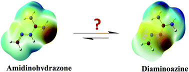
Molecular electrostatic potential (MESP) studies provide details regarding the nuclear and electronic charge distribution on the surface of the molecule, which is a result of the force experienced by a positive test probe. The colour contours provide a location of the positive (blue colour) and negative (red colour) electrostatic potentials. MESP calculation for the two tautomers clearly shows the difference in the electron density along the guanidino region wherein an electrostatic potential of around N1 is partially negative in hydrazone and partially positive in azine and vice versa at N3 (Fig. 4). There is a clear distinction in the surface properties of these two tautomers. Hence, treating the two tautomers individually is important for the proper estimation of their interactions with macromolecules.


image file: c5ra05574a-f4.tif
Fig. 4 MESP of hydrazone (5a) and azine (5b) tautomers of amidinohydrazone plotted onto a surface of constant electron density (0.004 e au−3).

Solvent effect

An implicit solvent study shows a gradual decrease in the energy difference between 5a and 5b as the dielectric constant increases (Table 3). This decrease is marginal from 4.66 to 3.78 kcal mol−1 from the gas phase to the water medium. This data indicates that under polar solvent conditions, the equilibrium between the two tautomeric states increases. Furthermore, microsolvation through water molecule was studied to examine the extent of the energy difference between the tautomers.
Table 3 Implicit solvent effect on relative Gibbs free energy difference (ΔG) in kcal mol−1 between 5a and 5b with 5b taken as a base
Phase Dielectric constant (ε) Relative ΔG (kcal mol−1)
Gas 4.66
Cyclohexane 2.0 4.21
THF 7.6 3.88
Ethanol 24.5 3.84
Acetonitrile 37.5 3.83
DMSO 46.7 3.87
Water 80.1 3.78


Water molecules were explicitly added to both systems. In the presence of explicit water molecules, the tautomer energy difference decreased to 3.38 kcal mol−1 with one water molecule, 2.40 kcal mol−1 with two water molecules and up to 1.75 kcal mol−1 with four water molecules (ESI). This analysis indicates that when the hydrogen atoms and the lone-pair electrons of these systems are stabilised by a network of hydrogen bonds in water, the difference between hydrazone and azine structures becomes less prominent (in all cases, azine tautomer being more stable). This could be one of the reasons as to why the distinction between the hydrazone and azine tautomers was not explicitly identifiable.

Substituent effect

The influence of various substituents on the energy difference between the two tautomers was estimated in the gas phase using B3LYP/6-311++G(d,p). Various substituents that are of importance in medicinal chemistry are considered in this study. In general, substitution at the R group tends to increase the preference for the azine tautomer (Table 4). Substituents that influence the π electron delocalization tend to more strongly stabilise the azine tautomer. The π electron-donating groups, such as NH2 and OH, as well as the π electron-withdrawing NO2 group increase the preference for the azine tautomer. On the other hand, substituents at the R2 position tend to decrease the preference for the azine tautomer. The tautomeric energy difference between 5a and 5b when substituted with electron-withdrawing groups, COPh and COCH3, at N1 was found to be lower than the unsubstituted form and appears to decrease the preference for the azine form. On the other hand, substituting with an electron-donating group such as CH3, the energy difference is slightly decreased by a marginal degree compared with the unsubstituted form (Table 4).
Table 4 Influence of substituents on the Gibbs free energy difference (ΔG) in kcal mol−1 between hydrazone and azine tautomers, with azine structure taken as a base

image file: c5ra05574a-u1.tif

R R1 R2 Relative Gibbs Ffree Eenergy, ΔG (kcal mol−1)
H H H 4.66
H H COCH3 2.25
H H COPh −0.67
H H CH3 4.12
CH3 H H 5.26
C2H5 H H 5.11
OH H H 10.76
NH2 H H 8.72
NO2 H H 5.25
CF3 H H 6.37
Cl H H 4.36
F H H 4.68
NH2 NH2 H 9.23
Cl Cl H 6.23
F F H 8.14


Tautomerism in various biologically active molecules

Several biologically active molecules containing the amidinohydrazone group were reported in their ‘hydrazone’ tautomeric form. Herein, we explored the possible tautomerism in such molecules and calculated the tautomeric energy difference in the gas phase using the B3LYP/6-31+G(d) level of quantum chemical calculations. In each case, the azine tautomer is found to be more stable than its corresponding hydrazone form; hence, the structures in Fig. 5 are drawn in their azine form. Careful observation of the tautomeric energy differences of these molecules suggests an “additive effect” wherein there is an increase of ∼6 kcal mol−1 for every unit of 1,1-diamino-2,3-diazabutadiene (azine) moiety in the molecular structure. For instance, the ΔG of guanabenz (containing a single azine unit) is 5.96 kcal mol−1, which increases to 11.53 kcal mol−1 in MGBG (containing two azine units) and further increases to 17.59 kcal mol−1 in VI (containing three azine units) (Fig. 5).
image file: c5ra05574a-f5.tif
Fig. 5 The Gibbs free energy difference (ΔG) in kcal mol−1 (numerical value written under each compound) between hydrazone and azine tautomers of various biologically active molecules with the azine tautomer being more stable than the corresponding hydrazone in each case.

Proton affinity

The drug molecule, guanabenz, is generally supplied as an acetate salt, mainly to improve oral bioavailability. The pKa of guanabenz is reported to be 8.1,68 proving it to be basic. To estimate the proton affinity of this class of species, quantum chemical analysis has been carried out. The most preferred site of protonation is N3 of amidinohydrazone moiety in guanabenz with a proton affinity value of 232.70 kcal mol−1.

It is important to note that in the protonated state, both hydrazone and azine representations lead to the same structure. This could be an additional reason for ignoring azine vs. hydrazone tautomeric preferences in the title compounds. However, considering the large energy difference, particularly in the cases of MGBG, V and VI (Fig. 5), it is advisable to correlate the tautomeric preferences (in the neutral state) with the experimental details while exploring the chemistry of these species.

Molecular docking analysis

An understanding of the tautomerism of drugs is essential in computer-aided drug discovery, so as to identify complementary pose of small molecules in the active sites of macromolecules. To demonstrate the importance of the tautomeric representation of amidinohydrazone, a molecular docking analysis was carried out. The docking of guanabenz has been performed in the active site of monoamine oxidase A (MAO-A), which was previously studied and performed by Ramsay and co-workers.45d The active site area is outlined by residues Tyr69, Tyr197, Phe208, Tyr407, Phe352, Tyr444 and the isoalloxazine ring of FAD. Molecular docking experiment has been performed using Glide59 module implemented in Maestro version 9.4 software package (by Schrodinger)60 on the crystal structure of MAO-A obtained from the Protein Data Bank (PDB ID: 2BXS).69 The docking protocol followed was as per that performed by Ramsay and co-workers.45d To reproduce the reported binding interaction, the validation of docking protocol was performed with agmatine on the MAO-A active site.69 The docking pose of agmatine was found to reflect the reported hydrogen bonds with Glu216, Tyr444, Tyr197 and Asn181. The docking pose of both the tautomers of guanabenz are clearly different. In the docked pose, the hydrazone tautomer prefers an almost planar arrangement (176°), whereas the azine tautomer undergoes a twist to about 127°. The amino ends of the diaminoazine tautomer formed hydrogen bonds with Ser209 and Glu216 unlike the hydrazone tautomer, which formed hydrogen bonds between the hydrogen of N5 with Arg206 and N3 with Ile207. This provides a clear indication on the importance of tautomerism in drug designing because the usage of improper tautomers may conceal certain important interactions with the receptor attributed to its activity. The docking score of the azine tautomer is found to be marginally better (−6.887) compared with that of the hydrazone tautomer (−6.575). Hence, the pharmacophoric feature varies with each tautomer and in turn affects the binding pattern or interactions with the receptor (Fig. 6).
image file: c5ra05574a-f6.tif
Fig. 6 Docking pose of (A) hydrazone tautomer (B) azine tautomer of guanabenz (C) agmatine in the active site (residues marked in green) of MAO-A (2BXS). Hydrogen-bond interactions with the nearby amino acids (residues marked in blue) are shown with black dotted lines, and hydrophobic interactions are shown with yellow dotted lines.

In several published reports of organic and medicinal chemistry, the title compounds were represented as hydrazones, presumably due to historic or convenience factors rather than a structural factor. Hence, such a representation potentially misleads the chemistry and surface information because these are different for each tautomer. This information often goes unnoticed by chemists during the interpretation of spectroscopic data or in molecular modelling analysis, which requires special attention in all future discussions of this class of compounds.

Conclusions

Quantum chemical calculations have been performed to establish the tautomeric preferences between azine and hydrazone tautomers of titled compounds. The azine tautomers are found to be about 4–6 kcal mol−1 more stable than the hydrazone tautomers. The electronic structure analysis clearly established the reason for this preference. Substituents and solvent effects influence the azine ⇌ hydrazone tautomerism, mostly preferring the azine tautomeric state. Many of the biologically active molecules have been represented in their hydrazone form, which is inappropriate. For example, in guanabenz, the azine form is more stable by 5.96 kcal mol−1, which showed a better docking score and interactions than its hydrazone form. This study established that azine tautomeric structure is more fundamental and the hydrazone tautomeric structure is the alternative in this class of compounds. Published reports gave importance to the hydrazone tautomer and ignored the energetically most preferred azine tautomeric state. The quantum chemical analysis reported in this study suggests that relatively more importance should be given to the azine tautomeric state, particularly while carrying out molecular modelling studies.

Acknowledgements

Authors thank DST, New Delhi, for financial support.

Notes and references

  1. (a) IUPAC Gold Book, http://goldbook.iupac.org/H02884.html, accessed, March, 2015 Search PubMed; (b) G. Verma, A. Marella, M. Shaquiquzzaman, M. Akhtar, M. R. Ali and M. M. Alam, J. Pharm. BioAllied Sci., 2014, 6, 69–80 CrossRef PubMed; (c) S. Rollas and S. G. Küçükgüzel, Molecules, 2007, 12, 1910–1939 CrossRef CAS.
  2. (a) IUPAC Gold Book, http://goldbook.iupac.org/A00557.html, accessed, March, 2015 Search PubMed; (b) J. Safari and S. Gandomi-Ravandi, RSC Adv., 2014, 4, 46224–46249 RSC.
  3. (a) M. A. Gouda, Synth. Commun., 2014, 44, 3347–3361 CrossRef CAS PubMed; (b) A. A. Fadda, S. Bondock, R. Rabie and H. A. Etman, Turk. J. Chem., 2008, 32, 259–286 CAS; (c) K. M. Dawood and M. A. Raslan, J. Heterocycl. Chem., 2008, 45, 137–141 CrossRef CAS PubMed; (d) S. Rollas, N. Gulerman and H. Erdeniz, Il Farmaco, 2002, 57, 171–174 CrossRef CAS; (e) F. A. Attaby and S. M. Eldin, Z. Naturforsch., A: Phys. Sci., 1999, 54b, 788–798 Search PubMed.
  4. (a) J. Galeta, S. Man and M. Potáček, ARKIVOC, 2009, vi, 245–259 CrossRef; (b) T. M. V. D. Pinho e Melo, Curr. Org. Chem., 2009, 13, 1406–1431 CrossRef CAS.
  5. (a) S. T. Chew, K. M. Lo, S. K. Lee, M. P. Heng, W. Y. Teoh, K. S. Sim and K. W. Tan, Eur. J. Med. Chem., 2014, 76, 397–407 CrossRef CAS PubMed; (b) P. G. Avaji, C. H. V. Kumar, S. A. Patil, K. N. Shivananda and C. Nagaraju, Eur. J. Med. Chem., 2009, 44, 3552–3559 CrossRef CAS PubMed; (c) G. Tamasi, L. Chiasserini, L. Savini, A. Sega and R. Cini, J. Inorg. Biochem., 2005, 99, 1347–1359 CrossRef CAS PubMed; (d) M. C. Rodrìguez-Argüelles, M. B. Ferrari, F. Bisceglie, C. Pelizzi, G. Pelosi, S. Pinelli and M. Sassi, J. Inorg. Biochem., 2004, 98, 313–321 CrossRef PubMed; (e) Z. H. Chohan and S. K. A. Sherazi, Synth. React. Inorg. Met.-Org. Chem., 1999, 29, 105–118 CrossRef CAS PubMed.
  6. B. Murukan and K. Mohanan, J. Enzyme Inhib. Med. Chem., 2008, 22, 65–70 CrossRef PubMed.
  7. (a) R. Kamal, V. Kumar, V. Bhardwaj, V. Kumar and K. R. Aneja, Med. Chem. Res., 2015 DOI:10.1007/s00044-014-1313-5; (b) R. Anbazhagan and K. R. Sankaran, Spectrochim. Acta, Part A, 2015, 135, 984–993 CrossRef CAS PubMed; (c) O. O. Ajani, C. A. Obafemi, O. C. Nwinyi and D. A. Akinpelu, Bioorg. Med. Chem., 2010, 18, 214–221 CrossRef CAS PubMed; (d) A. M. Pieczonka, A. Strzelczyk, B. Sadowska, G. Mlostoń and P. Stączek, Eur. J. Med. Chem., 2013, 64, 389–395 CrossRef CAS PubMed; (e) A. J. M. Rasras, T. H. Al-Tel, A. F. Al-Aboudi and R. A. Al-Qawasmeh, Eur. J. Med. Chem., 2010, 45, 2307–2313 CrossRef CAS PubMed.
  8. (a) H. S. N. Kumar, T. Parumasivam, F. Jumaat, P. Ibrahim, M. Z. Asmawi and A. Sadikun, Med. Chem. Res., 2014, 23, 269–279 CrossRef; (b) D. Sriram, P. Yogeeswari, D. R. K. Vyas, P. Senthilkumar, P. Bhat and M. Srividya, Bioorg. Med. Chem. Lett., 2010, 20, 4313–4316 CrossRef CAS PubMed; (c) M. J. Hearn, M. H. Cynamon, M. F. Chen, R. Coppins, J. Davis, H. Joo-On Kang, A. Noble, B. Tu-Sekine, M. S. Terrot, D. Trombino, M. Thai, E. R. Webster and R. Wilson, Eur. J. Med. Chem., 2009, 44, 4169–4178 CrossRef CAS PubMed; (d) D. Sriram, P. Yogeeswari and R. V. Devakaram, Bioorg. Med. Chem., 2006, 14, 3113–3118 CrossRef CAS PubMed; (e) B. K. Kaymakçıoğlu and S. Rollas, Il Farmaco, 2002, 57, 595–599 CrossRef; (f) M. T. Cocco, C. Congiu, V. Onnis, M. C. Pusceddu, M. L. Schivo and A. De Logu, Eur. J. Med. Chem., 1999, 34, 1071–1076 CrossRef CAS.
  9. (a) M. Sharma, K. Chauhan, R. K. Srivastava, S. V. Singh, K. Srivastava, J. K. Saxena, S. K. Puri and P. M. S. Chauhan, Chem. Biol. Drug Des., 2014, 84, 175–181 CrossRef CAS PubMed; (b) L. T. Thuy, H. X. Tien, V. D. Hoang and T. K. Vu, Lett. Drug Des. Discovery, 2012, 9, 163–168 CrossRef CAS; (c) P. Melnyk, V. Leroux, C. Sergheraert and P. Grellier, Bioorg. Med. Chem. Lett., 2006, 16, 31–35 CrossRef CAS PubMed.
  10. (a) S. Ulloora, R. Shabaraya, R. Ranganathan and A. V. Adhikari, Eur. J. Med. Chem., 2013, 70, 341–349 CrossRef CAS PubMed; (b) P. Kumar, B. Shrivastava, S. N. Pandeya, L. Tripathi and J. P. Stables, Med. Chem. Res., 2012, 21, 2428–2442 CrossRef CAS; (c) S. Kumar, S. Bawa, S. Drabu, R. Kumar and L. Machawal, Acta Pol. Pharm., 2010, 67, 567–573 CAS; (d) S. K. Sridhar, S. N. Pandeya, J. P. Stables and A. Ramesh, Eur. J. Pharm. Sci., 2002, 16, 129–132 CrossRef CAS.
  11. (a) Ö. D. Can, M. D. Altıntop, Ü. D. Özkay, U. I. Üçel, B. Doǧruer and Z. A. Kaplancıklı, Arch. Pharmacal Res., 2012, 35, 659–669 CrossRef PubMed; (b) K. N. de Oliveira, P. Costa, J. R. Santin, L. Mazzambani, C. Bürger, C. Mora, R. J. Nunes and M. M. de Souza, Bioorg. Med. Chem., 2011, 19, 4295–4306 CrossRef PubMed.
  12. (a) C. M. Moldovan, O. Oniga, A. Pârvu, B. Tiperciuc, P. Verite, A. Pîrnău, O. Crişan, M. Bojiţă and R. Pop, Eur. J. Med. Chem., 2011, 46, 526–534 CrossRef CAS PubMed; (b) M. A.-A. El-Sayed, N. I. Abdel-Aziz, A. A.-M. Abdel-Aziz, A. S. El-Azab, Y. A. Asiri and K. E. H. ElTahir, Bioorg. Med. Chem., 2011, 19, 3416–3424 CrossRef CAS PubMed; (c) W. B. Júnior, M. S. Alexandre-Moreira, M. A. Alves, A. Perez-Rebolledo, G. L. Parrilha, E. E. Castellano, O. E. Piro, E. J. Barreiro, L. M. Lima and H. Beraldo, Molecules, 2011, 16, 6902–6915 CrossRef PubMed; (d) S. M. Sondhi, M. Dinodia and A. Kumar, Bioorg. Med. Chem., 2006, 14, 4657–4663 CrossRef CAS PubMed.
  13. (a) T. Nasr, S. Bondock and M. Youns, Eur. J. Med. Chem., 2014, 76, 539–548 CrossRef CAS PubMed; (b) Z. Cui, Y. Li, Y. Ling, J. Huang, J. Cui, R. Wang and X. Yang, Eur. J. Med. Chem., 2010, 45, 5576–5584 CrossRef CAS PubMed; (c) R. Graeser, N. Esser, H. Unger, I. Fichtner, A. Zhu, C. Unger and F. Kratz, Invest. New Drugs, 2010, 28, 14–19 CrossRef CAS PubMed; (d) R. J. Abdel-Jalil, E. Q. El Momani, M. Hamad, W. Voelter, M. S. Mubarak, B. H. Smith and D. G. Peters, Monatsh. Chem., 2010, 141, 251–258 CrossRef CAS; (e) W. Y. Liu, H. Y. Li, B. X. Zhao, D. S. Shin, S. Lian and J. Y. Miao, Carbohydr. Res., 2009, 344, 1270–1275 CrossRef CAS PubMed; (f) M. Varache-Lembège, S. Moreau, S. Larrouture, D. Montaudon, J. Robert and A. Nuhrich, Eur. J. Med. Chem., 2008, 43, 1336–1343 CrossRef PubMed; (g) Y. Xia, C. D. Fan, B. X. Zhao, J. Zhao, D. S. Shin and J. Y. Miao, Eur. J. Med. Chem., 2008, 43, 2347–2353 CrossRef CAS PubMed.
  14. (a) R. C. Chelucci, L. A. Dutra, M. E. L. Pires, T. R. F. de Melo, P. L. Bosquesi, M. C. Chung and J. L. dos Santos, Molecules, 2014, 19, 2089–2099 CrossRef PubMed; (b) G. A. Silva, L. M. M. Costa, F. C. F. Brito, A. L. P. Miranda, E. J. Barreiro and C. A. M. Fraga, Bioorg. Med. Chem., 2004, 12, 3149–3158 CrossRef CAS PubMed; (c) A. C. Cunha, J. M. Figueiredo, J. L. M. Tributino, A. L. P. Miranda, H. C. Castro, R. B. Zingali, C. A. M. Fraga, M. C. B. V. de Souza, V. F. Ferreira and E. J. Barreiro, Bioorg. Med. Chem., 2003, 11, 2051–2059 CrossRef CAS.
  15. (a) K. Veena, M. Ramaiah, K. Shashikaladevi, T. S. Avinash and V. P. Vaidya, J. Chem. Pharm. Res., 2011, 3, 130–135 CAS; (b) J. Jayabharathi, V. Thanikachalam, A. Thangamani and M. Padmavathy, Med. Chem. Res., 2007, 16, 266–279 CrossRef CAS.
  16. M. Chandra, A. N. Sahay, D. S. Pandey, R. P. Tripathi, J. K. Saxena, V. J. M. Reddy, M. C. Puerta and P. Valerga, J. Organomet. Chem., 2004, 689, 2256–2267 CrossRef CAS PubMed.
  17. C. Liang, J. Xia, D. Lei, X. Li, Q. Yao and J. Gao, Eur. J. Med. Chem., 2013, 74, 742–750 CrossRef PubMed.
  18. E. F. Hahn, M. Carroll-Buatti and G. W. Pasternak, J. Neurosci., 1982, 2, 572–576 CAS.
  19. N. Latif and I. Fathy, J. Org. Chem., 1960, 25, 1614–1617 CrossRef CAS.
  20. V. J. Thiele and E. Dralle, Ann. Chem., 1898, 302, 275–299 CrossRef PubMed.
  21. (a) T. Baum, D. K. Eckfeld, N. Metz, J. L. Dinish, G. Rowles, R. van Pelt, A. T. Shropshire, S. P. Fernandez, M. I. Gluckman and W. F. Bruce, Experientia, 1969, 25, 1066–1067 CrossRef CAS; (b) S. Diamant, I. Agranat, A. Goldblum, S. Cohen and D. Atlas, Biochem. Pharmacol., 1985, 34, 491–498 CrossRef CAS.
  22. (a) M. Bianchi, P. Ulrich, O. Bloom, M. Meistrell III, G. A. Zimmerman, H. Schmidtmayerova, M. Bukrinsky, T. Donnelley, R. Bucala, B. Sherry, K. R. Manogue, A. J. Tortolani, A. Cerami and K. J. Tracey, Mol. Med., 1995, 1, 254–266 CAS; (b) D. Hommes, B. van den Blink, T. Plasse, J. Bartelsman, C. Xu, B. Macpherson, G. Tytgat, M. Peppelenbosch and S. van Deventer, Gastroenterology, 2002, 122, 7–14 CrossRef CAS PubMed; (c) M. B. Atkins, B. Redman, J. Mier, J. Gollob, J. Weber, J. Sosman, B. L. MacPherson and T. Plasse, Clin. Cancer Res., 2001, 7, 486–492 CAS; (d) S. Specht, S. R. Sarite, I. Hauber, J. Hauber, U. F. Görbig, C. Meier, D. Bevec, A. Hoerauf and A. Kaiser, Parasitol. Res., 2008, 102, 1177–1184 CrossRef PubMed.
  23. (a) J. Stanek, G. Caravatti, J. Frei, P. Furet, H. Mett, P. Schneider and U. Regenass, J. Med. Chem., 1993, 36, 2168–2171 CrossRef CAS; (b) U. Regenass, H. Mett, J. Stanek, M. Mueller, D. Kramer and C. W. Porter, Cancer Res., 1994, 54, 3210–3217 CAS.
  24. (a) A. Corti, C. Dave, H. G. Williamson-Ashman, F. Mihich and A. Schenone, Biochem. J., 1974, 139, 351–357 CAS; (b) J. Janne, L. Alhonen and P. Leinonen, Ann. Med., 1991, 23, 241–259 CrossRef CAS; (c) D. D. Von Hoff, Ann. Oncol., 1994, 5, 487–493 CrossRef CAS PubMed.
  25. D. D. Von Hoff, M. Rozencweig, W. T. Soper, L. J. Helman, J. S. Penta, H. L. Davis and F. M. Muggia, Cancer Treat. Rep., 1977, 61, 759–768 CAS.
  26. R. M. Soll, T. Lu, B. Tomczuk, C. R. Illig, C. Fedde, S. Eisennagel, R. Bone, L. Murphy, J. Spurlino and F. R. Salemme, Bioorg. Med. Chem. Lett., 2000, 10, 1–4 CrossRef CAS.
  27. F. Sielaff, M. E. Than, D. Bevec, I. Lindberg and T. Steinmetzer, Bioorg. Med. Chem. Lett., 2011, 21, 836–840 CrossRef CAS PubMed.
  28. M. Šekutor, K. Mlinarić-Majerski, T. Hrenar, S. Tomić and I. Primožič, Bioorg. Chem., 2012, 41–42, 28–34 CrossRef PubMed.
  29. B. Wu, H. Yang, Q. Lin, Z. Wang, C. Lu and G. Cheng, New J. Chem., 2015, 39, 179–186 RSC.
  30. (a) M. Sitzmann, W. D. Ihlenfeldt and M. C. Nicklaus, J. Comput.-Aided Mol. Des., 2010, 24, 521–551 CrossRef CAS PubMed; (b) P. Pospisil, P. Ballmer, L. Scapozza and G. Folkers, J. Recept. Signal Transduction, 2003, 23, 361–371 CrossRef CAS PubMed.
  31. B. J. Smith, M. T. Nguyen, W. J. Bouma and L. Radom, J. Am. Chem. Soc., 1991, 113, 6452–6458 CrossRef CAS.
  32. A. R. Katritzky, C. D. Hall, B. E.-D. M. El-Gendy and B. Draghici, J. Comput.-Aided Mol. Des., 2010, 24, 475–484 CrossRef CAS PubMed.
  33. (a) D. K. Dhaked and P. V. Bharatam, RSC Adv., 2014, 4, 45332–45344 RSC; (b) D. K. Dhaked and P. V. Bharatam, J. Phys. Chem. A, 2014, 118, 187–196 CrossRef CAS PubMed; (c) D. K. Dhaked and P. V. Bharatam, RSC Adv., 2013, 3, 25268–25277 RSC; (d) S. Bhatia, Y. J. Malkhede and P. V Bharatam, J. Comput. Chem., 2013, 34, 1577–1588 CrossRef CAS PubMed; (e) Z. P. Nathavad, S. Bhatia, D. K. Dhaked and P. V. Bharatam, Comput. Theor. Chem., 2013, 1023, 51–58 CrossRef CAS PubMed; (f) S. Bhatia, C. Bagul, Y. Kasetti, D. S. Patel and P. V. Bharatam, J. Phys. Chem. A, 2012, 116, 9071–9079 CrossRef CAS PubMed; (g) Y. Kasetti and P. V. Bharatam, Theor. Chem. Acc., 2012, 131, 1160–1171 CrossRef PubMed; (h) Y. Kasetti, N. K. Patel, S. Sundriyal and P. V Bharatam, J. Phys. Chem. B, 2010, 114, 11603–11611 CrossRef CAS PubMed; (i) A. Mehdi, L. Adane, D. S. Patel and P. V. Bharatam, J. Comput. Chem., 2010, 31, 1259–1267 CAS; (j) L. Adane and P. V. Bharatam, Int. J. Quantum Chem., 2008, 108, 1277–1286 CrossRef CAS PubMed; (k) P. V. Bharatam, D. S. Patel and P. Iqbal, J. Med. Chem., 2005, 48, 7615–7622 CrossRef CAS PubMed; (l) K. Lammertsma and P. V. Bharatam, J. Org. Chem., 2000, 65, 4662–4670 CrossRef CAS; (m) K. Lammertsma and P. V. Bharatam, J. Am. Chem. Soc., 1994, 116, 642–650 CrossRef CAS; (n) K. Lammertsma and P. V. Bharatam, J. Am. Chem. Soc., 1993, 115, 2348–2351 CrossRef CAS; (o) K. Suenobu, M. Nagaoka and T. Yamabe, J. Mol. Struct.: THEOCHEM, 1999, 461–462, 581–588 CrossRef CAS.
  34. (a) S. Bhatia and P. V. Bharatam, J. Org. Chem., 2014, 79, 4852–4862 CrossRef CAS PubMed; (b) M. Arfeen, D. S. Patel, S. Abbat, N. Taxak and P. V. Bharatam, J. Comput. Chem., 2014, 35, 2047–2055 CrossRef CAS PubMed; (c) D. S. Patel and P. V. Bharatam, J. Phys. Chem. A, 2011, 115, 7645–7655 CrossRef CAS PubMed; (d) P. V. Bharatam and P. Iqbal, J. Comput. Chem., 2006, 27, 334–343 CrossRef CAS PubMed; (e) M. Ahmed, S. Khanna and P. V. Bharatam, Indian J. Chem. Technol., 2005, 44B, 600–606 CAS; (f) P. V. Bharatam, P. Iqbal, A. Malde and R. Tiwari, J. Phys. Chem. A, 2004, 108, 10509–10517 CrossRef CAS; (g) P. V. Bharatam and S. Khanna, J. Phys. Chem. A, 2004, 108, 3784–3788 CrossRef CAS; (h) P. V. Bharatam, Amita and D. Kaur, Tetrahedron, 2002, 58, 10335–10339 CrossRef CAS; (i) P. V. Bharatam, Amita, P. Uppal and D. Kaur, Indian J. Chem. Technol., 2001, 40B, 181–186 CAS; (j) P. V. Bharatam, P. Uppal, A. Kaur and D. Kaur, J. Chem. Soc., Perkin Trans. 2, 2000, 43–50 RSC; (k) P. V. Bharatam, Indian J. Chem. Technol., 2000, 39A, 100–105 CAS.
  35. L. Adane, S. Bhagat, M. Arfeen, S. Bhatia, R. Sirawaraporn, W. Sirawaraporn, A. K. Chakraborti and P. V. Bharatam, Bioorg. Med. Chem. Lett., 2014, 24, 613–617 CrossRef CAS PubMed.
  36. T. W. Bell and A. T. Papoulis, Angew. Chem., Int. Ed. Engl., 1992, 31, 749–751 CrossRef PubMed.
  37. A. S. Morkovnik, L. N. Divaeva, A. I. Uraev, K. A. Lyssenko, R. K. Mamin, I. G. Borodkina, G. S. Borodkin, A. S. Burlov and A. D. Garnovskii, Russ. Chem. Bull., 2009, 57, 1496–1507 CrossRef.
  38. A. M. S. Silva, V. L. M. Silva, R. M. Claramunt, D. S. María, M. B. Ferraro, F. Reviriego, I. Alkorta and J. Elguero, Magn. Reson. Chem., 2013, 51, 530–540 CrossRef CAS PubMed.
  39. (a) I. Mutikainen, H. Elo and P. Lumme, J. Chem. Soc., Perkin Trans. 2, 1986, 291–293 RSC; (b) I. Mutikainen, H. Elo and P. Tilus, Z. Naturforsch., A: Phys. Sci., 1993, 48b, 1821–1827 Search PubMed.
  40. H. Elo, I. Mutikainen, L. Alhonen-Hongisto, R. Laine, J. Jänne and P. Lumme, Z. Naturforsch., A: Phys. Sci., 1986, 41c, 851–855 Search PubMed.
  41. M. Koskinen, I. Mutikainen, J. T. Koskinenb and H. Elo, Z. Naturforsch., A: Phys. Sci., 1997, 52b, 1114–1118 Search PubMed.
  42. M. Koskinen, I. Mutikainen and H. Elo, Z. Naturforsch., A: Phys. Sci., 1996, 51, 1161–1172 CAS.
  43. S. Demir, M. Dinçer and E. Sarıpınar, Acta Crystallogr., Sect. E: Struct. Rep. Online, 2006, 62, o4194–o4195 CAS.
  44. (a) W. Holzer and Z. Györgydeák, Monatsh. Chem., 1992, 123, 1163–1173 CrossRef CAS; (b) Z. Györgydeák, W. Holzer and K. Mereiter, Monatsh. Chem., 1999, 130, 899–913 Search PubMed.
  45. (a) S. Sparapani, S. Bellini, M. Gunaratnam, S. M. Haider, A. Andreani, M. Rambaldi, A. Locatelli, R. Morigi, M. Granaiola, L. Varoli, S. Burnelli, A. Leoni and S. Neidle, Chem. Commun., 2010, 46, 5680–5682 RSC; (b) K. Nikolic, S. Filipic and D. Agbaba, Bioorg. Med. Chem., 2008, 16, 7134–7140 CrossRef CAS PubMed; (c) A. G. Jobson, J. H. Cardellina II, D. Scudiero, S. Kondapaka, H. Zhang, H. Kim, R. Shoemaker and Y. Pommier, Mol. Pharmacol., 2007, 72, 876–884 CrossRef CAS PubMed; (d) T. Z. E. Jones, L. Giurato, S. Guccione and R. R. Ramsay, FEBS J., 2007, 274, 1567–1575 CrossRef CAS PubMed.
  46. M. J. Frisch, G. W. Trucks, H. B. Schlegel, G. E. Scuseria, M. A. Robb, J. R. Cheeseman, G. Scalmani, V. Barone, B. Mennucci, G. A. Petersson, H. Nakatsuji, M. Caricato, X. Li, H. P. Hratchian, A. F. Izmaylov, J. Bloino, G. Zheng, J. L. Sonnenberg, M. Hada, M. Ehara, K. Toyota, R. Fukuda, J. Hasegawa, M. Ishida, T. Nakajima, Y. Honda, O. Kitao, H. Nakai, T. Vreven, J. A. Montgomery, J. E. Peralta, F. Ogliaro, M. Bearpark, J. J. Heyd, E. Brothers, K. N. Kudin, V. N. Staroverov, R. Kobayashi, J. Normand, K. Raghavachari, A. Rendell, J. C. Burant, S. S. Iyengar, J. Tomasi, M. Cossi, N. Rega, J. M. Millam, M. Klene, J. E. Knox, J. B. Cross, V. Bakken, C. Adamo, J. Jaramillo, R. Gomperts, R. E. Stratmann, O. Yazyev, A. J. Austin, R. Cammi, C. Pomelli, J. W. Ochterski, R. L. Martin, K. Morokuma, V. G. Zakrzewski, G. A. Voth, P. Salvador, J. J. Dannenberg, S. Dapprich, A. D. Daniels, Ӧ. Farkas, J. B. Foresman, J. V. Ortiz, J. Cioslowski and D. J. Fox, Gaussian 09, Revision B.01, Gaussian, Inc., Wallingford, CT, 2009 Search PubMed.
  47. (a) J. A. Pople and D. L. Beveridge, Approximate Molecular Orbital Theory, McGraw-Hill Book, New York, 1970, ch. 1 and 2 Search PubMed; (b) W. J. Hehre, L. Radom, P. v. R. Schleyer and J. A. Pople, Ab Initio Molecular Orbital Theory, Wiley, New York, 1985 Search PubMed; (c) J. B. Foresman and A. E. Frisch, Exploring Chemistry with Electronic Structure Methods, Gaussian, Inc., Pittsburgh, PA, 1995 Search PubMed.
  48. (a) M. Head-Gordon, J. A. Pople and M. J. Frisch, Chem. Phys. Lett., 1988, 153, 503–506 CrossRef CAS; (b) S. Saebø and J. Almlöf, Chem. Phys. Lett., 1989, 154, 83–89 CrossRef; (c) M. J. Frisch, M. Head-Gordon and J. A. Pople, Chem. Phys. Lett., 1990, 166, 275–280 CrossRef CAS; (d) M. J. Frisch, M. Head-Gordon and J. A. Pople, Chem. Phys. Lett., 1990, 166, 281–289 CrossRef CAS; (e) M. Head-Gordon and T. Head-Gordon, Chem. Phys. Lett., 1994, 220, 122–128 CrossRef CAS.
  49. L. A. Curtiss, K. Raghavachari and J. A. Pople, J. Chem. Phys., 1993, 98, 1293–1298 CrossRef CAS PubMed.
  50. R. G. Parr and W. Yang, Density Functional Theory of Atoms and Molecules, Oxford University Press, New York, 1989 Search PubMed.
  51. (a) C. Lee, W. Yang and R. G. Parr, Phys. Rev. B: Condens. Matter Mater. Phys., 1988, 37, 785–789 CrossRef CAS; (b) J. P. Perdew and Y. Wang, Phys. Rev. B: Condens. Matter Mater. Phys., 1992, 45, 13244–13249 CrossRef; (c) A. D. Becke, J. Chem. Phys., 1993, 98, 5648–5652 CrossRef CAS PubMed.
  52. Y. Zhao and D. G. Truhlar, Theor. Chem. Acc., 2008, 120, 215–241 CrossRef CAS.
  53. (a) J. A. Montgomery Jr, M. J. Frisch, J. W. Ochterski and G. A. Petersson, J. Chem. Phys., 1999, 110, 2822–2827 CrossRef PubMed; (b) J. A. Montgomery Jr, M. J. Frisch, J. W. Ochterski and G. A. Petersson, J. Chem. Phys., 2000, 112, 6532–6542 CrossRef PubMed.
  54. (a) E. Cancès, B. Mennucci and J. Tomasi, J. Chem. Phys., 1997, 107, 3032–3041 CrossRef PubMed; (b) J. Tomasi, B. Mennucci and E. Cancès, J. Mol. Struct.: THEOCHEM, 1999, 464, 211–226 CrossRef CAS.
  55. (a) A. E. Reed, R. B. Weinstock and F. Weinhold, J. Chem. Phys., 1985, 83, 735–746 CrossRef CAS PubMed; (b) A. E. Reed, L. A. Curtiss and F. Weinhold, Chem. Rev., 1988, 88, 899–926 CrossRef CAS.
  56. A. D. Becke and K. E. J. Edgecombe, J. Chem. Phys., 1990, 92, 5397–5409 CrossRef CAS PubMed.
  57. T. Lu and F. Chen, J. Comput. Chem., 2012, 33, 580–592 CrossRef CAS PubMed.
  58. B. G. Johnson, P. M. W. Gill and J. A. Pople, Chem. Phys. Lett., 1993, 206, 239–246 CrossRef CAS.
  59. (a) R. A. Friesner, J. L. Banks, R. B. Murphy, T. A. Halgren, J. J. Klicic, D. T. Mainz, M. P. Repasky, E. H. Knoll, D. E. Shaw, M. Shelley, J. K. Perry, P. Francis and P. S. Shenkin, J. Med. Chem., 2004, 47, 1739–1749 CrossRef CAS PubMed; (b) T. A. Halgren, R. B. Murphy, R. A. Friesner, H. S. Beard, L. L. Frye, W. T. Pollard and J. L. Banks, J. Med. Chem., 2004, 47, 1750–1759 CrossRef CAS PubMed; (c) R. A. Friesner, R. B. Murphy, M. P. Repasky, L. L. Frye, J. R. Greenwood, T. A. Halgren, P. C. Sanschagrin and D. T. Mainz, J. Med. Chem., 2006, 49, 6177–6196 CrossRef CAS PubMed.
  60. Schrödinger, Release 2013-1, Maestro, version 9.4, Schrödinger, LLC, New York, NY, 2013 Search PubMed.
  61. P. H. Richter, A. Besch, D. Albrecht and K. Kasbohm, Pharmazie, 1992, 47, 911–914 CAS.
  62. A. Cousson, F. Nectoux, B. Bachet, B. Kokel and M. Hubert-Habart, Acta Crystallogr., Sect. C: Cryst. Struct. Commun., 1994, C50, 289–291 CrossRef CAS.
  63. A. Andreani, M. Rambaldi, A. Locatelli, R. Bossa, A. Fraccari and I. Galatulas, J. Med. Chem., 1992, 35, 4634–4637 CrossRef CAS.
  64. P. Ulrich and A. Cerami, J. Med. Chem., 1984, 27, 35–40 CrossRef CAS.
  65. P. Richter, M. Elsner and B. Vogt, US Pat., US5747508, 1998.
  66. M. Bianchi, A. Cerami, K. J. Tracey and P. Ulrich, US Pat., US5849794, 1998.
  67. Mondobiotech Interferon SA, Eur. Pat., EP1389480A1, 2004.
  68. W. O. Foye, T. L. Lemke and D. A. Williams, Foye's Principles of Medicinal Chemistry, Lippincott Williams & Wilkins, Philadelphia, 6th edn, 2008, p. 781 Search PubMed.
  69. L. De Colibus, M. Li, C. Binda, A. Lustig, D. E. Edmondson and A. Mattevi, Proc. Natl. Acad. Sci. U. S. A., 2005, 102, 12684–12689 CrossRef CAS PubMed.

Footnote

Electronic supplementary information (ESI) available: Table S1. Absolute Gibbs free energies (hartrees) in the gas phase of hydrazones and their corresponding azine tautomers mentioned in Scheme 1. Table S2. Absolute Gibbs free energies (hartrees) in implicit solvent phase conditions of hydrazone, 5a and the corresponding azine tautomer, 5b, of amidinohydrazone. Table S3. Absolute Gibbs free energies (hartrees) in explicit solvent phase conditions of hydrazone, 5a and the corresponding azine tautomer, 5b, of amidinohydrazone. Table S4. Absolute Gibbs free energies (hartrees) of substituted 5a and 5b in the gas phase. Table S5. Absolute Gibbs free energies (hartrees) of various biologically active molecules in the gas phase. See DOI: 10.1039/c5ra05574a

This journal is © The Royal Society of Chemistry 2015
Click here to see how this site uses Cookies. View our privacy policy here.