
Enhancement effect of essential oils from the fruits and leaves of Alpinia oxyphylla on skin permeation and deposition of indomethacin
Huanhuan Fengab,
Jiaoyang Luob,
Weijun Kongb,
Xiaowen Doub,
Yuting Wangc,
Xiangsheng Zhaod,
Wanping Zhang*a,
Qiong Lia and
Meihua Yang*bd
aShanghai Institute of Technology, 201400, Shanghai, China. E-mail: zhwanp@sit.edu.cn
bInstitute of Medicinal Plant Development, Chinese Academy of Medical Sciences, Peking Union Medical College, 100193, Beijing, China. E-mail: yangmeihua15@hotmail.com
cJiangsu University, 212013, Zhenjiang, China
dHainan Branch Institute of Medicinal Plant Development, Chinese Academy of Medical Sciences & Peking Union Medical College, Wanning 571533, China
First published on 20th April 2015
Abstract
Essential oils from plants are gaining increasing attention as potential chemical penetration enhancers. This study aimed to investigate the enhancement effect of essential oils from the fruits and leaves of Alpinia oxyphylla on skin permeation and deposition of indomethacin. In vitro permeation experiments were performed in Franz-type cells through rat skin, and the amount of drug passing through into the receptor phase was analyzed by ultra performance liquid chromatography-photodiode array (UPLC-PDA). Ultra fast liquid chromatography coupled with tandem mass spectrometry (UFLC-MS/MS) was used to analyze the plasma drug concentration of indomethacin to examine the enhancement effect of essential oils in an in vivo rat model of drug delivery. Both oils demonstrated a significant enhancement effect on drug delivery and skin deposition (p < 0.05). Particularly, at 3% concentration, enhancement ratios of fruit oil and leaf oil were 10.16 and 4.61, respectively, which were both significantly higher than that of the commonly used enhancer, Azone (2.04). Major constituents of both oils were identified by gas chromatography-mass spectrometry (GC-MS). It may be deduced that higher content hydrocarbon terpenes in the fruit oil contribute to the increased enhancement effect relative to leaf oil. The skin irritation test indicated that both oils at certain concentrations (1%, 3%, and 5%) did not cause obvious erythema or edema in rabbits. Considering their enhanced drug permeation and low skin irritation, essential oils from Alpinia oxyphylla could be novel penetration enhancers and have promising applications in transdermal drug delivery and cosmetics.
1 Introduction
Transdermal drug delivery (TDD) is often limited by the poor permeability of skin to drugs, which precludes their crossing the skin barrier at therapeutic rates.1,2 Many approaches have been developed to penetrate the layer of the stratum corneum with the goal of achieving therapeutically effective plasma concentrations, of which chemical penetration enhancers (CPEs) are used most widely.3–6 In ideal cases, CPEs should transiently and reversibly alter the prohibitive barrier of the stratum corneum thereby allowing drug molecules to permeate across the skin.5 However, the reported CPEs (e.g. pyrrolidones and sulphoxides) often express serious adverse effects such as skin erythema and edema.7,8 As a result, considerable research on the use of natural plant-based products such as essential oils and fatty acids as penetration enhancers is now in progress.9,10 Essential oils are considered clinically acceptable penetration enhancers as indicated by their high percutaneous enhancement ability, reversible effect on the lipids of the stratum corneum, and low cutaneous irritancy at low concentrations (1–5%).11 In addition, for their potential function in combining therapeutic efficacy with increased levels of permeation, essential oils from traditional Chinese medicinal plants have been regarded as promising enhancers.2,12–15Alpinia oxyphylla (Zingiberaceae, A. oxyphylla), a type of flowering plant of the ginger family, has been widely used to treat diarrhea and gastralgia, and has been shown to play a role in neuroprotection according to the Chinese Pharmacopoeia.16 In recent years, A. oxyphylla extracts were reported to possess antifungal, anti-angiogenic and anti-oxidative effects.17–19 Furthermore, A. oxyphylla cream was shown to have a protective effect against photoaging in mice.20 Few reports have examined the therapeutic use of essential oils isolated from fruits and leaves of A. oxyphylla by hydrodistillation as CPEs. Critically evaluating the potential of essential oils from A. oxyphylla would be highly significant for their wide applications in TDD and cosmetics.
The aim of this study was to investigate the enhancement effect of essential oils from fruits and leaves of A. oxyphylla on the skin permeation and deposition of the non-steroidal anti-inflammatory drug, indomethacin (IND). Oral therapy with IND is very effective, however, even regular dosage of the drug can trigger adverse effects on the gastrointestinal tract and central nervous system with a high incidence rate of 30–50%.21 Therefore, it is necessary to develop a transdermal drug delivery system for IND. In addition, IND has been widely used as the model drug in a number of studies on drug delivery.22–24 As such, we selected IND as the model drug to evaluate the penetration enhancement effect of essential oils. The in vitro permeation experiments were carried out utilizing Franz cells with intact rat skin as the membrane barrier. The amount of IND permeating into the receptor phase was quantified using UPLC-PDA. UFLC-MS/MS was used to analyze the plasma drug concentration of IND to examine the enhancement effect of essential oils in vivo. Major constituents of the oils used in these experiments were identified by GC-MS to correlate the relationship of the components with the permeation effects of essential oils from the various parts of A. oxyphylla. Finally, the skin irritation test was carried out with rabbits to evaluate the safety of both oils at varying concentrations.
2 Experimental
2.1 Chemicals, reagents and materials
IND (99.9%) was purchased from Sigma Chemical Co. (St. Louis, MO, USA). Diclofenac sodium (98.0%) was obtained from National Institutes for Food and Drug Control, Beijing, China. Carboxymethylcellulose (CMC) sodium was supplied by Whatman International Ltd., UK. PBS buffer (1 L) contains KH2PO4 (0.27 g), Na2HPO4 (1.42 g), NaCl (8.0 g) and KCl (0.2 g), which were obtained from Sinopharm Chemical Reagent Co., Ltd (Beijing, China). All samples of A. oxyphylla were collected from planting base in Hainan province, China. All samples were identified by Prof. Meihua Yang, and the voucher specimens were deposited at the analytical center of the Institute of Medicinal Plant Development, Chinese Academy of Medical Sciences, Beijing, China. Azone was purchased from Adamas Reagent Co. Ltd (Shanghai, China). Methanol of HPLC-grade was obtained from Fisher Co. Ltd. (Emerson, IA, USA). All other chemicals and solvents were of analytical grade.2.2 Preparation of essential oils
Preparation of the essential oil was in accordance with the protocol in the Chinese Pharmacopoeia for reproducibility.25,26 Detailed methodology is as follows: dry fruits or leaves of A. oxyphylla were crushed and passed through a 50-mesh sieve. Then a certain amount of sample powder was soaked in water (10-fold volume) for 8 h, and extracted through steam distillation for 6 h. Yields of fruit oil and leaf oil were 0.8–1.3%, 0.3–0.6%, respectively. The percentage of essential oil yield was calculated as the volume of essential oil divided by the weight of A. oxyphylla sample powder. All essential oils were dried with Na2SO4 before storing in the refrigerator.2.3 Preparation of skin membranes
Wistar rats, male, weighing 180–220 g, were obtained from Beijing HFK Bioscience Co., Ltd. (License no. SYXK 2009-0007). The protocol of the study was approved by the Ethical Committee of Animal Experimental Laboratory Institute of Medicinal Plant Development, Chinese Academy of Medical Sciences and Peking Union Medical College (no. SLXD-20140417). The animals were raised separately by gender and had unlimited access to food and water in an environmentally controlled breeding room (temperature 22 ± 2 °C, humidity 60–80%). The dorsal skin of rats, sacrificed by ether, was shaved by forceps and surgical scissors after the carefully removal of hair, and then clean the adhering subcutaneous fat and tissue. After flushing with right amount normal saline and washing with distilled water, the skin membranes were cut into proper pieces; afterwards, skins were either used immediately or individually stored in the −20 °C refrigerator with aluminum foil wrapped until required for use. The skin was defrosted at room temperature when required.2.4 In vitro permeation experiments
Diffusion experiments were performed in Franz-type diffusion cells. After being gently blotted dry with filter paper, the excised rat skin was fastened carefully between the donor and receptor chambers of the cell, with the dermal side in intimate contact with the reception medium. Each cell had a volume of 16.2 mL, and the skin surface available for permeation was 1.77 cm2. The donor cell and junction between the donor and the receptor chamber were covered with parafilm to avoid dispersion of volatile components. The receptor medium was ethanol/PBS buffer, pH 7.4 (1![[thin space (1/6-em)]](https://www.rsc.org/images/entities/char_2009.gif) :1, v/v), and 1 g of donor gel containing IND with or without enhancer pretreated was applied to the surface of the skin. The effective area of the rat skin was 1.77 cm2, and massage gently after the application to all skins. For preparing of the gel, a 2% (w/v) concentration of CMC sodium was added into about 9 mL pH 7.4 PBS buffer, after which the mixture was stirred continuously for 1 h. And 0.3% (w/v) IND in 1 mL ethanol was added into the mixture with continuous stirring for 1 h.
:1, v/v), and 1 g of donor gel containing IND with or without enhancer pretreated was applied to the surface of the skin. The effective area of the rat skin was 1.77 cm2, and massage gently after the application to all skins. For preparing of the gel, a 2% (w/v) concentration of CMC sodium was added into about 9 mL pH 7.4 PBS buffer, after which the mixture was stirred continuously for 1 h. And 0.3% (w/v) IND in 1 mL ethanol was added into the mixture with continuous stirring for 1 h.
During the experiments, the solution in the receptor phase was maintained at 37 °C and stirred by a magnetic stirrer at 600 rpm. At 1, 2, 4, 6, 8, 10, 12, and 24 h, samples (1 mL) were withdrawn from the receptor and immediately replaced with an equal volume of fresh buffer. All reagents and sample solutions were filtered through a 0.22 μm PTFE membrane prior to injection into the UPLC-PDA system. Samples that could not be analyzed immediately upon withdrawal were sealed in 2 mL vials with silicone/PTFE screw cap and stored at 4 °C. All experiments were carried out in triplicate.
For mass balance studies, the amounts of IND present in the donor compartment and skin were investigated at the end of the permeation experiment. To evaluate the amount of drug present in the donor compartment, the gel was removed, and the skin was washed with a cotton swab dipped in 50% (v/v) ethanol:PBS and then blotted with a dry cotton swab. The gel and cotton were collected in a bottle with 50% (v/v) ethanol:PBS, and then extracted by ultrasonication for 30 min. To investigate the amount of IND retained in the skin, the entire section of skin was weighted and cut into small pieces with scissors. The minced skin was boiled for 10 min, centrifuged at 13500 rpm for 20 min, and the supernatant was analyzed by UPLC-PDA.
2.5 Pharmacokinetic study in rats
A pharmacokinetic study was performed in healthy male SD rats (weight range 180–220 g) that had been fasted for 12 h in advance. Animals were obtained from Beijing HFK Bioscience Co., Ltd. (License no. SYXK 2009-0007). The protocol of the study was approved by the Ethical Committee of Animal Experimental Laboratory Institute of Medicinal Plant Development, Chinese Academy of Medical Sciences and Peking Union Medical College (no. SLXD-20150310). Rats had free access to water. Blood samples were obtained at 0.5, 1, 2, 4, 6, 10 and 16 h post-application of 1 g of gel with or without the pretreatment method. The donor gel and the application method were same with those used in the in vitro experiment. All samples were put into anti-coagulant tubes. Plasma was harvested by centrifugation at 4000 rpm for 15 min at 4 °C, and then supernatants were collected and kept frozen at −20 °C for subsequent analysis.The method validation (selectivity, matrix effect, linearity, lower limit of quantification, accuracy, precision, recovery and stability) was referred to a previous publication.27
2.6 UPLC-PDA conditions
Samples were analyzed using a Waters ACQUITY UPLC® H-Class system (Waters Corp., Milford, MA, USA) including quaternary solvent manager, sampler manager, column compartment and PDA detector, connected to Waters Empower 2 data station. An ACQUITY UPLC® BEH Shield RP 18 column (50 × 2.1 mm i.d., 1.7 μm; Waters Corp., Milford, MA, USA) was employed for the separation at 30 °C. The mobile phase consisted of 0.05% acetic acid:methanol (35:65, v/v) at a flow rate of 0.2 mL min−1. The volume injected was 1 μL and the samples were diluted when necessary. The detection wavelength was 268 nm.
2.7 UFLC-MS/MS conditions
Sample analysis was performed using an Applied Biosystems Sciex QTrap® 5500 MS/MS system (Foster City, CA, USA) equipped with an electrospray ionization (ESI) source coupled with an ultra-fast liquid chromatography system from Shiseido Technologies (Shimadzu, Kyoto, Japan). Applied Biosystems Analyst software (version 1.6) was used to control the UFLC-MS/MS system and for data acquisition and processing. An ACQUITY UPLC® BEH Shield RP 18 column (50 × 2.1 mm i.d., 1.7 μm; Waters Corp., Milford, MA, USA) was used for separation at 30 °C. The mobile phase consisted of 0.06% formic acid aqueous:acetonitrile (50:50, v/v) at a flow rate of 0.2 mL min−1.
Quantitation of IND was achieved by MS/MS detection in positive ion mode with an internal standard (IS, diclofenac sodium). The common parameters, viz. curtain gas, GS1 gas and GS2 gas were set at 35, 50 and 50 L min−1, respectively, whereas the collision-activated dissociation gas was set at medium. The compounds parameters, viz. declustering potential, collision energy, entrance potential and collision exit potential for IND and IS were 100, 24, 10, 13 V and 46, 29, 10, 8 V, respectively. Ion detection was performed in multiple reaction monitoring (MRM) mode, monitoring the transition of the m/z 358.4 precursor ion to the m/z 138.9 product ion for IND and m/z 296 precursor ion to the m/z 250 product ion for IS.
2.8 GC-MS analysis of essential oils
The oil composition was determined by GC-MS (Thermo Trace GC Ultra) interfaced with a Polaris Q ion trap mass spectrometer and AI/AS 3000 injector and Xcalibur software. Separation of components was carried out with a GC capillary column (TR-5MS, 30 m × 0.25 mm i.d., 0.32 μm). The temperature of the injection port was 250 °C, and 1.0 μL of the sample was injected with a splitless mode. The oven temperature was programmed as follows: initial temperature was held at 50 °C for 3 min, increased to 90 °C (6 °C min−1) for 5 min, raised to 140 °C by 6 °C min−1, then gradually increased to 145 °C at the rate of 0.2 °C min−1 maintaining for 2 min, then increased to 260 °C (15 °C min−1) and maintaining for 6 min. The mass conditions were set as follows: ion-source temperature, 250 °C; interface temperature, 250 °C; ionization mode with electron ionization (EI); scan time, 0.2 s per scan. All data were obtained by collecting the full-scan mass spectra within a scan range of 40.00–500.00. High-purity (over 99.999%) helium was selected as the carrier gas at a flow rate of 2.0 mL min−1.2.9 Skin irritation test
Male New Zealand pure big-ear white rabbits weighing 1500–2000 g were purchased from Vital River Laboratory Animal Technology Co., Ltd. The certificate of conformity is SCXK (Beijing) 2007-0003. The protocol of the study was approved by the Ethical Committee of Animal Experimental Laboratory Institute of Medicinal Plant Development, Chinese Academy of Medical Sciences and Peking Union Medical College (no. SLXD-20140916). The animals were raised separately and had unlimited access to food and water in an environmentally controlled breeding room (temperature 22 ± 2 °C, humidity 60–80%). Twenty-four hours after careful removal of hair, samples were applied to the left dorsal area, while the right dorsal area served as control. After 4 hours, samples were, wiped off with warm water and skin irritation was evaluated. Each group had four rabbits, and the above procedure was repeated for 7 consecutive days. Water and base oil was used as the blank and negative control group, respectively.The skin irritation was evaluated by visual scoring.28 The scores were assigned from 0 to 4 depending on the degree of erythema/edema as follows: no erythema/edema-0, slight erythema/edema-1, moderate erythema/edema-2, moderate to severe erythema/edema-3, and severe erythema/edema-4. Evaluation of skin irritation was expressed as: average score = ∑erythema score/N × D, where N represents the number of animals in each group, and D represents the number of days of treatment. An average score lower than 0.5 is regarded as no irritation, 0.5–2.0 (slight irritation), 2.0–6.0 (moderate irritation) and 6.0–8.0 (extreme irritation).
2.10 Data analysis
The cumulative amount of drug that transferred into the receptor side and permeation kinetics parameters were calculated. Permeation profiles of IND were constructed by plotting cumulative amount of drug permeated per unit area as a function of time. The X-intercept of the extrapolated linear region of the profile gave lag time (h) and steady state flux Js (μg cm−2 h−1) was calculated from the slope. The Kp was estimated from the flux and the donor drug concentration,29 expressed as eqn (1) where Cd is the saturated solubility of the drug in donor compartment.| Kp = Js/Cd | (1) |
Penetration-enhancing activities are expressed as Er which is calculated from eqn (2).30
| Er = Kp (with enhancer)/Kp (without enhancer) | (2) |
All data presented are the mean ± SD. Statistical significance between control and treated groups was tested by analysis of variance (ANOVA). p-values of less than 0.05 were considered to represent a statistically significant difference.
3 Results and discussion
3.1 Fruit oil and leaf oil enhance the permeation of IND
As previously reported, pretreatment with essential oils increases the skin flux values of drug compared with those obtained when the same essential oils were included in the transdermal devices.31 Moreover, the pretreatment method verified that essential oils act directly on the skin structure and avoid co-solvent effects on the thermodynamic activities of the model drug.32 Thus, in this study, we selected the pretreatment method to certify the enhancement effect of essential oils from leaves and fruits of A. oxyphylla.Permeation experiments of IND after pretreatment with varying essential oils were carried out and the results were presented in Fig. 1. The steady flux, lag time, permeability coefficient (Kp) and enhancement ratios (Er) of the drug through the skin are summarized in Table 1. As shown in Fig. 1, the essential oils acting as enhancers led to a significant increase in both flux (Fig. 1B) and cumulative amount (Fig. 1D) of the drug (p < 0.05). More specifically, as shown in Table 1, the IND flux values at steady state in control group were 2.01 ± 0.09 μg cm−2 h−1. Meanwhile all other treatment groups, with the exception of the 1% leaf oil group, led to a significant increase in IND flux compared with the control group (p < 0.05). The maximum transdermal flux value (20.42 ± 1.87 μg cm−2 h−1) was achieved by 3% fruit oil with an Er of 10.16 (Table 1), followed by 5% fruit oil (10.57 ± 0.27 μg cm−2 h−1) and 3% leaf oil (9.26 ± 0.40 μg cm−2 h−1) with Er of 5.25 and 4.61, respectively. Taken together, these results suggest that essential oils from leaves and fruits greatly enhanced the skin flux of IND. Moreover, these findings were in accordance with previously published results, which indicate that the enhancement effects of essential oils are concentration-dependent.33
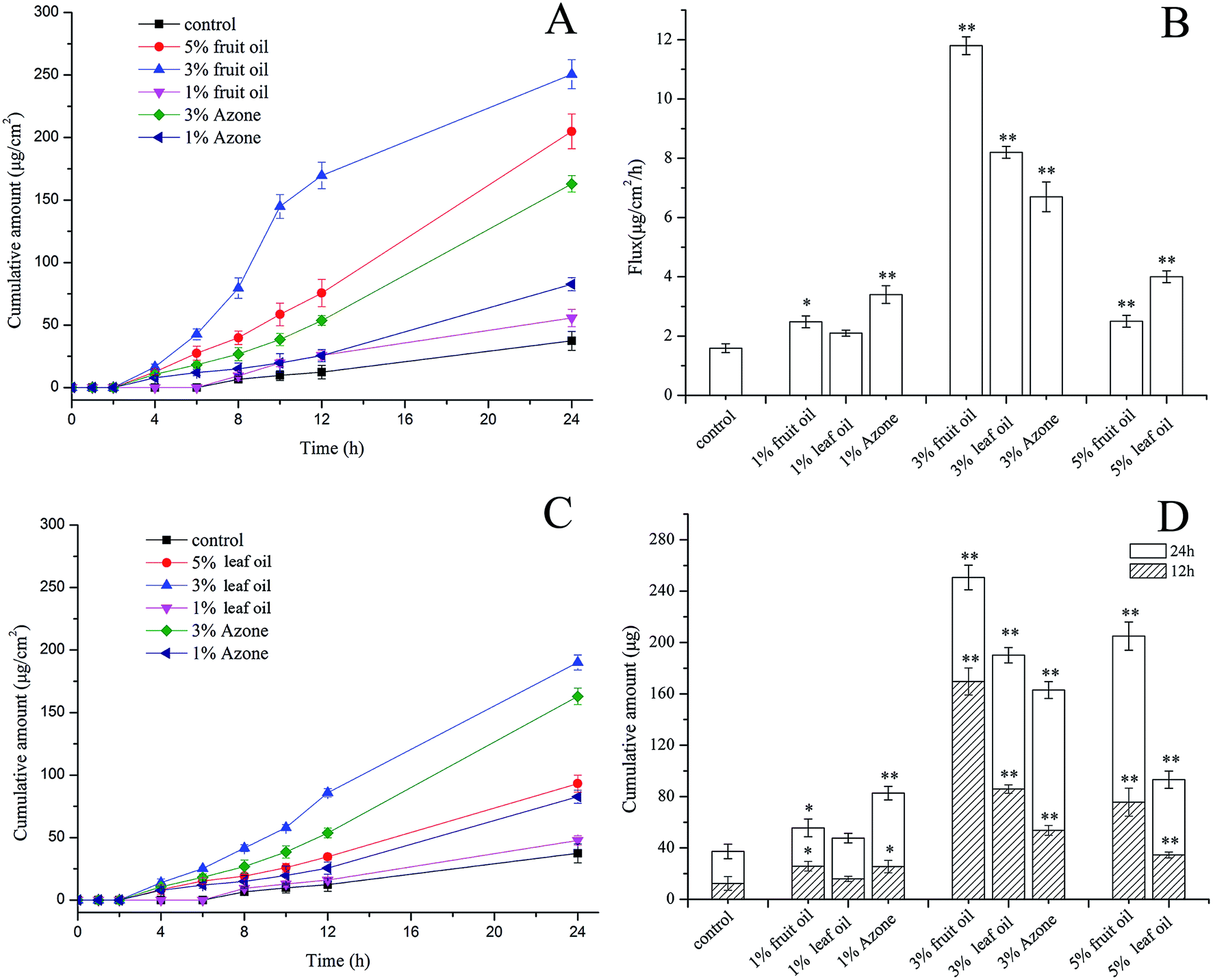 | ||
| Fig. 1 Comparison of the enhancing effect of fruit oil, leaf oil and Azone at various concentrations on the permeation of IND. (A) Cumulative amount of IND pretreated with fruit oil and Azone; (B) skin flux of IND after varying pretreatments; (C) cumulative amount of IND pretreated with leaf oil and Azone; (D) cumulative amount of IND after 12 h and 24 h. Data are represented as mean ± SD (n = 3). | ||
| Pretreatment | Steady flux (μg cm−2 h−1) | Lag time (h) | Kp | Er |
|---|---|---|---|---|
| — | 2.01 ± 0.09 | 5.52 ± 0.28 | 0.67 | — |
| 1% fruit oil | 2.74 ± 0.06 | 4.38 ± 0.88 | 0.85 | 1.27 |
| 3% fruit oil | 20.42 ± 1.87 | 3.56 ± 0.36 | 6.81 | 10.16 |
| 5% fruit oil | 10.57 ± 0.27 | 4.64 ± 0.14 | 3.52 | 5.25 |
| 1% leaf oil | 2.45 ± 0.14 | 4.73 ± 0.28 | 0.82 | 1.22 |
| 3% leaf oil | 9.26 ± 0.40 | 3.37 ± 0.01 | 3.09 | 4.61 |
| 5% leaf oil | 4.71 ± 0.15 | 4.32 ± 0.14 | 1.57 | 2.34 |
| 1% Azone | 4.10 ± 0.35 | 4.46 ± 0.35 | 1.37 | 2.04 |
| 3% Azone | 7.86 ± 0.57 | 4.08 ± 0.27 | 2.62 | 3.91 |
Azone was an excellent transdermal penetration enhancer, and for this reason was chosen as a positive control in this study,34 with its most effective concentration range 1–3%.7 Our results showed that the steady flux values of 1% and 3% Azone (4.10 ± 0.35 and 7.86 ± 0.57 μg cm−2 h−1, respectively) were significantly lower than that of 3% concentration in both oils (Table 1, and Fig. 1A and C). Furthermore, the Er of 3% fruit oil (10.16) was 2.6-fold higher than that of 3% Azone, which is generally regarded as the most effective concentration for many drugs. This result may be explained based on the partition coefficient between the hydrogel and the skin. Alternatively, it may because Azone did not penetrate into the deeper levels of the dermis thereby inhibiting its ability to enhance drug permeation.35 More importantly, in a previous report,32 the Er of fruit extracts with acetone only achieved 3.19 with IND as the model drug. Therefore, fruit oil in this study extracted by hydrodistillation has a greater enhancement effect than those extracts from organic solvents. The reason for this better performance, may be attributed to the synergistic effect between different constituents of essential oils. Overall, we concluded that at the same level, essential oils from A. oxyphylla are more effective than Azone in drug delivery of IND.
From Table 1, we also observed that 3% was the optimum concentration for both oils to reduce the lag time. The lag time to reach steady state flux in the control group was 5.52 ± 0.28 h, while it decreased to 3.56 ± 0.36 h and 3.37 ± 0.01 h after pretreatment with 3% fruit oil and leaf oil, respectively. Table 1 also shows that 5% level exhibited an increased lag time compared with 3% level in both oils. This can be due to a number of factors, among which the loss of cell functionality of the rat skin might be the cause.
3.2 Amount of drug retained in the donor and skin
At the end of the permeation experiment (48 h), the amounts of IND retained in the rat skin, as well as in the donor and receptor were calculated, and the results of several relative effective enhancers are shown in Table 2. Compared with the control group, the amount of IND retained in the rat skin was significantly enhanced after pretreatment with fruit oil and leaf oil (p < 0.05). Our results were consistent with a previous study which showed that essential oils affected the retention of drug in the skin.33 Table 2 showed that the amount of IND retained in the skin was 90 ± 22 μg for the control group, while after pretreatment, the values increased to 204 ± 35, 199 ± 85, 128 ± 72 and 142 ± 69 μg for 3% fruit oil, 3% leaf oil, 3% Azone and 5% fruit oil, respectively. According to the total amount of the drug in the receptor at the end of the experiment (48 h), we also found that the maximum penetration amount (997 ± 78 μg) was obtained in the 3% fruit oil group. Additionally, to avoid the influence of functionality loss of the rat skin cells, we discarded the data after 24 h in Fig. 1 to be more reasonable when observing the enhancement effects.| Amount of drug | Control | 3% fruit oil | 5% fruit oil | 3% leaf oil | 3% Azone |
|---|---|---|---|---|---|
| Retained in donor (μg) | 1965 ± 224 | 1450 ± 223 | 1604 ± 306 | 1707 ± 209 | 2051 ± 153 |
| Retained in skin (μg) | 90 ± 22 | 204 ± 35 | 142 ± 69 | 199 ± 85 | 128 ± 72 |
| Permeated into receptor (μg) | 378 ± 234 | 997 ± 78 | 765 ± 136 | 839 ± 102 | 663 ± 134 |
| Recovery (%) | 81.1 ± 16.0 | 88.4 ± 11.23 | 83.7 ± 17.0 | 91.5 ± 13.5 | 94.7 ± 11.9 |
While the enhancement effect of essential oils is dependent on the concentration used, there is no clear activity–concentration relationship.2 As a function of stratified skin structure, solubility, and the partition behaviour between vehicle and skin, many mechanisms for enhancers to affect skin permeation of drugs have been elucidated.36 According to the present study, one possible mechanism might be that essential oils increase the diffusion coefficient by disordering or altering the barrier property of the stratum corneum.37 Furthermore, the difference in permeation Er of essential oils may be attributed to variable molecular weights and boiling points of terpenes.33 The present study only utilized lipophilic IND as the model drug. As such, further studies are needed to determine whether other hydrophilic compounds are similarly enhanced by the essential oils.
3.3 Pharmacokinetic study
The most effective concentration of the essential oils (3%) was examined using the in vivo method. Profiles of the plasma concentration vs. time are shown in Fig. 2. The groups pretreated with fruit oil or leaf oil had higher plasma concentrations over time, which demonstrated that our essential oils have enhancement effect in rats.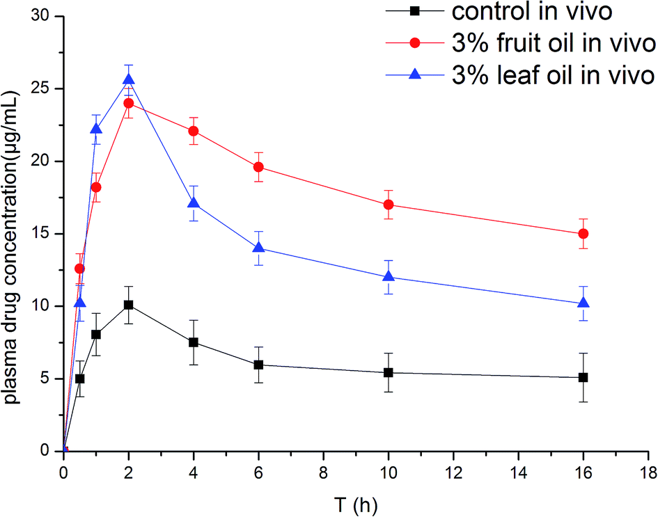 | ||
| Fig. 2 Plasma concentration over time of IND in rat plasma after application of gels with or without pretreatment (n = 3). | ||
The calibration curve was prepared according to a previous study.28 Results showed that the calibration curve was linear over the concentration range of 1–500 ng mL−1 in the rat plasma with a correlation coefficient of r = 0.9938. The resulting calibration curve of IND is Y = 0.002X + 0.0789. Fig. 3 shows MRM spectrum of IND and IS in blank rat plasma, rat plasma with IND and IS, and plasma sample obtained at 2 h after application of the donor gel. Blank rat plasma showed no significant interfering peaks at the retention times of IND and IS in the matrix. The retention time of IND and IS was 3.61 and 4.17 min, respectively. The total run time was 6.0 min. After 2 h, the plasma drug concentration were shown in Fig. 3C, it is obviously that the pretreated groups obtained higher concentrations of IND. Therefore, our essential oils definitely exhibited enhancement effect in the in vivo experiment.
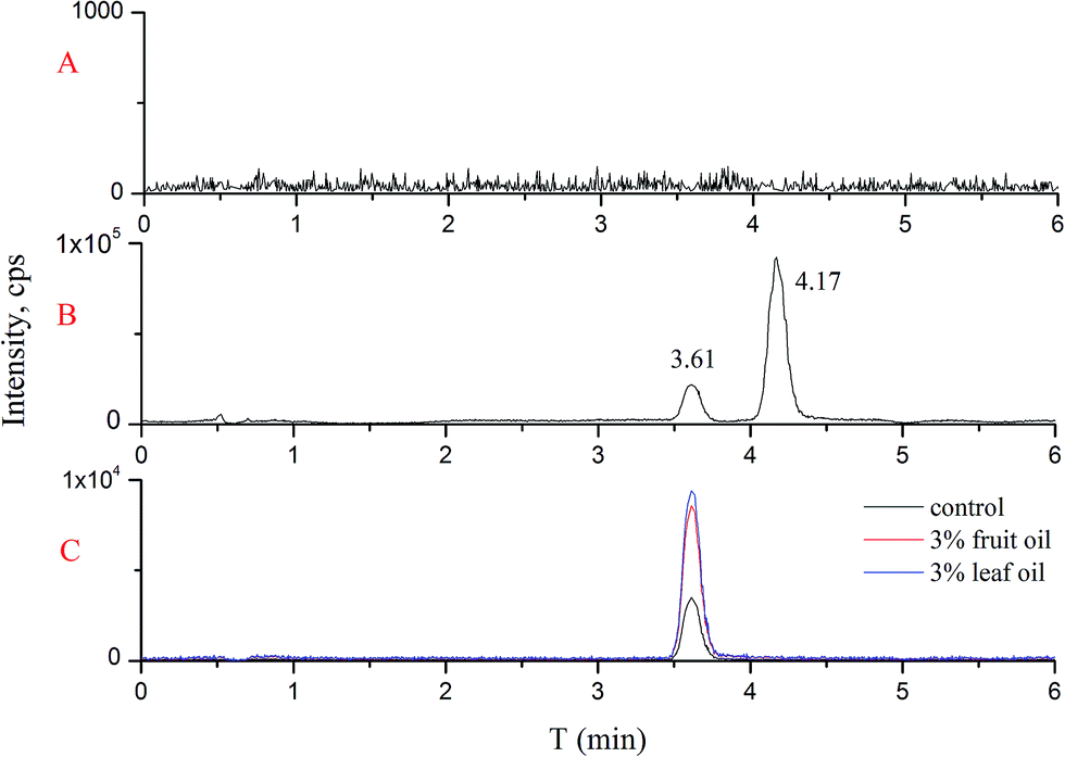 | ||
| Fig. 3 Typical MRM chromatograms of (A) blank rat plasma; (B) rat plasma with IND and IS; (C) plasma sample obtained at 2 h after application of the donor gel. | ||
3.4 Major constituents of essential oils from fruits and leaves
The major constituents of fruit oil and leaf oil were identified by GC-MS and the results are shown in Fig. 4. Fruit oil was found to contain 14 hydrocarbon terpenes and 15 oxygenated terpenes, representing 40.38% and 35.61%, respectively. Nootkatone (Rt = 33.54 min), accounting for 3.85% of the total content, has been shown to possess beneficial properties. The other major components with content exceeding 3% in fruit oil were: octahydro-1, 8-dimethyl-7-(1-methylethenyl)-naphthalene (Rt = 27.04 min, 7.65%) > o-cymene (Rt = 12.25 min, 7.44%) > globulol (Rt = 32.68 min, 6.46%) > caryophyllene (Rt = 25.34 min, 4.82%) > aromadendrene (Rt = 27.88 min, 4.24%) > alloaromadendrene oxide (Rt = 32.81 min, 3.94%).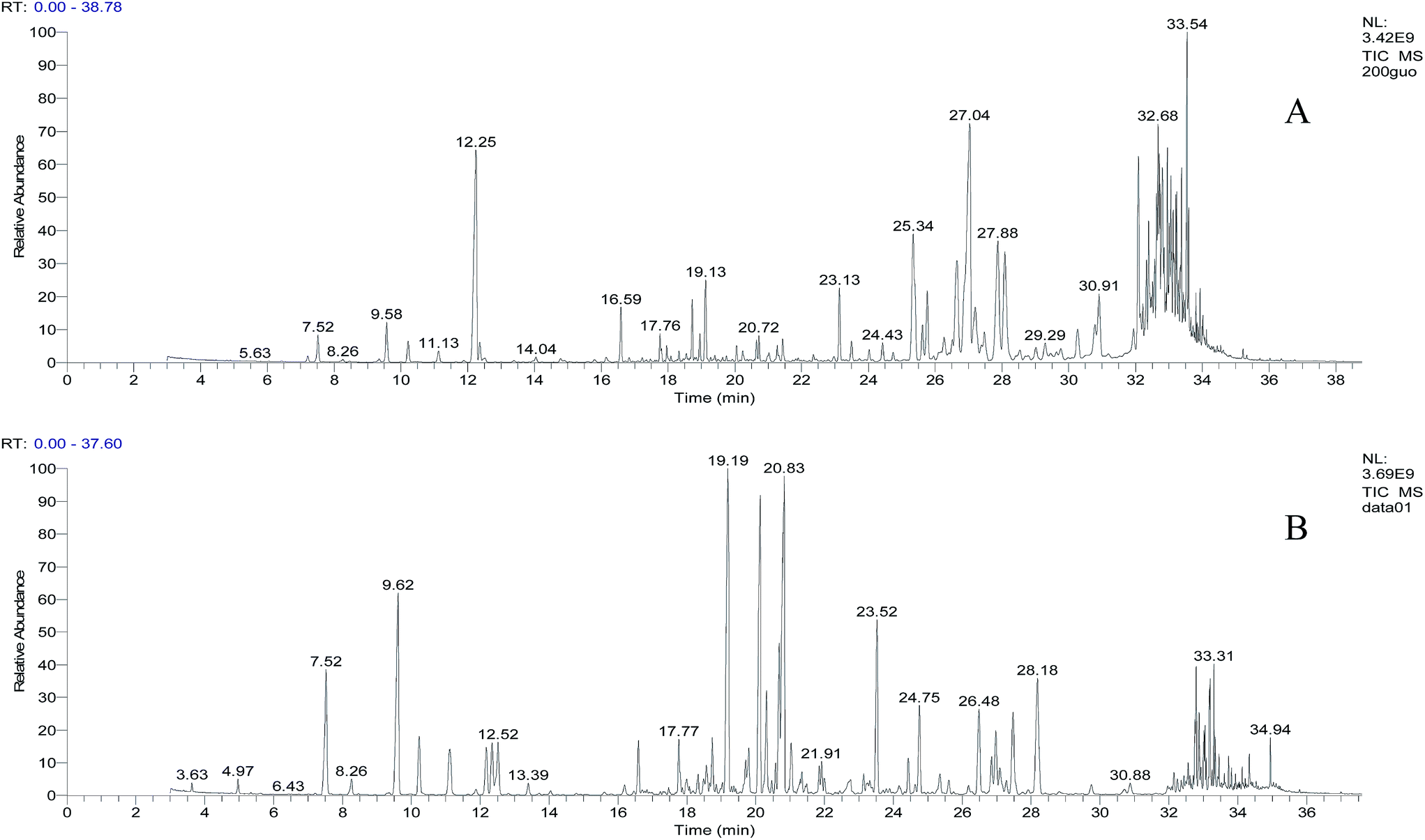 | ||
| Fig. 4 GC-MS spectrum of essential oils from fruits (A) and leaves (B). | ||
This is the first account of the identification of the major components in leaf oil. In contrast to fruit oil, leaf oil was found to contain 27.71% hydrocarbon and 50.77% oxygenated terpenes. However, we also found that it contained the same characteristic component nootkatone (1.39%) found in fruit oil. Therefore, given that leaves are often more readily available, they should be further studied as potential therapeutics. The major component with the highest content in leaf oil was myrtenal (Rt = 19.19 min, 10.25%), followed by α-citral (Rt = 20.83 min, 9.85%), β-citral (Rt = 20.13 min, 7.9%), β-pinene (Rt = 9.58 min, 6.14%), β-elemene (Rt = 23.52 min, 4.34%), β-sesquiphellandrene (Rt = 28.18 min, 3.91%), and α-pinene (Rt = 7.52 min, 3.37%).
It is widely known that essential oils and associated terpene constituents exhibit significant enhancement effects on various drugs.2 For example, α-pinene (hydrocarbon terpene) was reported to increase enhancement of the lipophilic drug, ketoprofen, as well as oxygen-containing monoterpenes (e.g., carvone and terpineol).38 Furthermore, nerolidol (alcohol terpenoid) was found to be an effective enhancer for the permeation of hydrophilic 5-fluorouracil through human abdominal skin.39 In the present study, we found that, at the same concentration, fruit oils outperformed leaf oils in enhancement effect on the permeation of lipophilic IND. This can be attributed to the higher content of hydrocarbon terpenes in the fruit oil (40.38%) relative to that in the leaf oil (27.71%). Importantly, leaf oil demonstrated a better performance in both steady flux and lag time (Table 1) than those of Azone. Hence, fruit oil and leaf oil are promising candidates as enhancers for lipophilic drugs. Future efforts will be put forth to study their effect on hydrophilic drugs.
In our previous study, the harvesting time and plant source were major factors that affected the chemical profiles of the resulting essential oils.40 Therefore, future studies will focus on the specific isolation of A. oxyphylla oils to confirm the chemical profiles in order to more systematically characterize the chemical constitution–activity relationship.
3.5 Skin irritation test
The comparison of the untreated and treated rabbit skins is presented in Fig. 5. Erythema and edema scores were evaluated by a visual scoring method. Warm water was used as the blank, and base oil, which was used to dilute the essential oils, was the negative control. Fig. 5 clearly shows that no visible erythema or edema present in the skin after 7 days treatment with all samples. Scores obtained from all rabbits demonstrated very subtle light pink skin discoloration and mild edema on the first two days after treatment with essential oils above 3% concentration. Importantly, this transient symptom did not cause any damage and disappeared over time, indicating that the slight changes caused by essential oils at high concentrations were reversible. We could conclude that essential oils from leaves and fruits of A. oxyphylla are safe to rabbits at the concentrations tested. However, for in-depth study of the toxicology of the essential oils, more investigations (e.g. metabolomics approaches) are still required in the following study.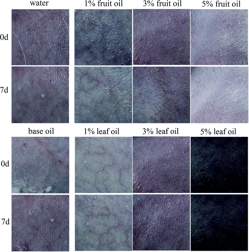 | ||
| Fig. 5 Comparison of untreated (0 day) and treated (7 days) rabbit skins. | ||
4 Conclusions
In this study, penetration enhancement effect of essential oils from the fruits and leaves of A. oxyphylla on skin permeation and deposition of indomethacin was evaluated. Results demonstrated that both oils had a significant enhancement effect on drug delivery and skin deposition of IND. Interestingly, the enhancement ratio of the fruit oil and leaf oil at 3% concentration, was almost 2.6-fold and 1.18-fold higher than that of Azone, a commonly used enhancer.Major constituents of both essential oils were identified using GC-MS to correlate the various components with the permeation effects of the essential oils isolated from different parts of A. oxyphylla. Both leaf and fruit-based oils were found to contain nootkatone. One major difference was that essential oil extracted from the fruit of A. oxyphylla had a higher content of hydrocarbon and oxygenated terpenes, which correlated with an increase in enhancement effect relative to leaf oil. Finally, the skin irritation test was carried out, and both oils were determined to be safe at the concentrations tested.
In conclusion, the penetration enhancement effect and safety assessment found in this study demonstrated that essential oils isolated from A. oxyphylla are promising candidates for chemical enhancers for use in transdermal drug delivery and cosmetics. However, further investigation is still required to decipher which specific components of these oils are eliciting this pharmacological effect. In addition, extensive pharmacodynamics and pharmacokinetic studies are needed to ascertain their safety as skin penetration enhancers in human volunteers.
Acknowledgements
This work was supported by the Major Science and Technology Project of Hainan Province (Grant no. ZDZX2013008).References
- K. Babiuch, M. Gottschaldt, O. Werz and U. S. Schubert, RSC Adv., 2012, 2, 10427–10465 RSC.
- A. Herman and A. P. Herman, J. Pharm. Pharmacol., 2015, 67, 473–485 CrossRef CAS PubMed.
- J. P. Wang, F. Guo, M. Ma, M. Z. Lei, F. P. Tan and N. Li, RSC Adv., 2014, 4, 45458–45466 RSC.
- S. Mutalik, P. K. Shetty, A. Kumar, R. Kalra and H. S. Parekh, Drug Delivery, 2014, 21, 44–54 CrossRef CAS PubMed.
- L. T. Fox, M. Gerber, D. P. Plessis and J. H. Hamman, Molecules, 2011, 16, 10507–10540 CrossRef PubMed.
- D. D. N’Da, Molecules, 2014, 19, 20780–20807 CrossRef PubMed.
- A. C. Williams and B. W. Barry, Adv. Drug Delivery Rev., 2012, 64, 128–137 CrossRef PubMed.
- I. B. Pathan and C. M. Setty, Trop. J. Pharm. Res., 2009, 8, 173–179 CAS.
- S. Aggarwal and S. Jalhan, Int. J. Pharma Bio Sci., 2013, 4, 857–868 CAS.
- Y. Lan, Q. Wu, Y. Q. Mao, Q. Wang, J. An, Y. Y. Chen, W. P. Wang, B. C. Zhao, N. Liu and Y. W. Zhang, J. Zhejiang Univ., Sci., B, 2014, 15, 153–164 CrossRef CAS PubMed.
- S. S. Jagannath, S. D. Manohar and S. R. Bhanudas, World J. Pharm. Pharm. Sci., 2014, 3, 1068–1080 CAS.
- A. Herman and A. Mlynarczyk, Isr. J. Plant Sci., 2015, 62, 1–2 Search PubMed.
- L. C. Zhang, L. H. Gao, J. H. Hu, C. X. Yan and Q. G. Zhu, Pharm. Care Res., 2006, 6, 413–416 CAS , in Chinese.
- G. X. Wang, H. Zhang, Z. L. Geng and Q. W. Wang, China J. Tradit. Chin. Med. Pharm., 2012, 27, 117–120 CAS , in Chinese.
- P. Yu, Q. Liang, H. Wang, L. M. Wu and D. Liu, China Med. Pharm., 2013, 3, 33–35 Search PubMed , in Chinese.
- Chinese Pharmacopoeia Commission, Pharmacopoeia of the People's Republic of China, China Medical Science Press, Beijing, 2010, pp. 273–274 Search PubMed.
- X. Chen, X. J. Liu, J. Wu, H. F. Dai and W. Q. Wang, Chin. Agric. Sci. Bull., 2010, 26, 366–371 CAS , in Chinese.
- Z. H. He, W. Ge, G. G. Yue, C. Lau, M. F. He and P. P. But, J. Ethnopharmacol., 2010, 132, 443–449 CrossRef PubMed.
- Y. Zheng, Y. Shao, A. H. Chen and N. N. Zhang, Food Sci., 2014, 35, 44–49 CAS.
- Y. L. Liu, X. Fang, L. Shi, Q. W. Yan, W. T. Feng, Y. H. Zhang, C. Y. Zhao, S. L. Lu and H. Liu, J. Hainan Norm. Univ., Nat. Sci., 2011, 24, 425–428 CAS , in Chinese.
- L. Li, L. J. Cai, J. R. Yang and Q. W. Zhang, Tianjin Med. J., 2011, 39, 165–167 CrossRef CAS PubMed.
- J. Y. Fang, Y. L. Leu, T. L. Hwang and H. C. Cheng, Biol. Pharm. Bull., 2004, 27, 1819–1825 CAS.
- F. Shakeel, N. Haq, F. K. Alanazi and I. A. Alsarra, Drug Dev. Ind. Pharm., 2014, 40, 1240–1245 CrossRef CAS PubMed.
- Y. H. Zhang, C. J. Wang, X. H. Jin, W. Zhang and S. Zhang, Prog. Mod. Biomed., 2013, 13, 6619–6623 CAS.
- Chinese Pharmacopoeia Commission, Pharmacopoeia of the People's Republic of China, China Medical Science Press, Beijing, 2010 Search PubMed.
- H. Uzuner, R. Bauer, T. P. Fan, D. A. Guo, A. Dias, H. E. Nezami, T. Efferth, E. M. Williamson, M. Heinrich, N. Robinson, P. J. Hylands, B. M. Hendry, Y. C. Cheng and Q. H. Xu, J. Ethnopharmacol., 2012, 140, 458–468 CrossRef PubMed.
- F. Qiu, X. P. Zhao, X. R. Lu, M. Y. Wang and M. X. Gong, RSC Adv., 2015, 5, 7260–7266 RSC.
- Hygienic Standard for Cosmetics, The Ministry of Health of PR China, 2007, 102–105.
- A. Martin, Diffusion and dissolution, in Physical Pharmacy: Physical Chemical Principles in the Pharmaceutical Sciences, 1993, pp. 877–898 Search PubMed.
- A. C. Williams and B. W. Barry, Int. J. Pharm., 1989, 57, 7–9 CrossRef.
- K. Cal, Arch. Dermatol. Res., 2006, 297, 311–315 CrossRef CAS PubMed.
- J. Y. Fang, Y. L. Leu, T. L. Hwang, H. C. Cheng and C. F. Hung, Eur. J. Pharm. Sci., 2003, 19, 253–262 CrossRef CAS.
- A. Nokhodchi, K. Sharabiani, M. R. Rashidi and T. Ghafourian, Int. J. Pharm., 2007, 335, 97–105 CrossRef CAS PubMed.
- K. Wang, Y. Yan, G. Zhao, W. Xu, K. Dong, C. Y. You, L. Zhang and J. F. Xing, Polym. Chem., 2014, 5, 4658–4861 RSC.
- M. Katz and B. J. Poulson, Absorption of Drugs through the Skin, New York, USA, 1971, pp. 103–162 Search PubMed.
- A. Arellano, S. Santoyo, C. Martin and P. Ygartua, Int. J. Pharm., 1996, 130, 141–145 CrossRef CAS.
- M. K. Das, A. Bhattacharya and S. K. Ghosal, Drug Delivery, 2006, 13, 425–431 CrossRef CAS PubMed.
- H. Bilek, N. Wonglertnirant, T. Ngawhirunpat, P. Opanasopit, M. K. Vollrath and M. Silpakorn, Univ. Sci. Technol. J., 2009, 3, 33–41 CAS.
- S. Songkro, T. Rades and G. Becket, Pharmazie, 2009, 64, 110–115 CAS.
- Q. Miao, W. J. Kong, X. S. Zhao, S. H. Yang and M. H. Yang, J. Pharm. Biomed. Anal., 2015, 102, 436–442 CrossRef CAS PubMed.
| This journal is © The Royal Society of Chemistry 2015 |