DOI:
10.1039/C5RA04843E
(Paper)
RSC Adv., 2015,
5, 44660-44665
High loading of doxorubicin into styrene-terminated porous silicon nanoparticles via π-stacking for cancer treatments in vitro†
Received
19th March 2015
, Accepted 12th May 2015
First published on 12th May 2015
Abstract
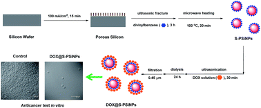
Porous silicon nanoparticles (PSiNPs) as nanocarriers for anticancer drug delivery have an important potential for cancer treatments, thus the development of PSiNPs-based delivery systems with efficient loading and controlled release of therapeutic drugs is necessary. Here, we present a novel strategy of incorporating doxorubicin (DOX) into styrene-terminated PSiNPs (S-PSiNPs) via π-stacking to form DOX@S-PSiNPs nanocomposites, which have a high-loading amount of DOX molecules. In addition, pH-controlled release of DOX molecules from the as-prepared DOX@S-PSiNPs nanocomposites was also observed. After cellular internalization of DOX@S-PSiNPs, the DOX molecules could be also efficiently released in the cytoplasm of cancer cells and then translocated into cellular nuclei, which prolonged their anticancer performance, compared with free DOX molecules.
1. Introduction
Doxorubicin (DOX) is an effective and widely used chemotherapeutic agent for the treatment of different cancers, such as acute leukemia, malignant lymphoma, and breast cancer.1 However, like most anticancer drugs, DOX has some drawbacks, e.g., limited solubility, nonspecific biodistribution, short blood half-life, high systemic toxicity to healthy tissue, and the development of drug resistance. To overcome these problems, various functional nanoparticles (NPs) (e.g., polymer NPs,2–7 mesoporous silica NPs,8–10 gold NPs,11,12 carbon nanotubes,13–16 graphene oxide NPs,17 iron oxide NPs,18 or silicon nanowires19,20) have been adopted as nanocarriers to deliver DOX in vitro or in vivo. The results demonstrated that these DOX delivery systems based on nanoparticles could significantly diminish the side effects, such as, prolong their blood half-life time, improve their targeted delivery efficiency, and lead to a sufficient drug concentration at the tumor sites.21 Besides, compared with free DOX, nanoparticles could also enter cells by an endocytosis pathway, independent from P-glycoprotein pathway, which could enhance drug concentration in intracellular region of tumor cells and efficiently reduce multidrug resistance factor.11,12,20
Porous silicon nanoparticles (PSiNPs) have emerged as excellent platform to deliver a wide variety of therapeutic and imaging agents in vitro or in vivo, which is attributed to their large surface area and pore volume, tunable pore size, and excellent biocompatibility, etc.22–26 In addition, PSiNPs could also exhibit tunable photoluminescence (PL), which resulted in easy tracking of their biodistribution in body by fluorescence signal.27,28 Benefiting from these advantages, DOX molecules have been successfully incorporated into PSiNPs by various methods. For example, Park et al. used oxidized PSiNPs to loading DOX molecules via electrostatic interactions, which obtained the loading amount of ∼44 μg mg−1.29 Wu et al. also reported that the DOX loading amount of carboxyl-terminated PSiNPs via covalent bonds of EDC/NHS chemistry was ∼39 μg mg−1.30 In addition, we designed a novel DOX delivery systems based on bovine serum albumin and porous silicon nanocomposites, which improved DOX loading capacity of PSiNPs to ∼240 μg mg−1.31 However, compared with others nanomaterials, DOX loading amount of PSiNPs has been low to date, which limited their clinical applications for cancer therapy. Notably, Liu et al. reported that the DOX loading capacity of graphene-oxide-based nanocarriers via π-stacking was ∼2350 μg mg−1, and they also found that single-walled carbon nanotubes had an ultrahigh loading capacity of DOX molecules via π-stacking (∼4000 μg mg−1), due to the aromatic nature of anthracyclines DOX and carbon nanotubes.16,17 In our previous work, we had fabricated styrene-terminated PSiNPs (S-PSiNPs) with near-infrared fluorescence for in vivo imaging, which could also provided enough aromatic groups on PSiNPs' surfaces.32 Herein, we adopted S-PSiNPs as nanocarriers to load DOX molecules via π-stacking (the details shown in Fig. 1a), which indicated that the loading amount was significantly increased to ∼660 μg mg−1. Moreover, DOX@S-PSiNPs remained highly stable in neutral buffer, in contrast, DOX could be significantly released in acidic buffer. Finally, after DOX@S-PSiNPs nanocomposites were cellular internalized, DOX molecules could be released inside human epithelial cervical cancer cells (Hela cells), which could provide a slower and prolonged DOX accumulation in cellular nuclei, compared to free DOX. Accordingly, we suggested that the delivery system of DOX@S-PSiNPs with continue anticancer behavior would be much favorable for cancer therapy.
 |
| Fig. 1 (a) Schematic diagram of microwave-assisted synthesis of S-PSiNPs, DOX loading and release, (b) CCD images of S-PSiNPs and DOX@S-PSiNPs incubated in a dual-phase solution of H2O/CH2Cl2, (c) loading kinetics of DOX into S-PSiNPs, and (d) release kinetics of DOX from DOX@S-PSiNPs under different pH conditions. | |
2. Results and discussion
2.1. Loading and release of DOX for S-PSiNPs
In our experiments, divinylbenzene was first grafted onto hydrogen-terminated PSiNPs via microwave-assisted hydrosilylation to form S-PSiNPs, and then S-PSiNPs were fully dispersed in DOX aqueous solution by ultrasonication (shown in Fig. 1a). As seen in Fig. 1b, after DOX encapsulation, hydrophobic S-PSiNPs were converted into hydrophilic DOX@S-PSiNPs with high water-dispersibility. Meanwhile, the loading kinetics of DOX into S-PSiNPs was monitored by UV-vis spectra. According to Fig. 1c and S4,† after ∼9 h of incubation in free DOX solution, the loading amount of DOX into S-PSiNPs were saturated, and then reached ∼660 μg mg−1 after 19 h. Compared with alkyl-terminated PSiNPs with the low loading amount of DOX (∼13 μg mg−1) even after 24 h in our previous work,31 the significant improvement of DOX loading into S-PSiNPs was mostly attributed to π-stacking from the aromatic nature of anthracyclines DOX and styrene groups. Release behavior of DOX from DOX@S-PSiNPs was also investigated under different pH conditions. The concentration of the released DOX were determined by measuring their PL intensity at 590 nm (λex = 480 nm), which was recorded in Fig. 1d and S1–3.† These results demonstrated that DOX molecules incorporated into DOX@S-PSiNPs remained stable at pH 7.5. Even after 25 h, only 14.3% of DOX molecules was released from DOX@S-PSiNPs. In contrast, when pH = 5.5, 52.1% of DOX molecules was fast released from DOX@S-PSiNPs after 5 h, and then DOX release reached at 56.3% after 25 h. These results indicated that DOX release from DOX@S-PSiNPs was pH-responsive, due to protonation and solubility of DOX under acidic conditions.13–16,19,20,31
2.2. Characterizations of DOX@S-PSiNPs composites
After excess DOX and large nanoparticles were removed using dialysis and filtration, DOX@S-PSiNPs nanocomposites could be prepared. Compared with yellowish PSiNPs alone, the color of DOX@S-PSiNPs was changed to reddish brown, due to DOX incorporation (shown in the insert of Fig. 2a). In Fig. 2a, two distinct emission peaks were found in PL spectra of DOX@S-PSiNPs, respectively. Under the excitation of 450 nm, the emission peak centred at ∼590 nm was attributed to DOX molecules. And under the excitation of 410 nm, the emission peak at ∼700 nm from S-PSiNPs was due to unique quantum confinement effects of PSiNPs alone.33–35 The mean hydrodynamic size and zeta potential of DOX@S-PSiNPs were also measured by dynamic-light-scattering (DLS) measurements, which was recorded in Fig. 2b. The results showed that DOX@S-PSiNPs held a high negative zeta potential (−23.1 ± 5.8 mV), which resulted in their excellent dispersibility and stability in aqueous solution. In addition, the mean hydrodynamic size of DOX@S-PSiNPs was ∼257 nm. Scanning electron microscopy (SEM) and transmission electron microscopy (TEM) measurements were further utilized to observe the size and morphology of DOX@S-PSiNPs. As seen in Fig. 2c and d, we found that the nanostructures in the range of 10–100 nm were not individual nanocrystals, but silicon nanoparticle domains were trapped in larger pieces of the porous silicon structure, similar to that of other types of PSiNPs.31–35 In addition, silicon nanoparticles domains (marked by blue circle) of S-PSiNPs could be obviously observed in Fig. S6,† however, they became blurry in DOX/S-PSiNPs (in Fig. 2d), due to the significant attachment of DOX layers, which could hinder electronic transmission.
 |
| Fig. 2 (a) PL spectra of DOX@S-PSiNPs, and its corresponding CCD images (in the insert), (b) the representative DLS histogram of DOX@S-PSiNPs in water, (c) SEM image of DOX@S-PSiNPs, and (d) TEM image of DOX@S-PSiNPs. | |
Moreover, X-ray photoelectron spectra (XPS) was adopted to determine atomic concentrations of elemental component of DOX@S-PSiNPs. The full XPS spectrum of DOX@S-PSiNPs was first displayed in Fig. 3a. From Fig. 3a, the signals of C 1s (285 eV), O 1s (532 eV), Si 2p (103 eV), and Si 2s (154 eV) were detected, which was consistent with the incorporation of organic molecules on PSiNPs. Additionally, the high-resolution XPS spectra of C 1s, O 1s, and Si 2p were also recorded in Fig. 3b–d, which provided the information of chemical bonds of these samples. Compared with that of bare PSiNPs and S-PSiNPs,32 (1) carbon concentration of DOX@S-PSiNPs increased from 28.02%, 51.85% to 62.92%, which was due to the loading of DOX molecules with high carbon elemental content. According to high-solution spectrum of C 1s in Fig. 3b, the peak at 284.4 eV was assigned to C–C and C–H, and the peak at 288.5 eV was assigned to C–carboxyl, which was due to rich carboxyl groups in DOX molecules; (2) oxygen concentration of DOX@S-PSiNPs increased from 14.94%, 24.01% to 31.24%, which demonstrated that significant oxidation happened during ultrasonication in DOX solution; (3) silicon and fluorine concentration of DOX@S-PSiNPs significantly reduced after DOX encapsulation. Because the increasing of thickness of surface coatings led to a decrease of the depth of silicon detected by X-ray, silicon and fluorine signals of DOX@S-PSiNPs would become much weaker, which was due to the formation of thick layers of DOX attached onto S-PSiNPs. According to above-mentioned results, we suggested that divinylbenzene had been successfully grafted onto hydrogen-terminated PSiNPs by microwave heating, and then DOX molecules were efficiently encapsulated onto S-PSiNPs via strong π-stacking to form DOX@S-PSiNPs nanocomposites.
 |
| Fig. 3 (a) Full XPS spectrum of DOX@S-PSiNPs, (b)–(d) high-resolution spectra of C 1s, O 1s, and Si 2p, and (e) the corresponding table of atomic concentrations. | |
2.3. Anticancer tests of DOX@S-PSiNPs in vitro
As a kind of broad-spectrum anticancer drug, DOX with high affinity towards DNA can efficiently accumulate in nucleus, intercalates DNA, and act as a cytostatic and apoptotic agent against tumor cells. Herein, the anticancer efficiency in vitro of DOX@S-PSiNPs was also investigated. In our experiments, Hela cells were chosen to be incubated with DOX@S-PSiNPs with the concentration of 20 μg mL−1 DOX in PBS solution (pH 7.4) at 37 °C for 24 h or 48 h, washed by PBS solution, and then observed by optical microscope, respectively (shown in Fig. 4a–c). These results exhibited that the significant inhibition of DOX@S-PSiNPs for Hela cells growth attached on Petri dishes. Compared with control cell samples (∼630 cell per cm2), the cell density was obviously reduced to about ∼80 cell per cm2 after 24 h of incubation with DOX@S-PSiNPs, and further decreased to ∼10 cell per cm2 after 48 h. In addition, MTT assays were also adopted to evaluate the viability of Hela cells incubated with free DOX and DOX@S-PSiNPs with different concentrations (2, 4, 6, 8, 10, and 20 μg mL−1, DOX). As seen in Fig. 4d, significant concentration-dependent anticancer performance of DOX@S-PSiNPs was found, similar with free DOX. Typically, after 24 h of incubation, DOX@S-PSiNPs at low concentration had little influence on the Hela cells, however, the cell viability was obviously reduced to 49.7% incubated with DOX@S-PSiNPs of high concentrations, and then reduced to 34.9% after 48 h, which was also in accordance with the results observed by optical microscope. However, compared with free DOX (26.2%, 24 h), DOX@S-PSiNPs (34.9%, 48 h) exhibited a slow and sustained anticancer effect.
 |
| Fig. 4 (a) Typical optical microscopy images of the growth of Hela cells attached on Petri dishes as a control, (b) Hela cells incubated with DOX@S-PSiNPs (20 μg mL−1, DOX) for 24 h, (c) for 48 h, and (d) cytotoxicity of free DOX and DOX@S-PSiNPs toward Hela cells. | |
To study the anticancer mechanism of DOX@S-PSiNPs, their cellular interactions were also monitored by laser scanning confocal microscopy (LSCM). Hela cells were incubated with free DOX or DOX@S-PSiNPs with the concentration of 20 μg mL−1 DOX in PBS solution at 37 °C for 24 h, washed by PBS solution, and then observed by LSCM, respectively. As seen in Fig. 5, with the excitation at 450 nm, an intense red fluorescence signal could be seen in intracellular compartments of Hela cells incubated with free DOX or DOX@S-PSiNPs, respectively. As a control, no significant fluorescence signal could be observed from Hela cells alone, which confirmed that red fluorescence signal was caused by cellular uptake of DOX molecules. When incubated with free DOX after 24 h, the fluorescence signal of Hela cells was mostly localized in intracellular nuclei, which demonstrated that nuclear DNA was the main subcellular site of DOX action for its antitumor activity. In contrast, according to the fluorescence signal of Hela cells incubated with DOX@S-PSiNPs, DOX molecules released from DOX@S-PSiNPs were mostly distributed in the cytoplasm after 24 h. And then DOX molecules could be translocated into intracellular nuclei was observed after 48 h (shown in Fig. S5†). Furthermore, these corresponding cell samples were also observed by TEM mode. As seen in Fig. 6, Hela cells as a control had intact cellular membranes and nuclei, in strike contrast, the damage of cellular membrane and the destruction of intracellular nuclei occurred in Hela cells after their interaction with free DOX or DOX@S-PSiNPs, due to cytotoxicity of DOX molecules. Compared with Hela cells interacted with free DOX, PSiNPs (marked by red arrow) could be clearly observed in cytoplasmas of Hela cells after the incubation with DOX@S-PSiNPs, which provided the direct evidence about cellular internalization of DOX@S-PSiNPs.
 |
| Fig. 5 (a) Typical LSCM images of the growth of Hela cells attached on Petri dishes as a control, (b) Hela cells incubated with of 20 μg mL−1 free DOX, and (c) Hela cells incubated with of DOX@S-PSiNPs (20 μg mL−1, DOX) (from left to right, fluorescence, bright-field, and overlay channel). | |
 |
| Fig. 6 (a) Typical TEM images of the growth of Hela cells attached on Petri dishes as a control, (b) Hela cells incubated with of 20 μg mL−1 free DOX, and (c) Hela cells incubated with of DOX@S-PSiNPs (20 μg mL−1, DOX). | |
According to the above-mentioned results, we suggested that free DOX could be passed through cellular and nuclear membranes by passive diffusion. However, DOX@S-PSiNPs was expected to enter cancer cells by endocytosis, and then DOX molecules could be efficiently released from DOX@S-PSiNPs in intracellular lysosome or endosomes, because of their acidic microenvironments. The transport process of DOX@S-PSiNPs into intracellular nuclei was slower than passive diffusion of free DOX, which could provide a slower and prolonged DOX accumulation in cellular nuclei and adequate DOX concentration to continually kill cancer cells, which would help overcome drug resistance of cancer cells.
3. Conclusions
In conclusion, we presented a method for fabricating S-PSiNPs with high loading of DOX (∼660 μg mg−1) via supermolecular stacking. DOX@S-PSiNPs remained highly stable in neutral buffer, in contrast, DOX could be significantly released in acidic buffer. After cellular uptake, DOX efficiently released from DOX@S-PSiNPs inside Hela cells, leading to an excellent anticancer effect. So DOX@S-PSiNPs would serve as a delivery platform to carry DOX for cancer therapy in future.
4. Experimental section
4.1. Preparation of S-PSiNPs
The single side polished, (100) oriented, and p-type silicon wafers (boron doped, 8–10 Ω cm resistivity, purchased from Hefei Kejing Materials Technology Co. Ltd, China) were boiled in 3
:
1 (v/v) concentrated H2SO4/30% H2O2 for 30 min and then rinsed copiously with Milli-Q water (≥18 MΩ cm resistivity). The porous silicon samples (1.54 cm2) were prepared by electrochemically etching in an ethanolic HF solution (40% HF/ethanol (1
:
1 v/v)) at 20 mA cm−2 for 45 min. And then freshly prepared porous silicon samples were immersed in pure divinylbenzene, followed by sonication and 20 min microwave heating at 100 °C. Using 30 min centrifugation at 1.2 × 10⁵ rpm, the samples were washed by ethanol to obtain S-PSiNPs.
4.2. Loading and release of DOX
3 mg S-PSiNPs were directly incubated in 3 mL free DOX solution. After centrifugation, UV-vis spectra of the supernatant of DOX solution were measured at different time point to track the loading kinetics of DOX. By centrifugal filtering of DOX@S-PSiNPs solution with pH 5.5 and 7.5, the fluorescence of DOX in supernatant was detected at each time point at 37 °C, respectively. According to the fluorescence of released DOX in solution at 590 nm (λex = 480 nm), the kinetics of DOX released from DOX@S-PSiNPs were measured.
4.3. Cellular interaction tests
Hela cells were plated onto 30 mm cell culture coverslips and incubated with 20 μg mL−1 DOX@S-PSiNPs for 24 h or 48 h, respectively. The attached nanoparticles or small molecules were washed three times with PBS solution (pH 7.4), and cells samples were monitored using LSCM (Leica TCS SP5, Germany) with the excitation at 450 nm. For TEM analysis of cell sections, the cells were seeded on a 6-well plate at a certain density (∼5 × 10⁵ cell per mL) and cultured for 24 h. Then the cells were incubated with DOX@S-PSiNPs. At a determined time, the cells were washed five times with PBS and trypsinized, centrifuged, and then fixed with 2.5% glutaraldehyde. After 2 h fixation at 4 °C, the samples were washed with PBS solution three times. Then the samples were fixed with 1% perosmic oxide for 2 h at 4 °C. After being washed in water, the samples were dehydrated in an alcohol series, embedded, and sliced with thickness from 50 to 70 nm.
4.4. Anticancer assay in vitro
Hela cells (∼3 × 10⁵ cell per mL) were dispersed within 96-well plates to a total volume of 100 μL per well and maintained at 37 °C in a 5% CO2/95% air incubator. Then the culture media was removed and the cells were incubated in culture medium containing the as-prepared DOX@S-PSiNPs or free DOX with different concentrations and washed with the culture medium. An amount of 100 μL of the new culture medium containing 3-(4,5-dimethylthiazol-2-yl)-2,5-diphenyltetrazolium bromide (MTT) (10 μL, 5 mg mL−1) was then added, followed by incubating for 4 h to allow the formation of formazan dye. After removing the medium, 150 μL DMSO was added to each well to dissolve the formazan crystals. Absorbance was measured at 570 nm in a microplate photometer. Cell viability values were determined (at least three times) according to the following formulae: cell viability (%) = the absorbance of experimental group/the absorbance of blank control group × 100%.
4.5. Instruments and methods
UV-vis adsorption spectra were recorded by a Shimadzu UV-2450 spectrophotometer. PL measurements were performed using a Perkin-Elmer LS55 fluorescence spectrometer. XPS were recorded using Kratos AXIS Ultra DLD system with a monochromatic Al Kα X-ray beam (1486.6 eV) at 150 W in a residual vacuum of <4 × 10⁻⁹ Pa. Analysis of nanoparticles size and surface charge was performed using Malvern Zetasizer Nano ZS DLS measurements. SEM images were taken by JEOL JSM-7600F scanning electron microscope with the accelerating voltage of 15 kV. TEM images were taken by JEOL JEM-2100 UHR transmission electron microscope with the accelerating voltage of 200 kV. Multiscan MK3 microplate photometer (Thermo Scientific) was used to monitor the absorbance of Hela cells during MTT assays.
Acknowledgements
This work is funded by the National Natural Science Foundation of China (no. 30930077 and no. 31000164), and Natural Science Foundation of Jiangsu Province (no. BK20130964). Thank Dr Huihua Min and Dr Jing Yang for TEM measurements, and Dr Yan Xuan for XPS measurements.
Notes and references
- G. Minotti, P. Menna, E. Salvatorelli, G. Cairo and L. Gianni, Pharmacol. Rev., 2004, 56, 185–229 CrossRef CAS PubMed.
- A. Kumari, S. K. Yadav and S. C. Yadav, Colloids Surf., B, 2010, 75, 1–18 CrossRef CAS PubMed.
- Z. Liu, Y. Jiao, Y. Wang, C. Zhou and Z. Zhang, Adv. Drug Delivery Rev., 2008, 60, 1650–1662 CrossRef CAS PubMed.
- X.-B. Xiong, Z. Ma, R. Lai and A. Lavasanifar, Biomaterials, 2010, 31, 757–768 CrossRef CAS PubMed.
- M. Shi, K. Ho, A. Keating and M. S. Shoichet, Adv. Funct. Mater., 2009, 19, 1689–1696 CrossRef CAS PubMed.
- J.-Z. Du, X.-J. Du, C.-Q. Mao and J. Wang, J. Am. Chem. Soc., 2011, 133, 17560–17563 CrossRef CAS PubMed.
- Z. Xu, K. Zhang, C. Hou, D. Wang, X. Liu, X. Guan, X. Zhang and H. A. Zhang, J. Mater. Chem. B, 2014, 2, 3433–3437 RSC.
- T. Lebold, C. Jung, J. Michaelis and C. Bräuchle, Nano Lett., 2009, 9, 2877–2883 CrossRef CAS PubMed.
- Q. He and J. Shi, J. Mater. Chem., 2011, 21, 5845–5855 RSC.
- H. Meng, M. Liong, T. Xia, Z. Li, Z. Ji, J. I. Zink and A. E. Nel, ACS Nano, 2010, 4, 4539–4550 CrossRef CAS PubMed.
- F. Wang, Y.-C. Wang, S. Dou, M.-H. Xiong, T.-N. Sun and J. Wang, ACS Nano, 2011, 5, 3679–3692 CrossRef CAS PubMed.
- M. Prabaharan, J. J. Grailer, S. Pilla, D. A. Steeber and S. Gong, Biomaterials, 2009, 30, 6065–6075 CrossRef CAS PubMed.
- S. Liu, A. C.-T. Ko, W. Li, W. Zhong and M. Xing, J. Mater. Chem. B, 2014, 2, 1125–1135 RSC.
- L. Meng, X. Zhang, Q. Lu, Z. Fei and P. J. Dyson, Biomaterials, 2012, 33, 1689–1698 CrossRef CAS PubMed.
- Z. Liu, A. C. Fan, K. Rakhra, S. Sherlock, A. Goodwin, X. Chen, Q. Yang, D. W. Felsher and H. Dai, Angew. Chem., Int. Ed., 2009, 48, 7668–7672 CrossRef CAS PubMed.
- Z. Liu, X. Sun, N. Nakayama-Ratchford and H. Dai, ACS Nano, 2007, 1, 50–56 CrossRef CAS PubMed.
- X. Sun, Z. Liu, K. Welsher, J. T. Robinson, A. Goodwin, S. Zaric and H. Dai, Nano Res., 2008, 1, 203–212 CrossRef CAS PubMed.
- M. Arruebo, R. Fernández-Pacheco, M. R. Ibarra and J. Santamaría, Nano Today, 2007, 2, 22–32 CrossRef.
- F. Peng, Y. Su, X. Wei, Y. Lu, Y. Zhou, Y. Zhong, S.-T. Lee and Y. He, Angew. Chem., Int. Ed., 2013, 52, 1457–1461 CrossRef CAS PubMed.
- F. Peng, Y. Su, X. Ji, Y. Zhong, X. Wei and Y. He, Biomaterials, 2014, 35, 5188–5195 CrossRef CAS PubMed.
- L. Brannon-Peppas and J. O. Blanchette, Adv. Drug Delivery Rev., 2012, 64, 206–212 CrossRef PubMed.
- E. J. Anglin, L. Y. Cheng, W. R. Freeman and M. J. Sailor, Adv. Drug Delivery Rev., 2008, 60, 1266–1277 CrossRef CAS PubMed.
- M. J. Sailor and E. C. Wu, Adv. Funct. Mater., 2009, 19, 3195–3208 CrossRef CAS PubMed.
- H. A. Santos, L. M. Bimbo, B. Herranz, M.-A. Shahbazi, J. Hirvonen and J. Salonen, J. Mater. Res., 2013, 28, 152–164 CrossRef CAS.
- H. A. Santos and H. Hirvonen, Nanomedicine, 2012, 7, 1281–1285 CrossRef CAS PubMed.
- D. J. Savage, X. Liu, S. A. Curley, M. Ferrari and R. E. Serda, Curr. Opin. Pharmacol., 2013, 13, 834–841 CrossRef CAS PubMed.
- K. Sato, H. Tsuji, K. Hirakuri, N. Fukata and Y. Yamauchi, Chem. Commun., 2009, 45, 3759–3761 RSC.
- Z. Kang, Y. Liu, C. H. A. Tsang, D. D. D. Ma, X. Fan, N.-B. Wong and S.-T. Lee, Adv. Mater., 2009, 21, 661–664 CrossRef CAS PubMed.
- J.-H. Park, L. Gu, G. V. Maltzahn, E. Ruoslahti, S. N. Bhatia and M. J. Sailor, Nat. Mater., 2009, 8, 331–336 CrossRef CAS PubMed.
- E. C. Wu, J. S. Andrew, A. Buyanin, J. M. Kinsella and M. J. Sailor, Chem. Commun., 2011, 47, 5699–5701 RSC.
- B. Xia, W. Zhang, J. Shi and S.-J. Xiao, J. Mater. Chem. B, 2014, 2, 5280–5286 RSC.
- B. Xia, B. Wang, J. S. Shi, W. Zhang and S.-J. Xiao, J. Mater. Chem. B, 2014, 2, 8314–8320 RSC.
- B. Xia, W. Zhang, W. Bao, C. Dong, J. Zhang and J. S. Shi, Phys. Status Solidi A, 2012, 209, 2247–2250 CrossRef CAS PubMed.
- B. Xia, W. Zhang, J. S. Shi and S.-J. Xiao, Analyst, 2013, 138, 3629–3632 RSC.
- B. Xia, W. Zhang, J. Shi and S.-J. Xiao, ACS Appl. Mater. Interfaces, 2013, 5, 11718–11724 CAS.
Footnote |
† Electronic supplementary information (ESI) available: PL and UV-vis spectra of DOX@S-PSiNPs incubated in aqueous solution under different conditions. See DOI: 10.1039/c5ra04843e |
|
This journal is © The Royal Society of Chemistry 2015 |
Click here to see how this site uses Cookies. View our privacy policy here.