
Morphological control of mesoporous CN based hybrid materials and their excellent CO2 adsorption capacity†
Kripal S. Lakhia,
Arun V. Baskara,
Javaid S. M. Zaidia,
Salem S. Al-Deyabb,
Mohamed El-Newehyb,
Jin-Ho Choyc and
Ajayan Vinu*a
aAustralian Institute for Bioengineering and Nanotechnology (AIBN), The University of Queensland, Brisbane, QLD, Australia. E-mail: a.vinu@uq.edu.au
bPetrochemical Research Chair, Department of Chemistry, College of Science, King Saud University, Riyadh 11451, Saudi Arabia
cDepartment of Chemistry and Nanoscience, EWHA Womans University, Seoul, South Korea
First published on 28th April 2015
Abstract
Highly ordered mesoporous carbon nitrides (MCN-1-Ts) with uniform rod shaped morphology have been synthesized by a hard templating technique using SBA-15 silicas prepared a under hydrothermal “static” condition at different temperatures as templates following a simple polymerization reaction between carbon tetrachloride (CTC) and ethylenediamine (EDA) inside the large pores of SBA-15. The static hydrothermal condition offers uniform rod shaped morphology for the template materials which has been completely replicated into the MCN nanostructures. The obtained materials were characterized with low angle XRD, N2 adsorption, high resolution transmission electron microscopy, high resolution scanning electron microscopy (FE SEM), Fourier transform infra-red (FT-IR), and X-ray photoelectron spectroscopy (XPS). The characterization results confirm the successful replication of the ordered structure, morphology and mesoporosity of the template material into carbon nitride. The FT-IR and XPS techniques confirm the presence of free –NH and –NH2 groups on the surface of MCN, which are critical for capturing CO2. Finally, these materials with high surface area and uniform morphology are used as adsorbents for high pressure CO2 adsorption at different temperatures of 0, 10 and 25 °C. It is found that the morphology of the materials which has a direct relation with the textural parameters plays a significant role in enhancing the amount of CO2 adsorption. The MCN with the uniform morphology and the highest surface area registers the highest CO2 adsorption capacity (16.5 mmol g−1) at 0 °C and 30 bar pressure, which is found to be higher than that of the previously reported 3D- cage type MCN, activated carbon, multiwalled carbon nanotubes and mesoporous silicas.
1. Introduction
The rapid increase in CO2 levels due to wide spread industrialization and human activities in both developed and developing countries is posing a potential threat to severe climatic conditions in the form of global warming, floods, melting of glaciers, drought and other natural events. CO2 is produced as a by-product in a number of processes but two prominent ones are burning of fossil fuels for power generation and the transportation industry. It is reported that CO2 alone contributes 60% to the total global warming caused by all the greenhouse gases put together.1 At the same time, CO2 has been viewed by the scientific community as a potential source for the production of useful chemicals and efficient transport fuels such as methanol, dimethyl ether and their useful derivatives.2 Therefore, researchers put a lot of effort into finding solutions not only to reduce CO2 emission into the atmosphere but also its conversion to value added products.Carbon capture and sequestration is being developed as a promising technology to reduce large scale CO2 emission particularly from coal fired power plants and all efforts are being made to make processes green and environmentally friendly. Although many physical and chemical absorption based technologies are available, the most common CO2 capture technology in use today is the absorption in liquid amine and ammonia solution. However, the liquid ammonia absorption process has a number of disadvantages such as high regeneration cost, corrosion of equipment, hazardous nature etc. A number of solid adsorbents including a wide range of porous materials such as metal organic frameworks (MOFs), activated carbon, porous carbon, amine-functionalized porous silica, functionalized porous carbons, zeolites, porous polymers have been tried as possible adsorbents for CO2.2–4 Among these adsorbents, porous carbon materials are considered as the best candidates due to their low cost, ease of preparation and regeneration, high surface area, excellent thermal and chemical stability. However, they suffer from poor CO2 adsorption capacities due to weaker adsorbate–adsorbent interaction resulting primarily from the hydrophobic and neutral surface charge.4 Therefore, much efforts have been given to the functionalization of porous carbon adsorbents with N atoms or basic molecules which enhances the adsorption of CO2 due to the acid–base interaction.5–7
Zhao et al. used the post-synthetic approach to incorporate N atoms on the 3D nanoporous carbon framework by treating with melamine precursor, which showed the maximum CO2 adsorption capacity of 8.5 mmol g−1 at 15 °C and 10 bar pressure8 whereas Zhu et al. reported the N-doped porous carbons from expensive ionic liquids, which registered the CO2 adsorption capacity of 4.39 mmol g−1 at 0 °C and 1 bar.9 Ahn et al. introduced polyethylenimine (PEI) inside the porous channels of mesoporous silicas such as SBA-15, SBA-16, KIT-6, MCM-48 and MCM-41 in order to enhance the amount of CO2 adsorption.10 However, these approaches require either additional steps or expensive nitrogen precursors for introducing the basic sites on the surface of the porous adsorbents which increases the cost of the materials and thereby presents a major hurdle for the practical applications.
Carbon nitride (CN) is a unique material with excellent electronic and electrical properties. The surface properties of CN are quite different from those of pure carbon materials as the incorporation of nitrogen atoms in the carbon nanostructure can enhance the mechanical, conducting, and semiconducting properties and further offer basic sites on the surface. Recently, Vinu et al. have introduced mesoporosity in the CN through a combined nanohard templating and a simple polymerization technique by mixing ethylenediamine and carbon tetrachloride inside the nanochannels of the mesoporous SBA-15 template.11,12 These materials have excellent properties such as strong basicity, inherent semiconducting nature, large surface area, high pore volume, controllable pore size and excellent thermal and mechanical stability, which may be utilized for various applications including adsorption, separation, and catalysis.11,12 Especially, mesoporous carbon nitride based hybrids (MCN) are particularly attractive for capturing acidic CO2 molecules because the free NH2 groups on its surface can selectively adsorb the CO2 molecules through acid–base interaction.13
Morphology and textural parameters of the mesoporous materials are extremely important for many applications including adsorption and catalysis as they dictate surface area and pore volume and offer easy access to the actives sites and support the inter-particle diffusion. In the nano-hard templating approach, in general, the structure of the template is inversely replicated and the particle morphology is retained into the desired materials. Therefore, it is important to find a suitable method to control the morphology of the template materials. For tuning the morphology of MCN, it is necessary to find a method to control the morphology of the template first as it directs the morphology of the replicated material. It has been widely reported that the morphology of the mesoporous silica materials can be controlled by altering the nature of the surfactants, cosolvents or the additives such as KCl or NH4F or (NH4)2SiF6.14–16 By varying the synthesis conditions, various morphologies of mesoporous silica including rod, sphere, fibre, platelet, gyroid, discoid, doughnut, sausage etc. have been prepared. For example, Kosuge et al. reported fibre and rod like SBA-15 mesoporous silica using the inexpensive sodium silicate which served both as a silica source as well as the salt.17 Although there are several methods available in the literature to control the morphology of the mesoporous silica materials, the use of “static” synthesis condition, reported by Sayari18 and his co-workers to prepare the mesoporous silica with a highly uniform and well-defined rod shaped morphology has been found to be the best as it is considered an easy, salt-free and inexpensive approach.
In the hard templating approach, the walls and the pores of the inorganic porous templates are converted into pores and walls of the final product, respectively. However, the final product retains the morphology of the inorganic template. Thus, it is important to control the morphology of the template in order to prepare the materials with different morphology using hard templating approach. With this approach, several nanoporous carbons with different pore diameters and particle morphology have been prepared.19 Previously, Vinu et al. have also used mesoporous silica with tunable pore diameters and/or particle morphology to fabricate a series of mesoporous carbon materials which showed excellent adsorption capacity for biomolecules including proteins, vitamins and aminoacids.19–26 However, there is no report available in the literature on controlling the particle morphology of the mesoporous carbon nitride materials.
In this work, we report on the synthesis of highly ordered MCNs with a high nitrogen content and controlled morphology using SBA-15 silica materials with uniform rod shaped morphology prepared under “static condition” as templates and ethylene diamine and carbon tetrachloride as the carbon and nitrogen sources, respectively. The materials exhibit a high structural order with well-defined cylindrical or rod shaped particle morphology and excellent textural parameters. We also show that the MCNs prepared with controlled morphology can be used as the adsorbents for CO2 molecules. The CO2 adsorption capacity was found to be ca. 726 mg g−1 (16.5 mmol g−1) of the adsorbent at 273 K which is much higher than that reported for other MCN,13 activated carbons, multi-walled carbon nanotubes, N-doped porous carbons8,9 and pure and functionalized mesoporous silica materials.10 The reason for the higher CO2 adsorption capacity of these MCN with controlled morphology, and the mechanism and the heat of adsorption have also been discussed in detail.
2. Experimental
2.1. Synthesis
The mesoporous SBA-15 silica template material was synthesized under static condition using a soft templating approach under strongly acidic condition. In a typical synthesis, 4 g of non-ionic surfactant Pluronic P-123 which is a triblock copolymer (EO20PO70EO20), avg. mwt ∼ 5800, Sigma-Aldrich was added to 30 g of water in a polypropylene (PP) bottle with a cap. After being stirred continuously for 4 h, 120 g of 2 M HCl was added to the solution. The temperature was simultaneously raised to 40 °C and the mixture was stirred for 2 h. Subsequently, 9 g of TEOS (tetraethyl orthosilicate, 98% Sigma-Aldrich) was added and the mixture was stirred for only 20 min after which stirring was stopped completely and the sample was left unagitated for the next 24 h with the temperature in the water bath maintained at 40 °C. The solution mixture was then transferred to a Teflon lined autoclave and kept in an oven at 100 °C for 48 h. The resulting product was filtered in hot, washed three times with a large amount of water, dried at 100 °C for 6 h and calcined at 540 °C for 12 h to remove the polymeric surfactant. Another set of mesoporous silica templates was prepared by using the above procedure except that autoclaving was done at 130 °C and 150 °C. The obtained samples were labeled as SBA-15-Ts where T is the hydrothermal synthesis temperature and s denotes static conditions.MCN was prepared using a hard templating approach involving a polymerization reaction between the carbon source, CCl4, and the nitrogen source, ethylenediamine, inside the mesopores of the SBA-15-Ts materials. In a typical synthesis, 0.5 g of SBA-15-Ts was mixed with 3 g of CCl4 and 1.35 g of ethylenediamine in a round bottom flask fitted with water cooled condenser. The mixture was refluxed at 90 °C for 6 h under constant stirring. The temperature was increased in steps of 10 °C from 60 to 90 °C. After 6 h of stirring at 90 °C, the unreacted CCl4 and ethylenediamine in the composite polymer was removed using the rotavapor at 55 °C. The sample was then dried at 100 °C for 6 h and then crushed into fine powder using a mortar and pestle. The crushed powder was then carbonized in a tubular furnace at 500 °C for 6 h under nitrogen flow. The carbonized sample was then treated with 5% HF and the sample was washed three times with excess ethanol and then kept for drying at 100 °C for 6 h. The samples were labelled as MCN-1-Ts where T denotes the synthesis temperature of the template.
2.2. Characterization
The powder X-ray diffraction measurements were carried out on a Bruker Advance D8-III diffractometer using Cu Kα radiation from a sealed tube source operating at 40 kV and 40 mA, a fixed divergence slit of 0.1° and a Lynxeye multi-pixel detector. The data were recorded in a 2θ range from 0.5 to 8° with a scan rate of 0.01° per sec. Nitrogen adsorption and desorption isotherms were measured at −196 °C on a Micromeritics ASAP 2420 surface area and porosity analyser. All the samples were separately degassed at 250 °C for 8 h under vacuum in the degas port of the adsorption analyser. The specific surface area was calculated using the BET model. Pore size distribution was obtained from both the adsorption and desorption branch of the nitrogen isotherms using the BJH model.X-ray photoelectron spectroscopy was carried out using a Kratos Axis ULTRA X-ray photoelectron spectrometer incorporating a 165 mm hemispherical electron energy analyser using monochromatic Al Kα X-rays (1486.6 eV) at 225 W (15 kV, 15 mA). Survey and multi-region spectra were recorded at C1s and N1s photoelectron peaks. Each spectra region of photoelectron of interest was scanned several times to obtain a good signal to noise ratios. Survey (wide) scans were taken at analyser pass energy of 160 eV and multiplex (narrow) high resolution scans at 20 eV. Survey scans were carried out over 1200–0 eV binding energy range with 1.0 eV steps and a dwell time of 100 ms. Narrow high-resolution scans were run with 0.05 eV steps and 250 ms dwell time. Atomic concentrations were calculated using the CasaXPS version 2.3.14 software and a Shirley baseline with Kratos library Relative Sensitivity Factors (RSFs). Peak fitting of the high-resolution data was also carried out using the CasaXPS software. FT-IR spectra were recorded on a Nicolet 5700 FTIR spectrometer fitted with a diamond attenuated total reflection (ATR) accessory by averaging 200 scans with a resolution of 2 cm−1 measured in absorbance mode.
High pressure CO2 adsorption was carried out on a Quantachrome Isorb HP1 equipped with temperature controlled circulator and a booster compressor. The CO2 adsorption was carried out in the pressure range of 0 to 30 bar and different analysis temperatures from 0 to 25 °C were used. Prior to CO2 adsorption, samples were degassed under vacuum at 250 °C for 8 h. The isosteric heat of adsorption was calculated using Clausius–Clapeyron equation. The structural morphology of MCN-1-Ts samples was observed on a JEOL FE SEM 7001 whereas the HR-TEM images were obtained using a Tecnai F20 FEG TEM equipped with EDAX EDS and GIF (Gatan Image Filter). The preparation of the samples for HR-TEM imaging involved sonication of 10–15 mg of the sample in ethanol for 5 to 8 min and deposition on a holey carbon film supported copper grid.
3. Results and discussion
Mesoporous silica materials with rod shaped morphology were prepared using polymeric surfactants under static conditions. The structure of the prepared template materials was first confirmed by powder X-ray diffraction analysis. Fig. 1a shows the low angle powder XRD patterns for the SBA-15-Ts template materials. All the XRD patterns exhibit several well-ordered peaks which can be indexed as (100), (110) and (200) reflection planes on a two-dimensional hexagonal lattice (p6mm) and are characteristic low angle peaks for the silica SBA-15 materials and consistent with the reports available in the literature.11,12,27 These results reveal that the static condition did not affect the structural order of SBA-15 materials prepared at different temperatures. Further, it is observed that diffraction peaks shifted to lower angles with increase of hydrothermal temperature, which indicates the increase in d spacing and the unit cell size. The values of cell constant and d100 spacing show an increasing trend as the synthesis temperature is increased from 100 through 150 °C (Table 1). Similar results were also reported previously for the SBA-15 samples prepared under dynamic conditions.11,12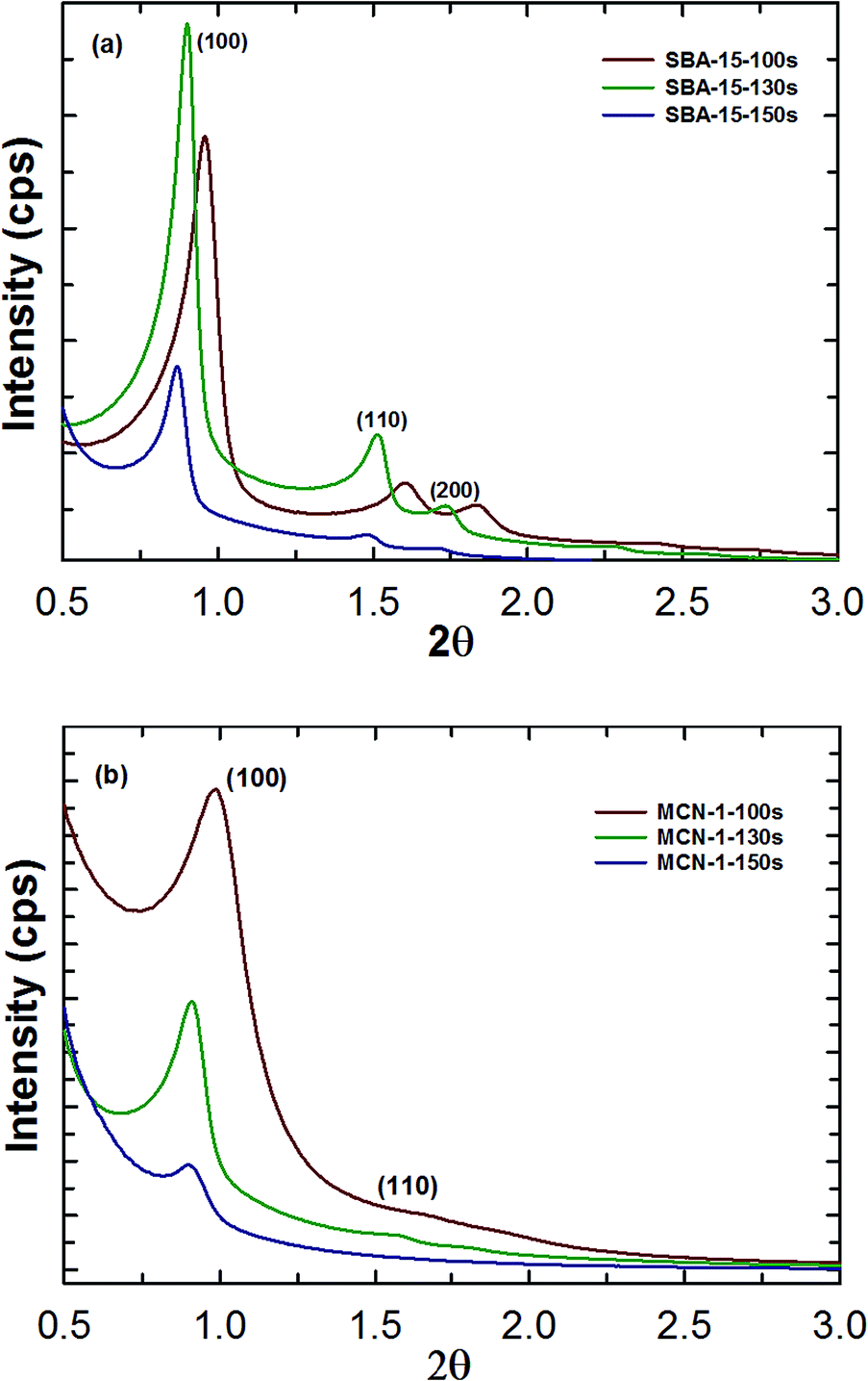 | ||
| Fig. 1 Low angle powder X-ray diffraction patterns of (a) SBA-15-Ts silica templates and (b) MCN-1-Ts samples. | ||
| Sample | a0 (nm) | d (nm) | S.A (m2 g−1) | PDa (nm) | PV (cm3 g−1) | CO2 adsorbedb (mmol g−1) |
|---|---|---|---|---|---|---|
| a Pore diameter calculated from the adsorption branch.b Adsorption measured at 0 °C and 30 bar using dry CO2 gas. | ||||||
| SBA-15-100s | 10.63 | 9.25 | 710 | 8.4 | 1.11 | 12.4 |
| SBA-15-130s | 11.32 | 9.85 | 506 | 11.25 | 1.24 | 10.1 |
| SBA-15-150s | 11.74 | 10.21 | 388 | 11.29 | 1.19 | 8.4 |
| MCN-1-100s | 10.38 | 9.03 | 621 | 3.76 | 0.67 | 12.9 |
| MCN-1-130s | 11.16 | 9.71 | 678 | 4.99 | 0.87 | 16.5 |
| MCN-1-150s | 11.32 | 9.85 | 518 | 5.94 | 0.76 | 12.3 |
| Activated carbon | — | — | 747 | 5.3 | 0.41 | 3.7 |
| MWCNT | — | — | 250 | 3.2 | 0.31 | 5.6 |
The increase of the unit cell constant of the samples with increasing synthesis temperature can be explained as follows: when the synthesis temperature is increased, dehydration of polyethylene oxide units surrounded by hydration spheres takes place. This would increase the length of the hydrophobic tail of the surfactant by coupling with the already existing hydrophobic polypropylene oxide moieties of the surfactant which is mainly responsible for creating the large mesoporosity in the samples. Moreover, it is known that the nonionic surfactant P123 is stable up to 130 °C and starts to decompose into smaller fragments at temperature higher than 130 °C. These small polymeric fragments also contribute to the enlargement of pores and the unit cell constant as they act as swelling agents and expand the micelles.
Fig. 1b shows the low angle powder XRD plots for MCN-1-Ts samples which also exhibit a sharp peak and two low angle peaks. These peaks can be indexed to the (100), (110) and (200) reflections which are associated with the hexagonally ordered mesostructures with a space group of p6mm. The presence of multiple peaks at lower angles clearly confirms the successful replication of the structure of SBA-15 silica template into MCNs. Interestingly, the peaks are shifted towards lower angle as the synthesis temperature or pore size of the SBA-15-Ts is increased. As observed in case of silica template material, the peak shift is more pronounced in MCN-1-150s as compared to the other two samples. Further, the intensity of the higher order peaks for MCN-1-150s is much lower as compared to that for MCN-1-100s and MCN-1-130s which indicates a slight structural disorder in the sample. For MCN-1-150s, the lower structural order may be attributed to the incomplete filling of the large mesopores of the SBA-15-150s with the carbon and nitrogen sources. Fig. 1S† shows the high angle XRD patterns for MCN-1-Ts samples. These samples exhibit a broad peak at 2 theta values close to 24.8°, confirming the turbostratic ordering of carbon and nitrogen atoms in the graphene layer.11
Fig. 2a shows the N2 adsorption isotherms of the silica template SBA-15-Ts whereas the corresponding textural parameters are given in Tables 1 and 1S.† All the template materials show type IV adsorption isotherm with a sharp capillary condensation step and a H1 hysteresis loop, which is typical of mesoporous materials with well-ordered large and cylindrical type pores. As the synthesis temperature is increased, the capillary condensation step is shifted from lower relative pressure to higher relative pressure, revealing the huge expansion of the pore size. The pore diameter of the samples increases from 8.4 to 11.29 nm upon increasing the synthesis temperature from 100 to 150 °C. On the other hand, the specific surface area of the materials decreases with increasing the synthesis temperature. The pore volumes of the samples are in the range of (1.11 to 1.29 cm3 g−1) which is quite similar to that of the samples prepared under dynamic conditions12 (Table 1). Among the materials studied, SBA-15-130s exhibits the highest pore volume which might be attributed to the excellent structural order and the large pore diameter.
 | ||
| Fig. 2 Nitrogen adsorption isotherms for (a) SBA-15-Ts, and (b) MCN-1-Ts samples. | ||
The N2 adsorption–desorption isotherms of MCN-1-Ts samples are shown in Fig. 2b. It is clear from the isotherms of MCN-1-Ts samples that these materials also exhibit type IV isotherm with a capillary condensation step at the higher relative pressure, which indicates the presence of mesopores in the sample and further reinforces our claims about the successful replication of the structure of the templates to MCN-1-Ts. It can be seen from Table 1 that the pore diameter of the samples increases in the following order: MCN-1-100s > MCN-1-130s > MCN-1-150s (ESI Fig. 2S†). The utilization of the template with the large pore diameter offers the material with a large pore diameter. The specific surface area and the pore volume of the MCN-1-130s are much higher than those of other samples studied in this work, which may be attributed to the excellent structural order and better textural parameters of the SBA-15-130s template.
One of the highlights of this work is that the morphology of the materials can be easily controlled by varying the synthesis temperature and the stirring conditions. The morphology and the mesostructure of the MCN-1-Ts samples were studied by using HRSEM and HRTEM measurements. Fig. 3(a–e) show the SEM images for MCN-1-Ts samples prepared using the template synthesized at different temperatures. These images clearly demonstrate the presence of uniform rectangular morphology with small particle sizes for the MCN-1-Ts. These morphologies are similar to those of the parent silica template prepared by the static condition but much more uniform as compared to those of SBA-15 prepared under dynamic condition.12 From the shape and size of the particles of all the samples prepared, it is quite clear that the morphology of the templates is replicated into the MCN-1-Ts samples and static condition is playing a crucial role in controlling the particle morphology including the size and shape. It should be noted that MCN-1-100s consists of uniform but bent rod shaped morphology which are cross-linked and thinner (Fig. 3a and b) as compared to MCN-1-130 (Fig. 3c and d) and MCN-1-150s (Fig. 3e and f). On the other hand, MCN-1-130s exhibits uniform rod shaped particles with almost equal dimensions and are highly dispersed. Among the three samples, MCN-1-130s stands out in terms of uniformity of particle morphology. Interestingly, the size of the particles increases with increasing the synthesis temperature of the templates. This could be due to the fact that the higher temperature always reduces the surface curvature of the micelles and further enhances the interaction between the micelles and the silica species due to the quick condensation, which significantly increases the particle size. Fig. 4(a–e) show the HR-TEM images of MCN-1-100s, MCN-1-130s and MCN-1-150s samples. From the TEM images, it is clear that all the MCN-1-Ts samples exhibit well-ordered mesopores with a rectangular rod shaped morphology which is consistent with the data obtained from the HRSEM. It should be noted however that MCN-1-150s exhibits ordered mesopores but at a lower level compared to the samples MCN-1-100s and MCN-1-130s (Fig. 4c and d), which is again supported by the data obtained from XRD and nitrogen isotherm.
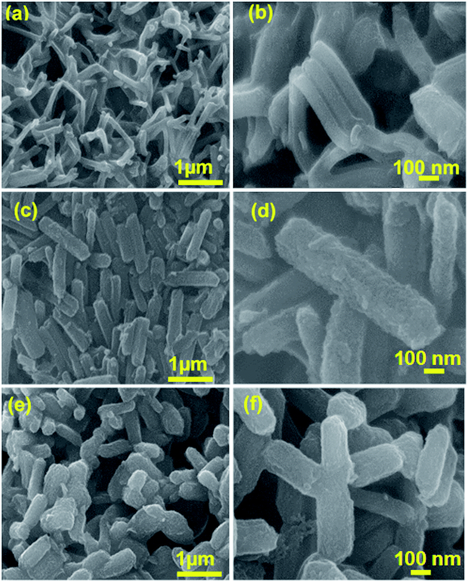 | ||
| Fig. 3 HRSEM images of (a and b) MCN-1-100s, (c and d) MCN-1-130s, and (e and f) MCN-1-150s. | ||
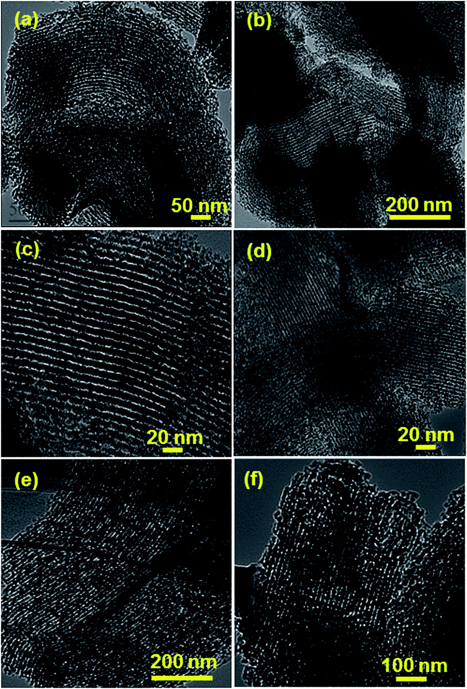 | ||
| Fig. 4 HRTEM images of (a and b) MCN-1-100s, (c and d) MCN-1-130s, and (e and f) MCN-1-150s. | ||
The nature and co-ordination of C and N atoms in the MCN-1-Ts samples were characterized by using XPS and FT-IR spectroscopic techniques (ESI†). The XPS survey spectra of the MCN-1-Ts samples reveal that the samples are composed of C and N with a very small quantity of oxygen (ESI Fig. 3Sa†). The presence of small quantity of oxygen could be attributed to the ethanol wash or adsorption of moisture or CO2 by the samples. No shift in the binding energy values of C and N was observed for the MCN-1-Ts samples with different particle size and diameters. The surface atomic composition for all the three samples is shown in Table 2. The absence of the Si in the XPS survey spectra confirms that the template was completely removed during the HF treatment. The chemistry of C and N in MCN-1-Ts samples was further investigated using high resolution core level C1s and N1s scans and the data are shown in (ESI Fig. 3Sb and Sc† respectively). The C1s spectrum was deconvoluted into four peaks with B.Es as 289.1, 287.6, 285.3 and 284.5 eV. The low energy peak at 284.5 eV is assigned to pure graphitic sites in amorphous CN matrix whereas the peak at 287.6 is assigned to sp3 hybridized carbon atom, which may come from the ethanol that was used for the washing of MCN. On the other hand, the peak at 285.3 is attributed to sp2 carbon atoms bonded to N atom inside the aromatic structure while the peak at 289.1 is assigned to sp2 hybridized carbon in the aromatic ring attached to NH2 groups.11,12,27
| Sample | C (%) | N (%) | O (%) |
|---|---|---|---|
| MCN-1-100s | 80.33 | 17.06 | 2.62 |
| MCN-1-130s | 77.4 | 18.88 | 3.73 |
| MCN-1-150s | 78.95 | 17.11 | 3.73 |
The N1s high resolution scan after deconvolution shows three well resolved peaks with B.Es as 398.2, 400.6 and 401.8 eV. The peak at the highest binding energy (401.8) is associated with terminal amino functions (C–H–N) whereas the peak at 398.2 eV is ascribed to nitrogen sp2 bonded to carbon. The peak at 400.6 eV could be attributed to N atoms trigonally bonded to all sp2 carbons, or to two sp2 carbon atoms and one sp3 carbon atom in an amorphous C–N network.11–13,27 However, interestingly, the amount of nitrogen content in the sample MCN-1-130s is much higher than that of the other samples, which reveals that the structural order and fine morphology helps to avoid the thermal decomposition of N species from the CN framework in the wall structure of MCN-1-130s (Table 2). These results also reveal that MCN-1-130s has more number of free NH2 groups that are required for enhanced adsorption of CO2 molecules as compared to that of MCN-1-100s and MCN-1-150s. The nature of the functional group on the surface of MCN-1-Ts samples was also analyzed using FT-IR spectroscopy (ESI Fig. 4S†). The FT-IR scans of all the three samples show peaks that are consistent with the previous reported results of MCN-1 samples prepared using the SBA-15 templates.11,12
3.1. CO2 adsorption
The CO2 adsorption capacity of the pore tuned and morphology controlled MCN-1-Ts samples was evaluated at high pressures up to 30 bar and different analysis temperatures of 0, 10 and 25 °C. The choice of MCN as an adsorbent for CO2 was motivated by a number of reasons. CO2 being a Lewis acid is highly likely to interact with any Lewis base in an acid base neutralization reaction. The successful incorporation of highly basic nitrogen atoms in the mesostructures of MCN-1-Ts make it an ideal candidate for capture of an acidic molecule like CO2 through physisorption and chemisorption. Physisorption of CO2 takes place on the surface sites and mesoporous channels whereas the chemisorption is favored by the nitrogen or NH2 functionalities on the surface of the CN walls. Pore tuning, together with morphology control and better textural parameters also affect the CO2 adsorption capacity besides the number of NH2 groups on the surface. The nitrogen containing groups such as NH2 that are present on the surface of the MCN also act as Lewis basic sites thereby enable easy neutralization reaction with acidic CO2.Fig. 5 shows the CO2 adsorption isotherms of the MCN-1-Ts samples measured at 0 °C at different pressures. It can be seen from the Fig. 5 that MCN-1-130s registered the maximum CO2 adsorption of 16.5 mmol g−1 whereas MCN-1-100s and MCN-1-150s samples showed the CO2 adsorption capacities of 12.9 and 12.3 mmol g−1, respectively under identical conditions. The higher adsorption capacity of MCN-1-130s could be attributed to the better structural and morphological order, higher surface area and pore volume and nitrogen contents as compared to those of MCN-1-150s and MCN-1-100s. For the purpose of comparison, we recorded the CO2 adsorption isotherms of commercially available activated carbon, multi-walled carbon nanotubes (MWCNT), and large pore mesoporous silica SBA-15, MCN-1-130d (prepared under dynamic conditions) and mesoporous carbon nitride with 3D structure (MCN-7-130) at 0 °C and 30 bar (Fig. 6).13 The CO2 adsorption was found to be in the following order: MCN-1-130s (16.5 mmol g−1) > MCN-1-130d (14.6 mmol g−1) > MCN-7-130 (13.5 mmol g−1) > SBA-15-130s (10.1 mmol g−1) > MWCNT (5.6 mmol g−1) > AC (3.7 mmol g−1). It is interesting to note that even though MCN-7-130 (ref. 13) has 3D structure and much better textural parameters than the MCN-1-130s, the latter showed much better adsorption capacity. This could be attributed to the uniform and rectangular shaped morphology of the MCN-1-130s, revealing the role of morphology of the adsorbent on the adsorption capacity. Table 3 shows the CO2 adsorption capacities of other reported materials such as polyethyleneimine (PEI) functionalized mesoporous capsules, PEI functionalized mesoporous silica KIT-6, zeolite like metal organic framework and MCN-7 measured under identical conditions.10,13,28,29 Based on the values indicated in Table 3, it is evident that MCN-1-130s sample has superior CO2 adsorption capacity under identical temperature and pressure conditions. It is clear from all these results that overall CO2 adsorption capacity of a material is dictated not just by structure or structural parameters or functional moieties alone but the morphology also plays a key role in determining the adsorption capacity of the adsorbents. In order to understand the role of temperature on the adsorption of CO2 over MCN-1-Ts materials, the adsorption experiments were conducted at different temperatures.
 | ||
| Fig. 5 CO2 adsorption isotherms of MCN-1-Ts samples measured at 0 °C and pressure up to 30 bar. | ||
 | ||
Fig. 6 Comparison of CO2 adsorption capacities of different materials with MCN-1-130s sample measured at 0 °C and pressure up to 30 bar: ( ) activated carbon, ( ) activated carbon, ( ) MWCNT, ( ) MWCNT, ( ) SBA-15-130s, ( ) SBA-15-130s, ( ) MCN-7-130, ( ) MCN-7-130, ( ) MCN-1-130d, and ( ) MCN-1-130d, and ( ) MCN-1-130s. ) MCN-1-130s. | ||
| Sample | T (°C) | Pressure (bar) | CO2d (mmol g−1) | Ref. |
|---|---|---|---|---|
| a PEI: polyetheyleneamine.b MC: mesoporous silica capsules.c ZMOF: zeolite like metal organic framework.d Dry CO2 gas.e MWCNT: multi-walled carbon nanotubes; PW: present work. | ||||
| PEI-KIT-6a | 75 | 1.0 | 3.07 | 10 |
| MC400/10PEI%83b | 75 | 0.101 | 4.91 | 29 |
| Sod-ZMOFc | 25 | 1.0 | 1.15 | 28 |
| MCN-7-130 | 0 | 30.0 | 13.5 | 13 |
| MCN-7-130 | 25 | 30.0 | 5.9 | 13 |
| MCN-7-130 | 25 | 1.0 | 1.4 | 13 |
| MCN-1-100s | 0 | 30.0 | 12.9 | PW |
| MCN-1-130s | 0 | 30.0 | 16.5 | PW |
| MCN-1-130s | 25 | 30.0 | 7.31 | PW |
| MCN-1-130s | 25 | 1.0 | 1.61 | PW |
| MCN-1-150s | 0 | 30.0 | 12.3 | PW |
| MWCNTe | 0 | 30.0 | 5.63 | PW |
| Activated carbon | 0 | 30.0 | 3.66 | PW |
Fig. 7(a–c) show the adsorption isotherms of MCN-1-100s, MCN-1-130s and MCN-1-150s respectively at 0, 10 and 25 °C. We found that as the adsorption temperature was increased from 0 to 25 °C, the CO2 adsorption capacity of the materials decreased drastically. This clearly suggests that CO2 adsorption process is exothermic in nature. Further the strength of the interaction between the adsorbate and adsorbent was studied by calculating the heat of adsorption. As MCN-1-130s shows the highest adsorption capacity among the three samples, adsorption isotherms of MCN-1-130s samples were recorded at 0, 10 and 25 °C and 30 bar (Fig. 7b). The isosteric heat of adsorption (Fig. 8) of MCN-1-Ts sample was calculated from the Clausius–Clayperon equation using the three isotherms recorded at different temperatures and shown in Table 4. The samples register higher isosteric heat of adsorption at lower loading but decreases as the loading is increased. This observation can be explained based on the fact that at lower CO2 loadings, active nitrogen sites are mainly responsible for adsorption whereas other active sites including carbon sites and well-ordered structure facilitate higher adsorption at a higher pressure. Similar trend in variation of isosteric heat with CO2 loading was observed with MCN-7 with 3D structure.13
 | ||
| Fig. 7 CO2 adsorption isotherms at 0, 10 and 25 °C and pressure up to 30 bar for (a) MCN-1-100s, (b) MCN-1-130s, and (c) MCN-1-150s. | ||
 | ||
| Fig. 8 Variation of isosteric heat of adsorption with CO2 loading for MCN-1-Ts samples and their comparison with MCN-7-130 sample.13 | ||
Interestingly, MCN-1-150s shows the highest heat of adsorption followed by MCN-1-130s and MCN-1-100s which indicates that the adsorbate–adsorbent interaction is the strongest for MCN-1-150s sample, followed by MCN-1-130s and MCN-1-100s. This trend could be explained on the basis of easy accessibility of CO2 molecules to the large pores of MCN-1-150s and high N% per unit surface area which favor the multilayer adsorption and strong adsorbent–adsorbate interaction respectively. In addition, the large micropore volume of the sample also support the strong interaction between the CO2 molecules. However, the pore volume and the specific surface area of the MCN-1-150s, which are the key parameters to enhance the CO2 adsorption capacity, are lower than those of MCN-1-130s (Tables 1 and 1S†). Therefore, highest CO2 adsorption capacity was achieved for MCN-1-130s. It is interesting to note that the heat of adsorption of MCN-1-Ts samples is higher than that of MCN-7-130 although the later has a 3D cage type large pores which is expected to show a strong interaction with the CO2 molecules. The higher heat of adsorption of MCN-1-Ts could be attributed to the uniform particle size and shape together with the large mesopores volume, which would allow the CO2 molecules adsorb strongly with the surface and even the active sites at the deep internal part of the pores.
Noteworthy, CO2 adsorption capacity of MCN-1-Ts samples depends strongly on the surface area and pore volume of the samples when other factors are kept the same. Sample with higher BET surface area and pore volume gives the highest adsorption keeping the adsorption temperature and pressure same (Fig. 9). This result is expected as higher surface would mean larger number of basic sites with free NH2 groups resulting in greater adsorption. In order to check the recyclability, the samples were regenerated by heating under vacuum between 200–250 °C for 6–8 h. To our surprise, the regenerated samples did not show any structural collapse or loss of adsorption capacity. These results prove that the prepared MCN samples are promising adsorbents for CO2 capture.
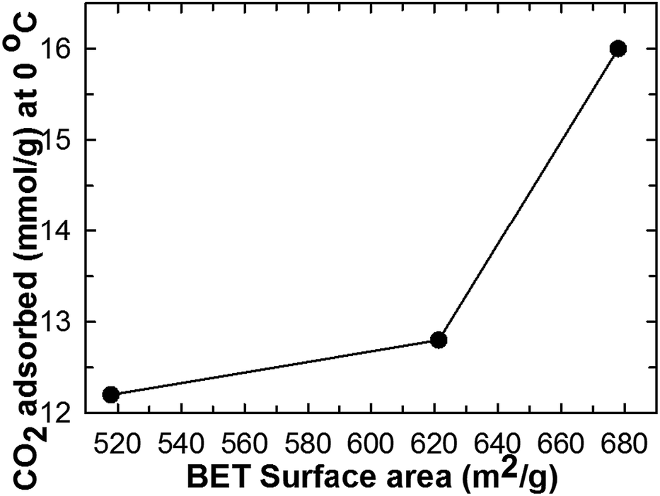 | ||
| Fig. 9 Effect of BET specific surface area on the quantity of CO2 adsorption over MCN-1-Ts samples. | ||
4. Conclusions
In this paper, we have reported the synthesis and CO2 adsorption performance of highly ordered mesoporous carbon nitride with uniform particle morphology and tunable pore diameters from the SBA-15 template prepared by static method. The prepared materials exhibited high specific surface area, large pore volume and well-ordered porous structure with highly uniform particle morphology. The pore size of the samples can be tuned from 3.76 to 5.94 with the simple adjustment of the pore diameter of the template. With this strategy, a variety of mesoporous carbon nitride with different structure and pore diameters can be easily fabricated. These materials were employed as adsorbents for CO2 molecules and the results were compared with other mesoporous materials including 3D mesoporous carbon nitride. In comparison to the 3D cage type MCN-7-130 (13.5 mmol g−1 at 0 °C and 30 bar), MCN-1-130s sample showed much higher CO2 adsorption capacity (16.5 mmol at 0 °C and 30 bar), which is higher than other mesoporous and porous samples. The higher adsorption capacity was related with the well-ordered structure with uniform particle morphology, and higher specific surface area and large pore volume. We also demonstrated that a combination of low CO2 adsorption temperature and high adsorption pressure is a favorable condition for higher CO2 adsorption. At higher temperature, CO2 adsorption capacity reduced significantly possibly due to the physically adsorbed CO2 molecules. It has also been found that these materials possess excellent recyclability and could be re-generated by heating under vacuum between 200–250 °C for 6–8 h without suffering any structural collapse or loss of adsorption capacity and thus promising to be an excellent adsorbent for large scale industrial application involving capture and conversion of CO2 molecules.Acknowledgements
One of the authors A. Vinu is grateful to ARC for the award of future fellowship and the University of Queensland for the start-up grants. The authors also acknowledge the facilities, and the scientific and technical assistance, of the Australian Microscopy and Microanalysis Research facility at the centre for microscopy and microanalysis (CMM), the University of Queensland. A. Vinu is also grateful to the CINBM for the award of visiting professorship, which was supported by a National Research Foundation of Korea (NRF) Grant funded by the Korean Government (MSIP) (2005-0049412). The project was also financially supported by King Saud University, Vice Deanship of Scientific Research Chairs.Notes and references
- C.-H. Yu, C.-H. Huang and C.-S. Tan, Aerosol Air Qual. Res., 2012, 12, 745 CAS.
- D. Qian, C. Lei, E.-M. Wang, W.-C. Li and A.-H. Lu, ChemSusChem, 2014, 7, 291 CrossRef CAS PubMed.
- K. Ahmad, O. Mowla, E. M. Kennedy, B. Z. Dlugogorski, J. C. Mackie and M. Stockenhuber, Energy Technol., 2013, 1, 345 CrossRef CAS PubMed.
- Y. Zhao, K. X. Yao, B. Teng, T. Zhang and Y. Han, Energy Environ. Sci., 2013, 6, 3684 CAS.
- Y. Jing, L. Wei, Y. Wang and Y. Yu, Microporous Mesoporous Mater., 2014, 183, 124 CrossRef CAS PubMed.
- R. Sanz, G. Celleja, A. Arencibia and E. S. Sanz-Perez, Appl. Surf. Sci., 2010, 256, 5323 CrossRef CAS PubMed.
- R. Sanz, G. Calleja, A. Arencibia and E. S. Sanz-Perez, Microporous Mesoporous Mater., 2012, 158, 309 CrossRef CAS PubMed.
- Z. Wu, P. A. Webley and D. Zhao, J. Mater. Chem., 2012, 22, 11379 RSC.
- X. Zhu, P. C. Hillesheim, S. M. Mahurin, C. Wang, C. Tian, S. Brown, H. Luo, G. M. Veith, K. S. Han, E. W. Hagaman, H. Liu and S. Dai, ChemSusChem, 2012, 5, 1912 CrossRef CAS PubMed.
- W.-J. Son, J.-S. Choi and W.-S. Ahn, Microporous Mesoporous Mater., 2008, 113, 31 CrossRef CAS PubMed.
- A. Vinu, K. Ariga, T. Mori, T. Nakanishi, S. Hishita, D. Golberg and Y. Bando, Adv. Mater., 2005, 17, 1648 CrossRef CAS PubMed.
- A. Vinu, Adv. Funct. Mater., 2008, 18, 816 CrossRef CAS PubMed.
- K. S. Lakhi, W. S. Cha, S. Joseph, B. J. Wood, S. S. Aldeyab, G. Lawrence, J.-H. Choy and A. Vinu, Catal. Today, 2015, 243, 209 CrossRef CAS PubMed.
- Y. Wang, F. Zhang, Y. Wang, J. Ren, C. Li, X. Liu, Y. Guo, Y. Guo and G. Lu, Mater. Chem. Phys., 2009, 115, 649 CrossRef CAS PubMed.
- C. Yu, J. Fan, B. Tian, D. Zhao and G. D. Stucky, Adv. Mater., 2002, 14, 1742 CrossRef CAS.
- P. S. Winkel, P. Yang, D. I. Margolese, B. F. Chmelka and G. D. Stucky, Adv. Mater., 1999, 11, 303 CrossRef.
- K. Kosuge, T. Sato, N. Kikukawa and M. Takemori, Chem. Mater., 2004, 16, 899 CrossRef CAS.
- A. Sayari, B.-H. Han and Y. Yang, J. Am. Chem. Soc., 2004, 126, 14348 CrossRef CAS PubMed.
- L.-C. Sang, A. Vinu and M.-O. Coppens, J. Mater. Chem., 2011, 21, 7410 RSC.
- A. Vinu, C. Sterb, V. Murugesan and M. Hartmann, J. Phys. Chem. B, 2003, 107, 8297 CrossRef CAS.
- A. Vinu, M. Miyahara and K. Ariga, J. Phys. Chem. B, 2005, 109, 6436 CrossRef CAS PubMed.
- M. Hartmann, A. Vinu and G. Chandrasekar, Chem. Mater., 2005, 17, 829 CrossRef CAS.
- A. Vinu, M. Miyahara, V. Sivamurugan, T. Mori and K. Ariga, J. Mater. Chem., 2005, 15, 5122 RSC.
- A. Vinu, K. Z. Hossian, G. S. Kumar and K. Ariga, Carbon, 2006, 44, 530 CrossRef CAS PubMed.
- L.-C. Lang, A. Vinu and M.-O. Coppens, Langmuir, 2011, 27, 13828 CrossRef PubMed.
- A. Vinu, M. Miyahara and K. Ariga, J. Nanosci. Nanotechnol., 2006, 6, 1510 CrossRef CAS PubMed.
- Q.-F. Deng, L. Liu, X.-Z. Lin, G. Du, Y. Liu and Z.-Y. Yuan, Chem. Eng. J., 2012, 203, 63 CrossRef CAS PubMed.
- C. Chen, J. Kim, D.-A. Yang and W.-S. Ahn, Chem. Eng. J., 2011, 168, 1134 CrossRef CAS PubMed.
- G. Qi, Y. Wang, L. Estevez, X. Duan, N. Anako, A.-H. A. Park, W. Li, C. W. Jones and E. P. Giannelis, Energy Environ. Sci., 2011, 4, 444 CAS.
Footnote |
| † Electronic supplementary information (ESI) available. See DOI: 10.1039/c5ra04730g |
| This journal is © The Royal Society of Chemistry 2015 |