Preclinical appraisal of terbutaline analogues in precipitation of autism spectrum disorder

Neha Sharmaa, Swetlana Gautama, Uma Devib, Manjari Singha, Jitendra K. Rawata, Nikunj Sethic, Shubhini A. Sarafa and Gaurav Kaithwas*a
aDepartment of Pharmaceutical Sciences, School of Biosciences and Biotechnology, Babasaheb Bhimrao Ambedkar University, Vidya Vihar, Raebareli Road, Lucknow 226 025, U.P., India. E-mail: gauravpharm@hotmail.com; Tel: +91-9670204349
bDepartment of Pharmaceutical Sciences, Faculty of Health Medical Sciences Indigenous and Alternative Medicine, SHIATS-Deemed to be University Formerly Allahabad Agricultural Institute, Naini, Allahabad, U.P., India
cDivision of Toxicology, CSIR-Central Drug Research Institute, Lucknow-226 031, U.P., India

Received 10th March 2015 , Accepted 13th April 2015

First published on 13th April 2015


Abstract

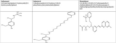
Terbutaline is a β2 agonist used in the clinical management of asthma and as a tocolytic agent during pregnancy. In the recent past, the preterm use of terbutaline has been shown to hasten autistic-like symptoms in offsprings. For this reason, the present study was carried out to understand the effects of pharmacological analogues of terbutaline (salbutamol, salmeterol and montelukast) in the progression of ASDs in experimental animals. Pregnant rats were treated with salbutamol (10 mg kg−1, sc), salmeterol (10 μg kg−1, sc) and montelukast (10 mg kg−1, sc) and the offsprings were scrutinized for behavioral, biochemical, neuro-inflammatory and histopathological changes. The offsprings from the rats treated with salbutamol and montelukast were determined to be closely associated with various symptoms of ASDs.


Introduction

Autism spectrum disorders (ASDs) are a cluster of neurodevelopmental disorders in the category of pervasive developmental disorders, delineated by impaired social interaction, communication, and restricted and repetitive behavior. ASDs are perceived across the population spectrum with a prevalence of 6 cases per 1000. Furthermore, approximately four times as many males are diagnosed than females.1 As evidenced through literature, pathophysiological changes in the brain begin during fetal development in autistic individuals.2 Contemporary studies prompted that maternal exposure to illness, medication, environmental toxins and psychological stress are in agreement with the prevalence of ASDs.3

Terbutaline is a β2 adrenergic receptor (B2AR) agonist used in the treatment of asthma and it is also used as a tocolytic agent during preterm labor. Terbutaline readily crosses placenta and it can disrupt the replication and differentiation of developing neurons. The progeny of pregnant women treated with terbutaline are manifested with impaired academic performance, cognitive dysfunction and inflated incidences of psychiatric disorders. A recent study conducted at Duke University exemplified the augmented risk of brain damage and cognitive defects in infants whose mothers were exposed to terbutaline during pregnancy.4 Preclinical studies have also identified terbutaline as a neuroteratogen that alters neuroprotein markers and architectural organization, in addition to neuronal and glial cell distributions in the cerebellum, hippocampus and somatosensory cortex.4 Following the same strategic path, Zerrate et al. validated innate neuroinflammation after terbutaline treatment, as delineated by microglial activation and behavioral anomalies.5

In 2011, FDA began necessitating a black-box warning for terbutaline, stating that the drug should not be administered during pregnancy. Terbutaline actuates B2AR receptors and dysregulates adenylate cyclase, leading to an anomalous generation of cyclic adenosine monophosphate in various stages of neonatal development.3 Mature cells are protected from such overstimulation due to their competence to uncouple receptors from the production of cAMP.6 Exaggerated cAMP either through B2AR stimulation or other stress-related factors also create inflated oxidative stress, which exacerbates the toxicity.

In consideration to the complications associated with terbutaline during pregnancy and recent directions issued by the FDA, salbutamol and salmeterol (both B2AR agonists) are the drugs of choice to be used for the management of preterm labor and/or asthma during pregnancy. In addition to the aforementioned substances, montelukast (a leukotriene receptor antagonist) is also currently used for the management of asthmatic conditions during pregnancy (Fig. 1). Nonetheless, to date neither direct nor indirect scientific data exists, which can define the safety profile of salbutamol/salmeterol/montelukast in expecting mothers. Eventually, the present study was initiated to investigate the neuroinflammatory, biochemical and behavioral alterations in experimental animals after salbutamol/salmeterol/montelukast exposure.


image file: c5ra04213e-f1.tif
Fig. 1 Chemical structures of salmeterol, salbutamol and montelukast.

Animals

Pregnant albino rats (Wistar strain) were acquired from central animal house, United Institute of Pharmacy, United Group of Institution, Naini, Allahabad, (U.P), India. The animals were housed in polypropylene breeding cages under standard temperature conditions (25 ± 1 °C) with 12 h light-and-dark cycles and free access to a diet of commercial pellets and water ad libitum. The experimental protocol was endorsed by “Institutional Animal Ethical Committee (IAEC)” (UIP/IAEC/2014/FEB./04). The study was supervised as per the guidelines established by the Department of Animal Welfare, Government of India.

Drugs

Salbutamol was received as a gift sample from Yash Pharma Laboratories Private Limited, Bhaddi, India. Montelukast (Montair, Cipla Global Limited, Mumbai, India), Salmeterol (Esiflow, Lupin, Laboratories Limited, Mumbai, India) and DPT vaccine (Comvac 3: Bharat Biotech International Limited, Hyderabad, India) were solicited from a local market.

Chemicals

Ovalbumin, aluminum hydroxide, DTNB (5,5-dithiotris-2-nitrobenzoic acid) and donepezil solution were obtained from Himedia Laboratories, Mumbai, India. Thiobarbituric acid (TBA), trichloroacetic acid (TCA), potassium dihydrogen phosphate and di-sodium hydrogen phosphate were supplied by SD Fine Chemicals Limited, Mumbai, India. All the other chemicals used were of analytical grade and were procured from Hi Media Laboratories, Mumbai, India.

Experimental protocol

Pregnant rats were selected and relocated to the laboratory and allowed to acclimatize in laboratory conditions. Twenty pregnant rats were divided into five groups at random (n = 4). Groups I and II served as sham and toxic controls, respectively. Groups III, IV and V were treated with salbutamol (10 mg kg−1, sc), salmeterol (10 μg kg−1, sc) and montelukast (10 mg kg−1, sc), respectively. The pregnant females were treated daily for seven days with respective dosages, 30 min before the OVA aerosol challenge. The sham and toxic control pregnant rats were instead treated with the subsequent volume of normal saline.

OVA aerosol challenge

Groups II, III, IV and V were sensitized with 1 ml (sc) of saline containing 1 mg of ovaalbumin and 3.5 mg of aluminium hydroxide, in addition to 0.5 ml (sc) of Bordetella pertussis vaccine containing 2 × 10¹⁰ heat-killed organism, which was administered as an adjuvant. The sensitized rats were exposed to an aerosol of 1% ovaalbumin daily from 14 to 21 days, whereas the control rats were exposed only to filtered air. Pups (n = 8) from the pregnant dams were examined for postnatal growth and behavioral alteration up to 45 PND. On the 46th day, the animals were sacrificed using light ether anesthesia followed by heart perfusion for the removal of total blood from the brain. Brains were extracted without damage. Collected brain tissues were evaluated on the prototypes of biochemical, oxidative embodiments, inflammatory markers and histopathological changes.

Postnatal growth and maturation development

Pups were evaluated for weight gain on the 5th, 15th, 25th, 35th and 45th PNDs, and for eye opening once on the 9th and daily from 12th to 15th PNDs and were rated in the following manner: 0, both eyes closed; 1, one eye open; and 2, both eyes open.7

Behavioral tests

Negative geotaxis. Negative geotaxis was observed once on 7th, 9th and 11th PNDs. Pups were timed for completing a 180° turn when placed in a head-down position on a 25° inclined surface. Negative geotaxis reflects vestibular function and motor development.8
Swimming performance. An aquarium filled with water (28–29 °C) was used for swimming test on the 15th, 20th and 25th PNDs. Each animal was placed at the center of the aquarium and observed for 10 s. Swimming performance was determined according to the position of nose and head (angle) on the surface of water. The angle of swimming was classified in the following manner: 0, head and nose below the surface; 1, nose below the surface; 2, nose and top of head at or above the surface but ears still below the surface; 3, same as 2 except that water line was at mid-ear level; and 4, same as 3 except that water line was at the bottom of the ears. Swimming is a combination of motor development and consolidation of coordinated reflex responses.9
Locomotor activity. It was documented individually for each animal by numbers of photo-beam crossings in an actophotometer in an interval of 10 min.10
Olfactory discrimination. The test was conducted on the 10th, 12th, and 15th PNDs. The apparatus consisted of a plastic container 20 × 8 × 8 cm3 (l × w × h), two small bins, and a clear plastic cover, which was placed over the bins and container. One end of the container encompassed a bin filled with new bedding, whereas the other end had a bin filled with home cage bedding. A 3 cm2 area was demarcated at the center of the screen. A line was drawn on the screen above each bin. Each pup was allocated in the centrally marked-off region on the screen, and the latency to enter the home bedding side by crossing the delineated line with the front paws and head was timed. The central placement of the pup was balanced by altering the position of the pup to or away from the experimenter. The age of the home bedding was balanced across the groups and was averaged to be 3 days old at testing. The test reflects a nest-seeking response arbitrated by the olfactory system.11
Nociception. Nociceptive effect was observed using tail-flick test on the 10th, 15th and 25th PNDs using a tail-flick analgesiometer. The animal was gently restrained, and radiant heat was focused onto its tail. The cut-off time was 9 s. Tail-flick measurements were taken three times at the intervals of 30 s.12
Elevated plus maze test (EPM). EPM was implemented on the 14th, 22nd and 30th PNDs. The apparatus consisted of two open (30 × 5 cm) and two closed arms (30 × 5 × 25 cm) in perpendicular positions. The open and closed arms were connected by a transparent acrylic sheet. The floor was made of black acrylic sheet. The maze was positioned 45 cm above the floor. The rat was placed at the center of the plus maze with its nose in the direction of one of the closed arms and observed for 5 min to measure the following parameters: number of entries in the open (NEOA) and closed (NECA) arms.13

Biochemical changes

The animals were sacrificed using light-ether anesthesia followed by heart perfusion on 46th PND. The brains were evacuated instantly, rinsed with ice-cold saline, dried on a filter paper, and 10% w/v tissue homogenates were prepared in ice-cold 0.15 M KCI using a tissue homogenizer. The post-nuclear fraction obtained by centrifuging the homogenate at 10[thin space (1/6-em)]000 rpm for 10 min at 4 °C in a cooling centrifuge were used for the spectrophotometric estimation of acetylcholinesterase (AchE), thiobarbituric acid-reactive substances (TBARS), catalase, superoxide dismutase (SOD), protein carbonyl and tissue glutathione using the previously established methods in our laboratory.13–16

Inflammatory markers

The level of interleukins IL-1β (catalogue no. K0331212P), IL-2 (catalogue no. K0332100P), IL-4 (catalogue no. K0332133P), IL-6 (catalogue no. K0331229P), and IL-10 (catalogue no. K0332134) were enumerated in the brain tissue using an Elisa plate reader (Alere AM 2100 microplate reader) from commercially available kits procured from Koma Biotech, Australia.

Morphological evaluation

Brain tissues were analysed histopathologically using hematoxylin and eosin staining. The tissues were fixed overnight in paraformaldehyde, followed by 70% isopropanol overnight. The tissues were further exposed to the augmenting concentrations of isopropanol (70%, 90%, and 100%), followed by dehydration with 100% xylene. The tissues were embedded in paraffin wax, and 5 μm sections were prepared using a microtome, followed by hematoxylin and eosin staining.17

Statistical analysis

All data were presented as mean ± SD and analyzed by one-way ANOVA, followed by a Bonferroni test for the possible significance identification among the various groups. c/*P < 0.05, b/**P < 0.01, and a/***P < 0.001 were considered as statistically significant. Statistical analysis was performed using Graph Pad Prism software (3.2), San Diego, California.

Results

Postnatal growth and maturation

Compelling delays in postnatal growth and maturation were observed in all the groups when correlated to sham control (Fig. 2A and B).
image file: c5ra04213e-f2.tif
Fig. 2 Effect of prenatal exposure of terbutaline analogues on maturation development and behavioural changes.

Behavioral tests

The pups from the dams treated with the drugs in question and ovaalbumin exhibited momentous subsidence in locomotor activity, which was successively headed towards normalization with progression of age. No significant variation in the locomotor activity could be observed between the test groups and ovaalbumin. Similar patterns of restoration were examined in pups on a negative geotaxis prototype with the drugs in question giving significant cutback in time to align on a negative geotaxis paradigm in comparison to ovaalbumin treatment (Fig. 2C and F). Control group animals exhibited a downturn in innate escape response with an upturn in growth. Similar cutback patterns were ascertained in the toxic and test groups. Nonetheless, the salbutamol- and montelukast-treated groups were perceived to exhibit a considerably higher innate escape response compared to the control. Thermal nociception was momentously increased in test groups, compared with the sham control; nonetheless, detainment in the response was observed with the progression of age. Treatment with salbutamol and montelukast was observed to afford significant protection against the same, when compared to the ovaalbumin treatment on PND 25 (Fig. 2D). The ontogeny of swimming behavior was significantly delayed in toxic animals. The drugs in question significantly restored swimming performance scores with montelukast demonstrating to be most efficacious on PND 25 (Fig. 2E). When perceived for anxiety-like behavior through the plus maze, adolescent as well adult pups manifested diminished open-arm entries after the ova challenge. Resultant treatment with salbutamol, salmeterol and montelukast further illustrated abatement in the number of open-arm entries (Fig. 2G). The nest-seeking behavior arbitrated through olfactory clues was adversely affected in the toxic group. In addition, successive treatments with salbutamol and montelukast suppressed the nest-seeking behavior (Fig. 2H).

Biochemical tests

Brain tissues were also appraised for the antioxidant defense through embodiments of peroxidation and antioxidant enzymatic defenses. Momentous augmentation in TBAR generation and protein carbonyl formation was observed in toxic control, which was alleviated after the administration of the drugs in question. The enzymatic activities of SOD and catalase were notably truncated in toxic treatment, which furthermore diminished after treatment with test drugs. When an additional investigation for AchE activity was carried out, we observed a momentous increase in enzymatic activity in the toxic group. The subsequential administration of test compounds slightly restored the enzymatic activity of AchE (Table 1).
Table 1 Effect of prenatal exposure of terbutaline analogues on physiological antioxidant defence in experimental pupsa
Groups TBARS (nm of MDA/mg of protein) GSH × 10−4 (mg%) SOD (unit of SOD/mg of protein) Catalase (nM of H2O2 per min per mg of protein) Protein carbonyl (nm ml−1 unit) Acetylcholinesterase (nm per min per ml unit)
a (Values are mean ± SD); comparisons were made on the basis of one-way ANOVA, followed by a Bonferroni test. All groups were compared with the control group (Group 1) (*p < 0.05, **p < 0.01, ***p < 0.001). Groups 3, 4 and 5 were compared with Group 2 (cp < 0.05, bp < 0.01, ap < 0.001).
I 2.16 ± 0.01 0.25 ± 0.01 0.88 ± 0.01 81.32 ± 2.19 77.42 ± 0.13 0.48 ± 0.03
II 2.73 ± 0.33** 0.14 ± 0.02*** 0.77 ± 0.03*** 70.15 ± 1.72*** 142.35 ± 0.26*** 1.07 ± 0.11***
III 4.03 ± 0.20***a 0.19 ± 0.01***a 0.75 ± 0.01*** 56.03 ± 2.96***a 146.97 ± 0.52***a 0.62 ± 0.04a
IV 2.87 ± 0.04*** 0.19 ± 0.002***a 0.73 ± 0.01***c 62.85 ± 3.34***b 172.65 ± 0.35***a 0.64 ± 0.05a
V 2.28 ± 0.04c 0.16 ± 0.01*** 0.77 ± 0.01*** 52.05 ± 1.77***a 164.17 ± 0.47***a 0.67 ± 0.11b


Brain tissues were further scrutinized for the presence of cytokines pertaining to inflammation and adaptive immunity. As a result, IL-2 and IL-4 tissue levels were determined to be inflated after the treatment with test drugs. The levels of pro-inflammatory cytokines, IL-1β and IL-6 were observed to have compelling upsurge, whereas the IL-10 (anti-inflammatory) levels were curtailed after the administration of the drugs in question (Table 2).

Table 2 Effect of prenatal exposure of terbutaline analogues on inflammatory and adaptive immunity markersa
Group IL-1β (pg ml−1) IL-6 (pg ml−1) IL-10 (pg ml−1) IL-2 (pg ml−1) IL-4 (pg ml−1)
a (Values are mean ± SD); each group contains atleast 8 animals. Comparisons were made on the basis of one-way ANOVA followed by a Bonferroni test. All groups were compared with the control group (Group 1) (*p < 0.05, **p < 0.01, ***p < 0.001). Groups 3, 4 and 5 were compared with Group 2 (cp < 0.05, bp < 0.01, ap < 0.001).
I 386.15 ± 10.34 1579.10 ± 175.83 1288.9 ± 51.98 789.5 ± 223.11 708.32 ± 28.56
II 92.69 ± 4.8** 898.50 ± 136.21** 724.95 ± 33.37*** 357.62 ± 9.43* 232.68 ± 6.23**
III 294.67 ± 131.58c 1965.30 ± 518.47a 209.03 ± 8.23***a 859.10 ± 135.64b 461.06 ± 151.05
IV 279.75 ± 7.49c 1469.20 ± 768.90b 199.94 ± 15.56***a 857.89 ± 59.73b 365.27 ± 145.50*
V 292.93 ± 7.8c 2978.30 ± 194.03***a 1166.8 ± 61.34*a 1180.1 ± 22.63*a 443.36 ± 11.88


Histopathology

Histopathological studies could confirm severe neuronal degeneration and neuronal loss were observed in the test groups when treated with salbutamol and montelukast. However, salmeterol exhibited only slight variability, compared to the control (Fig. 3).
image file: c5ra04213e-f3.tif
Fig. 3 Histopathological changes in animals treated with salbutamol, salmeterol and montelukast: (A) control; (B) toxic control; (C) salbutamol-treated; (D) salmeterol-treated; (E) montelukast-treated.

Discussion

Autism is a neurodevelopmental disorder symbolized by aberrant development, abnormality in social interaction, restricted interest, and stereotypical repetitive behavior, in addition to other neuroanatomical, biochemical and inflammatory disturbances. The present probe reports the effect of B2ARs agonist (salbutamol and salmeterol) and leukotriene antagonist (montelukast) on the amelioration of autistic spectrum disorder in the offspring of pregnant Wistar rats. To mimic the clinical condition, the rats were challenged with ovaalbumin and subjected to treatment with salbutamol, salmeterol and montelukast. Subsequently, the pups delivered were observed for maturation, sensory function, behavioral changes, biochemical changes, inflammatory markers and brain morphology.

We perceived no change in the delay of maturation between the toxic and subsequent treatments with the drugs in question. The experimental animals exhibited lower sensitivity to spinal pain, exaggerated time to align on a negative geotaxis paradigm (delayed reflexes, motor skills and cerebral integration) and attenuation of coordinated reflexes (swimming performance) when treated with ovaalbumin and the test drugs. It would be apropos to acknowledge that ovaalbumin exhibited a more striking deterioration of motor coordination (swimming performance and negative geotaxis), which was restored to nearly normal by the drugs in question. The lower pain sensitivity and diminished reflexes in autistic subjects in extensively deliberated phenomenon in clinical and preclinical subjects.5,7,18,19

Exploratory behavior is one the basic forms of behavioral activity, and its prenatal disruption can cause CNS developmental damages and autism.7,20 A momentous downturn in locomotor activity was documented in the ovaalbumin- and test drug-treated animals, with more a conspicuous effect by salbutamol. The aforementioned decline in locomotor activity is a well-established phenomenon in distinct experimental autistic models.21,22 Diminution in locomotor activity could be attributed either to the reduced number of purkinje fiber in vermal lobules (associated with reduced exploration) or marked changes in the neural structures of the cortex and amygdale.23,24 The pups were also observed to exhibit delayed nest-seeking response (entries in the old husk, olfactory discrimination) manipulated to the ova challenge and test drugs, contributing to socially deficient development. The aforementioned findings are in agreement with the antecedent clinical and preclinical reports.25,26 The agreement between prenatal exposure with the drugs in question and autism was further investigated on the basis of EPM, considering NEOA as a measure of anxiety-like behavior. The pups manifested an augmented anxiety/lower exploratory behavior in EPM with fewer entries into the open arm when treated with ovaalbumin. The exploratory behavior was further diminished by the drugs in question with a selectively more pronounced effect by montelukast.

The behavioral abnormalities observed in the current experiment were pronounced with the ovaalbumin challenge, which was restored to some extent by the drugs in question. It would be justifiable to state that salbutamol affected the thermal nociception and motor coordination much more adversely then the ova challenge. On the contrary, montelukast augmented much more pronounced detrimental effects on the exploratory behavior and social deficiency paradigm, compared with the control or ova challenge.

Prima facie, the behavioral abnormalities were more extensive in the ova challenge, suggesting the rise of abnormal phenotypes by ovaalbumin, which was further deteriorated by salbutamol and montelukast, in comparison to salmeterol.

Various mechanisms, such as oxidative stress, membrane lipid anomalies, changes in membrane fluidity, changes in immune and inflammatory response, and mitochondrial dysfunction, in addition to abnormal energy metabolism, have been observed to aid in ASD pathogenesis.27–29 The antioxidant defense mechanism in ASD may be down- or up-regulated, suggesting an altered antioxidant defense mechanism. The triple-regulated enzymatic defenses of SOD/catalase/GSH represent cellular oxidative stress or successive compensatory mechanisms.30,31 Similar results were observed in the present study, as observed with the slackened enzymatic activity of SOD and catalase with consecutive ovaalbumin treatments, which was further slackened by the salbutamol and montelukast treatment. Treatment with ovaalbumin afforded a significant decline in GSH levels, which were restored to some extent by the drugs in question, except for montelukast. In contrast to enzymatic defense, the oxidative markers of protein and lipid peroxidation are much more evidently defined both clinically and preclinically, when considered for ASDs.32–34 Similar results were arbitrated in the current study after the ova challenge, which was more conspicuously pronounced by salbutamol (TBARS) and salmeterol (protein carbonyl). Notwithstanding, significantly up-regulated protein carbonyl content was observed after salbutamol, salmeterol and montelukast treatment when compared with the ova challenge.

Several cholinergic abnormalities have been reported in autistic subjects, including reduced nAchR binding, reduced M1 receptor binding and intensified activity of brain-derived neurotropic factors. In fact, the role of cholinergic systems in the development of autism is well established.35 The basal forebrain, which is involved in attention span, largely resides with cholinergic neurons and is believed to be immensely abnormal in autistic subjects.36 The present study also corroborated with previous reports and demonstrated momentous upsurge in Ache activity by the group subjected to the ova challenge. However, sententious restoration in the Ache activity was available with test drug treatments.

Cytokines play a key role in the governance of inflammatory/immune responses in neurological circuits and act as key regulators for systemic communication. Cytokines affect the developmental and functional aspects of the nervous system, and dysregulated cytokine production/signaling/regulation is involved in a wide range of neurological disorders together with ASDs.37,38 In the current study, we evaluated pro-inflammatory (IL-1β and IL-6), anti-inflammatory (IL-10 and IL-4) and immunoregulatory (IL-2) cytokines to give a wider perspective to the study.39 We observed noteworthy diminution in the levels of anti-inflammatory (IL-10 and IL-4) cytokines after the ova challenge, which is in agreement with the forgoing preclinical and clinical studies.40 Treatment with the drugs in question further diminished the levels of anti-inflammatory cytokines with more pronounced diminution by salbutamol and salmeterol in comparison to montelukast. The current consideration could be accounted to the fact that the focal brain inflammation was most adversely affected by salmeterol and salbutamol. However, when scrutinized for pro-inflammatory cytokines (IL-1β and IL-6), remarkable downregulation in the levels of IL-6 and IL-1β was observed by the ovaalbumin. However, treatment with the drugs in question afforded momentous increase in the levels of IL-6, suggesting the precipitation of inflammatory reaction by the test drugs. It should be noted that montelukast and salbutamol exhibited a much higher increase in the levels of IL-6 in comparison to salmeterol. On the contrary, an unusual decline in the levels of IL-1β was accredited to the test drugs, which could be accredited to the fact that IL-1β is expressed very early in immune response, and considering the long duration of the study, one may expect a compensatory decline in the levels of IL-1β to counteract inflammation.

In addition, when the level of IL-2 was investigated, we observed a down-regulation after the ova challenge. IL-2 is a signaling molecule of the immune system because of its direct effect on T cells.41,42 Diminished levels of IL-2 may reflect the utmost immunocompromised state of the experimental animals following the ova challenge. Treatment with the drugs in question helped to restore deregulated immune systems, with the most conspicuous effect demonstrated by the montelukast. In view of the dysregulated cytokine profile after the consequent treatment with the test drugs and considering the reported inflammatory/immunological disturbance affiliated with ASDs, the authors suggest that all the test drugs have the potential to participate in the progression of ASDs to varying degrees.

Neuronal degeneration is an undisputed but barely deliberated phenomenon in terms of autism. Recently, Kern et al. summarized the diversified aspects related to neurodegeneration in autism, and concluded that neuronal cell loss, activated microglia, pro-inflammatory cytokines and oxidative stress accompany the neurodegenerative changes, accounting for breakthroughs in autism research.43 When studied histopathologically, salbutamol- and montelukast-treated groups were found to have conspicuous neuronal loss (fewer numbers of neuronal cells) and neuronal degeneration.

As discussed above, authors suggest that inflammatory dysregulation and oxidative stress launched by the ova challenge in the experiment could cause various behavioral abnormalities associated with autism. In addition, it can be observed that treatment with test drugs does not affect the maturation and helped to restore a few of the endpoints evaluated in the study. However, while analysing behavioral, biochemical and histopathological findings, it can be inferred that the all the test drugs can have varying effects on the neurological circuits of a developing brain. Salbutamol, montelukast, and to some extent, salmeterol can exacerbate various behavioral symptoms pertaining to autism. However, the case of salmeterol needs additional investigation.

It would be appropriate to state that all the test drugs are amply lipophilic in nature, which can cross the physiological blood–brain and placental barriers; they have the proficiency to adversely affect the fetus if administered prenatally. Nonetheless, the antecedent report by Manchee and colleagues enumerated the low transfer of salmeterol across the placental barrier, which could be attributed to the diminished toxicological effects as elaborated in the current study.44

It should be noted that monoamine oxidase (MAO) inhibitors have been implicated in disorders like Parkinson's disease, which are observed to have a close confinement with oxidative stress.45–47 Considering this, it can be suggested that exploiting MAO inhibitors in ASDs could be a viable study for future research.

As discussed above, the authors suggest that salbutamol and montelukast can stand forth in a broad spectrum of biochemical, inflammatory, behavioral and neurological changes when administered prenatally and can hasten the autistic-like symptoms in experimental animals. The result also suggests the existence of a wide range of pathological concordance between the salbutamol- and montelukast-treated animals and previously vested animal models for autism. With the observation obtained through the present study, we would like to hypothesize that the precipitation of autistic symptoms is not the sole prerogative of drug usage during pregnancy; rather the inflammatory and immunological disturbances during pregnancy and their subsequent modification by various drugs could be one of the reasons for the precipitation of such a complex behavioral disorder. In spite of this, much additional research is vital to elucidate the molecular basis and clinical implications of salbutamol and montelukast treatments.

References

  1. C. R. Marshall, A. Noor and S. W. Scherer, Structural variation of chromosome in Autistic Spectrum Disorder, Am. J. Hum. Genet., 2008, 82, 477–488 CrossRef CAS PubMed.
  2. S. Baron-Cohen, B. Auyeung, B. Nørgaard-Pedersen, D. M. Hougaard, M. W. Abdallah, L. Melgaard, A. S. Cohen, B. Chakrabarti, L. Ruta and M. V. Lombardo, Elevated fetal steroidogenic activity in autism, Mol. Psychiatry, 2015, 20(3), 369–376 CrossRef CAS PubMed.
  3. L. A. Croen, S. L. Connors and A. W. Zimmerman, Prenatal exposure to β2-adrenergic receptor agonists and risk of autism spectrum disorders, J. Neurol. Dev. Disord., 2011, 3, 307–315 CrossRef PubMed.
  4. M. C. Rhodes, F. J. Seidler, A. A. Rahman, C. A. Tate, A. Nyska, H. L. Rincavage and C. A. Slotkin, Terbutaline is a developmental neurotoxicant: Effects on neuroproteins and morphology in cerebellum, hippocampus and somatosensory cortex, J. Pharmacol. Exp. Ther., 2004, 308, 529–537 CrossRef CAS PubMed.
  5. M. C. Zerrate, M. Pletnikov, S. L. Connors, D. L. Vargas, F. J. Seidler, A. W. Zimmerman, T. A. Slotkin and C. A. Pardo, Neuroinflammation and behavioral abnormalities after neonatal terbutaline treatment in rats: Implication for autism, J. Pharmacol. Exp. Ther., 2007, 322, 16–22 CrossRef CAS PubMed.
  6. T. A. Slotkin, J. T. Auman and F. J. Seidler, Ontogenesis of β-Adrenoceptor Signaling: Implications for Perinatal Physiology and for Fetal Effects of Tocolytic Drugs, J. Pharmacol. Exp. Ther., 2003, 306, 1–7 CrossRef CAS PubMed.
  7. T. Schneider and R. Przewłocki, Behavioral Alterations in Rats Prenatally Exposed to Valproic Acid: Animal Model of Autism, Neuropsychopharmacology, 2005, 30, 80–89 CrossRef CAS PubMed.
  8. J. Altman and K. Sudarshan, Postnatal development of locomotion in the laboratory rats, Anim. Behav., 1975, 23, 896–920 CrossRef CAS.
  9. S. Schapiro, M. Salas and K. Vinkovich, Hormonal effects on ontogeny of swimming ability in rats: Assessment of central nervous system development, Science, 1970, 168, 147–151 CAS.
  10. U. A. Bhosale, R. Yegnanarayan, P. D. Pophale, M. R. Zambare and R. S. Somani, Study of central nervous system depressant and behavioral activity of an ethanol extract of Achyranthesaspera (Agadha) in different animal models, Int. J. Appl. Basic Med. Res., 2011, 1(2), 104–108,  DOI:10.4103/2229-516x.91154.
  11. E. H. Gregory and D. W. Pfaff, Development of olfactory-guided behavior in infant rats, Physiol. Behav., 1971, 6, 573–576 CrossRef CAS.
  12. G. Kaithwas, A. Mukherjee, A. K. Chaurasia and D. K. Majumdar, Antiinflammatory, analgesic and antipyretic activities of Linum usitatissimum L. (flaxseed/linseed) fixed oil, Indian J. Exp. Biol., 2011, 49(12), 932 CAS.
  13. G. Kaithwas, K. Dubey, D. Bhatia, A. D. Sharma and K. K. Pillai, Reversal of sodium nitrite induced impairment of spontaneous alteration by aloevera gel: involvement of cholinergic system, Pharmacologyonline, 2007, 3, 428–437 Search PubMed.
  14. G. Kaithwas, K. Dubey and K. K. Pillai, Effect of aloe vera (Aloe barbadensis Miller) gel on doxorubicin-induced myocardial oxidative stress and calcium overload in albino rats, Indian J. Exp. Biol., 2011, 49(4), 260 Search PubMed.
  15. G. Kaithwas and D. K. Majumdar, In vitro antioxidant and in vivo antidiabetic, antihyperlipidemic activity of linseed oil against streptozotocin-induced toxicity in albino rats, Eur. J. Lipid Sci. Technol., 2012, 114(11), 1237–1245 CrossRef CAS PubMed.
  16. K. G. Sukesh, S. Gautum, J. K. Rawat, M. Singh, S. A. Saraf and G. Kaithwas, Efficacy of variable dosage of aspirin in combating methotrexate-induced intestinal toxicity, RSC Adv., 2015, 5, 9354–9360 Search PubMed.
  17. A. Z. Reznick and L. Packer, Oxidative damage to proteins: spectrophotometric method for carbonyl assay, Methods Enzymol., 1994, 233, 357–363 CAS.
  18. B. Belur and N. Kandaswamy, Laboratory techniques in histopathology, in Medical laboratory technology-A procedure manual for routine diagnostic tests, ed. K. L. Mukherjee, Tata Mc Graw Hill Co. Ltd, 1990, vol. 4, pp. 1124–1188 Search PubMed.
  19. K. A. Foley, K. P. Ossenkopp, M. Kavaliers and D. F. Macfabe, Pre and neonatal exposure to lipopolysaccharide or the enteric metabolite, propionicacid, alters development and behavior in adolescent rats in a sexually dimorphic manner, PLoS One, 2014, 9(1), e87072,  DOI:10.1371.
  20. J. Piven, J. Harper, P. Palmer and S. Arndt, Course of behavioral change in autism: a retrospective study of high-IQ adolescents and adults, J. Am. Acad. Child Adolesc. Psychiatry, 1996, 35(4), 523–529 CAS.
  21. T. Schneider, J. Turczak and R. Przewłocki, Environmental enrichment reverses behavioral alterations in rats prenatally exposed to valproic acid: issues for a therapeutic approach in autism, Neuropsychopharmacology, 2006, 31(1), 36–46 CAS.
  22. L. Olexová, T. Senko, P. Štefánik, A. Talarovičová and L. Kršková, Habituation of exploratory behaviour in VPA rats: animal model of autism, Interdiscip. Toxicol., 2013, 6(4), 222–227 Search PubMed.
  23. G. J. Grover, C. Dunn, N. H. Nguyen, J. Boulet, G. Dong, J. Domogauer, P. Barbounis and T. S. Scanlan, Pharmacological profile of the thyroid hormone receptor antagonist NH3 in rats, J. Pharmacol. Exp. Ther., 2007, 322(1), 385–390 CrossRef CAS PubMed.
  24. P. M. Rodier, J. L. Ingram, B. Tisdale and V. J. Croog, Linking etiologies in humans and animal models: studies of autism, Reprod. Toxicol., 1997, 11, 417–422 CrossRef CAS.
  25. L. Sui and M. Chen, Prenatal exposure to valproic acid enhances synaptic plasticity in the medial prefrontal cortex and fear memories, Brain Res. Bull., 2012, 87(6), 556–563 CrossRef CAS PubMed.
  26. L. Bennetto, E. S. Kuschner and S. L. Hyman, Olfaction and taste processing in autism, Biol. Psychiatry, 2007, 62(9), 1015–1021 CrossRef PubMed.
  27. P. T. Tsai, C. Hull, Y. Chu, E. Greene-Colozzi, A. R. Sadowski, J. M. Leech, J. Steinberg, J. N. Crawley, W. G. Regehr and M. Sahin, Autistic-like behaviour and cerebellar dysfunction in Purkinje cell Tsc1 mutant mice, Nature, 2012, 488(7413), 647–651 CrossRef CAS PubMed.
  28. A. Chauhan and V. Chauhan, Oxidative stress in autism, Pathophysiology, 2006, vol. 13, no. 3, pp. 171–181 Search PubMed.
  29. A. Ghanizadeh, M. Berk, H. Farrashbandi, A. A. Shoushtari and K. A. Villagonzalo, Targeting the mitochondrial electron transport chain in autism, a systematic review and synthesis of a novel therapeutic approach, Mitochondrion, 2012, vol. 13, no. 5, pp. 515–519 Search PubMed.
  30. C. A. Heberling, P. S. Dhurjati, and M. Sasser, Hypothesis for a systems connectivity model of autism spectrum disorder pathogenesis: link to gut bacteria, oxidative stress, and intestinal permeability, Medical Hypotheses, 2013, vol. 80, no. 3, pp. 264–270 Search PubMed.
  31. S. S. Zoroglu, F. Armutcu, S. Ozen, A. Gurel, E. Sivasli, O. Yetkin and I. Meram, Increased oxidative stress and altered activities of erythrocyte free radical scavenging enzymes in autism, Eur. Arch. Psychiatry Clin. Neurosci., 2004, 254(3), 143–147 CrossRef PubMed.
  32. Y. Al-Gadani, A. El-Ansary, O. Attas, and L. Al-Ayadhi, Metabolic biomarkers related to oxidative stress and antioxidant status in Saudi autistic children, Clinical Biochemistry, 2009, vol. 42, no. 10-11, pp. 1032–1040 Search PubMed.
  33. A. Gvozdjáková, J. Kucharská, D. Ostatníková, K. Babinská, D. Nakládal and F. L. Crane, Ubiquinol Improves Symptoms in Children with Autism, Oxid. Med. Cell. Longevity, 2014, 6, 798957 Search PubMed.
  34. A. Ghezzo, P. Visconti, P. M. Abruzzo, A. Bolotta, C. Ferreri, G. Gobbi, G. Malisardi, S. Manfredini, M. Marini, L. Nanetti, E. Pipitone, F. Raffaelli, F. Resca, A. Vignini and L. Mazzanti, Oxidative Stress and Erythrocyte Membrane Alterations in Children with Autism: Correlation with Clinical Features, PLoS One, 2013, 8, 6 Search PubMed.
  35. A. K. El-Ansary, A. Ben Bacha and M. Kotb, Etiology of autistic features: the persisting neurotoxic effects of propionic acid, J. Neuroinflammation, 2012, 9, 74 CrossRef CAS PubMed.
  36. S. I. Deutsch, M. R. Urbano, S. A. Neumann, J. A. Burket and E. Katz, Cholinergic abnormalities in autism: Is there a rationale for selective nicotinic agonist interventions?, Clin. Neuropharmacol., 2010, 33, 114–120 CrossRef CAS PubMed.
  37. K. Koves and A. Heinzlmann, Neurotransmitters and neuropeptides in autism, Nova science publication, 2007, ISBN-978-1-60021-936-8, p. 57 Search PubMed.
  38. P. Goines, L. Haapanen, R. Boyce, P. Duncanson, D. Braunschweig, L. Delwiche, R. Hansen, I. Hertz-Picciotto, P. Ashwood and J. Van de Water, Autoantibodies to cerebellum in children with autism associate with behavior, Brain, Behav., Immun., 2011, 25(3), 514–523 CrossRef CAS PubMed.
  39. C. Onore, P. Krakowiak, A. Enstrom, R. Hansen, I. Hertz-Picciotto, J. Van de Water and P. Ashwood, Decreased cellular IL-23 production in children with autism spectrum disorders, J. Neuroimmunol., 2009, 216(1–2), 126–129 CrossRef CAS PubMed.
  40. P. E. Goines and P. Ashwood, Cytokine dysregulation in autism spectrum disorders (ASD): Possible role of the environment, Neurotoxicolteratol, 2013, 67–81 CrossRef CAS PubMed.
  41. C. A. Molloy, A. L. Morrow, J. Meinzen-Derr, K. Schleifer, K. Dienger and P. Manning-Courtney, et al., Elevated cytokine levels in children with autism spectrum disorder, J. Neuroimmunol., 2006, 172, 198–205 CrossRef CAS PubMed.
  42. W. Liao, J. X. Lin and W. J. Leonard, IL-2 family cytokines: new insights into the complex roles of IL-2 as a broad regulator of T helper cell differentiation, Curr. Opin. Immunol., 2011, 23(5), 598–604 CrossRef CAS PubMed.
  43. T. R. Malek and I. Castro, Interleukin-2 receptor signaling: at the interface between tolerance and immunity, Immunity, 2010, 33, 153–165 CrossRef CAS PubMed.
  44. J. K. Kern, D. A. Geier, L. K. Sykes and M. R. Geier, Evidence of neurodegeneration in autism spectrum disorder, Transl. Neurodegener., 2013, 2(1), 17 CrossRef PubMed.
  45. M. Pavlin, J. Mavri, M. Repič and R. Vianello, Quantum-chemical approach to determining the high potency of clorgyline as an irreversible acetylenic monoamine oxidase inhibitor, J. Neural Transm., 2013, 120(6), 875–882 CrossRef CAS PubMed.
  46. P. Riederer and L. Lachenmayer, Selegiline's neuroprotective capacity revisited, J. Neural Transm., 2003, 110(11), 1273–1278 CrossRef CAS PubMed.
  47. N. J. Ives, R. L. Stowe, J. Marro, C. Counsell, A. Macleod, C. E. Clarke, R. Gray and K. Wheatley, Monoamine oxidase type B inhibitors in early Parkinson's disease: meta-analysis of 17 randomised trials involving 3525 patients, BMJ, 2004, 329(7466), 593 CrossRef CAS PubMed.

This journal is © The Royal Society of Chemistry 2015