Automated nucleic acid extraction from whole blood, B. subtilis, E. coli, and Rift Valley fever virus on a centrifugal microfluidic LabDisk

O. Strohmeierab, S. Keila, B. Kanata, P. Pateld, M. Niedrigd, M. Weidmanne, F. Hufertf, J. Drexlerg, R. Zengerleabc and F. von Stetten*ab
aHSG-IMIT-Institut für Mikro- und Informationstechnik, Georges-Koehler-Allee 103, 79110 Freiburg, Germany. E-mail: felix.von.stetten@hsg-imit.de
bLaboratory for MEMS Applications, IMTEK-Department of Microsystems Engineering, University of Freiburg, Georges-Koehler-Allee 103, 79110 Freiburg, Germany
cBIOSS – Centre for Biological Signalling Studies, University of Freiburg, 79110 Freiburg, Germany
dCentre for Biological Threats and Special Pathogens 1, Robert Koch-Institute, Nordufer 20, 13353 Berlin, Germany
eInstitute of Aquaculture, University of Stirling, Pathfoot Building, FK9 4LA Stirling, UK
fInstitute of Microbiology and Virology, Brandenburg Medical School Theodor Fontane, Campus Senftenberg, Germany
gQIAGEN Lake Constance GmbH, Jacques-Schiesser-Straße 3, 78333 Stockach, Germany

Received 24th February 2015 , Accepted 26th March 2015

First published on 27th March 2015


Abstract

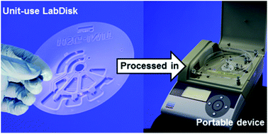
We present total nucleic acid extraction from whole blood, Gram-positive Bacillus subtilis, Gram-negative Escherichia coli, and Rift Valley fever RNA virus on a low-cost, centrifugal microfluidic LabDisk cartridge processed in a light-weight (<2 kg) and portable processing device. Compared to earlier work on disk based centrifugal microfluidics, this includes the following advances: combined lysis and nucleic acid purification on one cartridge and handling of sample volumes as large as 200 μL. The presented system has been validated for logarithmic dilutions of aforementioned bacteria and viruses from various sample matrices including blood plasma and culture media and extraction of human DNA from whole blood. Recovered DNA and RNA concentrations in the eluate were characterized by quantitative PCR to: 58.2–98.5%, 45.3–102.1% and 29.5–34.2% versus a manual reference for Bacillus subtilis, Escherichia coli and Rift Valley fever virus, respectively. For extraction of human DNA from whole blood, similar results for on-disk ((10.1 ± 7.6) × 104 DNA copies) and manual reference extraction ((10.2 ± 6.3) × 104 DNA copies) could be achieved. Eluates from on-disk extraction show slightly increased ethanol concentrations of 4.1 ± 0.3% to 5.5 ± 0.2% compared to a manual reference (2.0 ± 0.5% to 3.6 ± 0.6%). The complete process chain for sample preparation is automatically performed within ∼30 minutes, including ∼15 minutes lysis time. It is amenable to concatenation with downstream modules for multiplex nucleic acid amplification as recently demonstrated for panel testing of various pathogens at the point of care.


Introduction

Nucleic acid extraction comprises cell lysis and nucleic acid purification and is a crucial front end process chain in molecular biological analysis,1 aiming at the isolation, purification and concentration of target nucleic acids, DNA and RNA, for subsequent analysis e.g. via polymerase chain reaction (PCR). To make target nucleic acids accessible, the process chain of nucleic acid extraction mostly starts with enzymatic, chemical or mechanical lysis of the host cell (eukaryotic and prokaryotic) or of viral particles (solely enzymatic or chemical but not mechanical lysis). The multiplicity of sample matrices including blood, blood plasma or bacterial cultures to name just a few, and their different physical and bio-chemical properties along with the requirement to lyse different forms of cells and pathogens render nucleic acid extraction labour intensive and time consuming.2

Centrifugal microfluidics3–6 recently demonstrated its potential to automate complex process chains in nucleic acid analysis including polymerase chain reaction (PCR),7–11 nested PCR,12 digital PCR13 and isothermal recombinase polymerase amplification (RPA).14 Disadvantageously, these systems required already purified nucleic acid samples which were prepared off-chip.

Suitable process chains for upstream target-unspecific nucleic acid extraction from intact, non-lysed cells have not been presented so far. To the best of our knowledge, all presented solutions within centrifugal microfluidics are limited to perform either cell lysis15–17 or purification without chip-integrated lysis.18–21 Mechanical lysis on centrifugal microfluidic cartridges has been demonstrated using collision and friction of glass beads in a rimming flow15 or agitation of beads by an integrated magnetic element.16,17,22 Afterwards, the obtained eluates had to be post treated prior to PCR analysis. In another approach, antibody labelled beads were used for off-chip pathogen specific capturing and concentration of pathogens and subsequent on-chip thermal lysis.23 Purification of RNA and DNA from virus lysates and pre-lysed whole blood, respectively, has been demonstrated using sol–gel,20 glass beads21 or silica membranes as solid phase.18 The most comprehensive approach showed the microfluidic integration of pathogen specific DNA extraction from Hepatitis B DNA viruses and Gram-negative E. coli bacteria from a whole blood sample using anti-body coated magnetic beads. After separation, captured pathogens were thermally lysed by laser irradiation.1 However, the need for target-specific antibody coating does not allow the detection of a certain pathogen from a broad panel of causative agents.

In this work, we demonstrate a target independent, comprehensive and generic approach for total nucleic acid extraction from whole blood, Gram-positive Bacillus subtilis and Gram-negative Escherichia coli bacteria, and the RNA Rift Valley fever virus (RVFV) on a centrifugal microfluidic “LabDisk” platform. The application of magnetic beads as solid phase allows for a concentration of the nucleic acids from sample volume (200 μL) to elution volume (100 μL). DNA recovery and eluate purity of the extracted DNA and RNA were characterised by quantitative real-time PCR, residual ethanol- and spectro-photometric A260/280 measurements, respectively. Compared to our previous work, we report the following advances: (1) microfluidic integration of sample lysis prior to nucleic acid purification; (2) handling of extended sample (200 μL) and reagent volumes (up to 500 μL) comparable to standard bench top extraction protocols; and (3) validation for logarithmic dilutions of different target organisms including Gram-positive and Gram-negative bacteria, RNA viruses and human cells. The developed LabDisk cartridges can be processed in a small and light weight (<2 kg) centrifugal processing device, suitable for application at the point-of-care. After loading of sample and extraction reagents and starting of a predefined frequency protocol, no further manual interaction is required. The presented process chain for total nucleic acid extraction was designed to be combinable with downstream microfluidic modules for multiplex nucleic acid analysis by PCR7–9,12 for future sample-to-answer pathogen panel testing in one unit use cartridge. Here, the pathogen-unspecific extraction principle would allow extraction of DNA and/or RNA from a group of pathogens related to a certain disease.

Functional principle of bead based nucleic acid extraction

Microfluidic implementation of lysis and binding

In this work, nucleic acid extraction is based on a bind-wash-elute protocol2 and automated by means of centrifugal microfluidics. After the LabDisk is placed in the portable processing device, sample and extraction reagents (lysis, binding, 2× washing and elution buffer) are pipetted to the corresponding inlets (Fig. 1a). The predefined frequency protocol is started (ESI). In a first step, the LabDisk cartridge is accelerated to 10 Hz. Due to the resulting centrifugal forces, the sample and all extraction reagents except the binding buffer are gated radially outwards into microfluidic structures for nucleic acid extraction. Sample and lysis buffer merge in a first chamber where they rehydrate an air-dried pellet of magnetic beads, prestored during manufacturing of the LabDisk cartridge (Fig. 1b). Mixing of sample and lysis buffer is enhanced by the periodic deflection of magnetic beads within the liquid at alternating spin frequencies. At 6 Hz, the beads are attracted by the collection- and transport-magnet radially inwards while they are centrifuged radially outwards at 10 Hz.24 The LabDisk cartridge is then stopped and the binding buffer primes the siphon channel due to capillary forces. To transfer the binding buffer into the microfluidic structure for nucleic acid extraction, the LabDisk is re-accelerated to 17 Hz. The binding buffer is merged with the lysate and the released nucleic acids bind to the surface of the magnetic beads (Fig. 1c). Processing device and LabDisk cartridges are depicted in the ESI section 3.
image file: c5ra03399c-f1.tif
Fig. 1 Schematic of the microfluidic structure for nucleic acid extraction. Radial position of the transport magnet is r1 = 42 mm and of the collection magnet r2 = 52.5 mm (see also ESI Fig. S1). Azimuthal distance between both magnets is 42°. Sample liquid and nucleic acid extraction reagents are pipetted to the inlets (a) and the disk is rotated, transferring the liquids radially outwards (b). After a short halt in the frequency protocol and capillary priming of the siphon, the disk is rotated again and the binding buffer is merged with the lysed sample (c).

Transport of magnetic beads between large microfluidic chambers

The transport of magnetic beads between different microfluidic chambers is based on our recently presented unit operation for bead transport exploiting an interplay between magnetic forces and azimuthal positioning of the LabDisk.19 However, the increased sample volume of 200 μL and the application of reagent volumes similar to standard bench top extraction protocols (lysis-buffer: 300 μL; binding-buffer: 450 μL; 2× washing buffer each 500 μL) require large microfluidic chambers for liquid handling. During magnetic attraction by one external magnet at position 2 in Fig. 2a, the beads would have to travel from different positions x and y to the liquid air interface depending on their location within the chamber. Hence, they arrive at the interface at different times and are magnetically attracted out of the liquid in portions as soon as the volume of beads Vcluster at the interface exceeds the critical volume Vcrit at which magnetic forces Fmag (eqn (1))25 overcome retaining forces from surface tension Fsurface (eqn (2)). Smaller bead clusters (Vcluster < Vcrit) would be retained by the surface and wouldn't further contribute to the extraction. To increase the yield of transported beads, a second magnet “collection magnet” was integrated, that collects the magnetic beads first within the liquid and in a second step, the beads are pulled over the interface as one cluster. The step-by-step bead transport with two magnets is described in ESI and in a supporting video file. The dependency of the critical cluster volume Vcrit on the distance d between magnet and liquid–air interface is depicted in Fig. 2b.
 
image file: c5ra03399c-t1.tif(1)
 
Fsurface = 61/3·π2/3σliquid·Vcluster1/3 (2)

image file: c5ra03399c-f2.tif
Fig. 2 (a) Section of the implemented microfluidic structure depicting the designated path of the magnetic beads (red arrow), the lysis-/bind chamber (green; volume ∼ 1 mL) and the adjacent washing chamber (blue; volume 500 μL). A set-up with only one transport magnet (2) would attract the beads along the dashed arrows (x and y) to the interface. Due to the difference in distance yx the beads arrive at the interface at different times and are attracted over the liquid air interface in portions everytime the bead cluster exceeds the critical volume while smaller volumes of beads are retained by the surface and are not transported into the next chamber. To solve this problem, a second collection magnet (1) is implemented that collects the beads within the liquid phase before the collected bead cluster is pulled over the interface. (b) Estimation on Fsurface and different Fmag depending on the distance magnet–interface d. The critical bead cluster volume is defined by the intersection of Fmag and Fsurface.

χmag and χliquid are the magnetic volume susceptibilities of the magnetic beads and the surrounding liquid, σ is the surface tension of the surrounding liquid and (grad(BB) is the strength and gradient of the magnetic field.

Materials and methods

LabDisk fabrication and preparation

All LabDisk cartridges were fabricated from 188 μm thick cyclo olefine polymer foils (COP ZF 14, Zeon Chemicals, USA) by microthermoforming as recently described7,19,26 (HSG-IMIT Lab-on-a-Chip Design & Foundry Service, Germany). All surfaces on the microthermoformed part which later come in contact with magnetic beads during bead transport were hydrophobically coated with 250 μL 0.5% (w/w) Teflon AF (DuPont, USA) dissolved in Fluorinert FC-770 (3M, USA) using a pipette for dispensing. After drying, these surfaces were coated a second time with 150 μL Teflon solution. Finally, a pellet of magnetic beads (MAG Suspension M, Analytik Jena, Jena, Pelleting process described ESI) was placed in the lysis-/binding chamber and the microthermoformed parts were sealed with pressure sensitive adhesive foil (#900 320, HJ Bioanalytik, Germany). Holes for positioning and fixation were lasered into these LabDisk cartridges using a CO2 laser (PLS 3.60, Universal Laser Systems, Austria).

Preparation of human blood, bacterial and viral samples for characterisation

Human whole blood was donated by a healthy lab member. EDTA containers with whole blood were stored at +4 °C and used within 2 days.

Gram-positive Bacillus subtilis (DSM 4451, Leibniz-Institute DSMZ – German collection of Microorganisms and Cell Cultures, Germany) and Gram-negative Escherichia coli DH5αZ1 (provided by Prof. Gescher, Karlsruhe Institute of Technology, Germany) were cultivated over night (30 °C and 37 °C respectively) in LB media. Afterwards, the B. subtilis and E. coli stocks were cooled down on ice and a log[thin space (1/6-em)]10 dilution series was prepared. For preservation of the bacteria during storage at −80 °C, each dilution was mixed with glycerol (99.5%, Carl Roth, Germany) for a final glycerol concentration of 35%. The mixture was aliquoted into 250 μL subvolumes and stored at −80 °C. Cell count of the undiluted B. subtilis and E. coli glycerol stock solution was determined by plate-counting to 1.07 × 10⁸ colony forming units (CFU) mL−1 and 1.45 × 10⁸ CFU mL−1, respectively.

Inactivated RVFV samples with a stock concentration of 3.3 × 10⁶ genome equivalents mL−1 (Robert Koch Institute, Germany) were diluted logarithmically in DI-water, aliquoted into 20 μL aliquots and stored at −80 °C.

Preparation of manual reference and on-disk extraction

Genomic DNA from human EDTA whole blood samples was extracted using reagents from the QiaAMP DNA blood mini Kit (QIAGEN, Germany). Prior to each experiment, 200 μL whole blood aliquots were taken from the fridge, mixed with 20 μL protease and subjected to manual reference or automated on-disk extraction. For manual extraction, 300 μL lysis buffer AL and the blood sample were transferred to a standard 1.5 mL tube, mixed by vortexing (5 seconds) and incubated for 10 minutes at room temperature for chemical lysis. After lysis, 450 μL ethanol and a pellet of magnetic beads were added to the lysate and mixed again (5 seconds vortexing). Via magnetic bead separation (i.e. pulling the beads to the wall of the tube using a magnet, while discarding the liquid), the mixture of blood, lysis buffer and ethanol was replaced by 500 μL washing buffer AW 1. The beads were resuspended (5 seconds vortexing) and via bead separation, washing buffer AW 1 was replaced by washing buffer AW 2. After a brief resuspension (5 seconds vortexing) washing buffer AW 2 was replaced by 100 μL RNAse free water to elute the purified DNA from the beads surface. Finally, beads were fixed by the magnet and the eluate with the purified DNA was transferred to a fresh tube and stored at −20 °C. Additionally, gold-standard manual reference experiments were conducted using QIAGEN spin columns according to manufacturer's protocol (please see ESI).

For automated on-disk extraction, the whole blood sample, pre-mixed with protease was added to the sample inlet port. Lysis buffer AL (300 μL), ethanol for DNA binding (450 μL), washing buffers AW 1 and AW 2 (2× 500 μL) and RNAse free water (100 μL) were loaded to the corresponding inlets of the disk and the predefined spin protocol was started. Prestored silica coated magnetic beads served as the solid phase.

For DNA and RNA extraction from the bacterial (B. subtilis and E. coli) and viral (RVFV) targets, extraction reagents from a commercially available magnetic bead based nucleic acid extraction kit (innuPREP MP Basic Kit A, Analytik Jena AG, Germany) were used. For a single extraction experiment for B. subtilis or E. coli, a 200 μL aliquot of the bacterial stock dilution was taken from the freezer, thawed on ice and shaken for homogenisation. Each 200 μL aliquot was then split into two 100 μL portions, one for manual reference and one for automated on-disk extraction. 100 μL portions of bacterial stock dilution were then mixed with 100 μL pure LB media. RVFV samples were composed from 20 μL virus stock spiked in 180 μL blood plasma. Bacterial and viral samples were afterwards mixed with 5 μL lambda phage DNA and 20 μL Proteinase K. Manual reference extractions from these bacterial and viral samples were conducted as described for the genomic DNA extraction from whole blood but using reagents from the innuPREP MP Basic Kit (300 μL lysis buffer RL, 450 μL binding buffer RBS, 2× 500 μL washing buffer HS and LS and 100 μL RNAse free water for elution). For automated on-disk extraction, the sample, and equivalent volumes of lysis, binding, washing and elution buffer were added to the corresponding inlet of the LabDisk. Magnetic beads were already prestored in the disk as dry pellet and the pre-defined spin protocol was started.

Quantification by real-time PCR amplification

DNA and RNA yield from manual reference and on-disk extraction were quantified by real-time PCR and real-time reverse transcriptase PCR, respectively, in triplicates per sample. For quantification, PCR standards from at least three different DNA or RNA concentrations were processed along with each PCR run. Parallel, no-template controls ensured the absence of cross contaminations in the used reagents.

Extracted DNA from human whole blood was quantified on a Rotor-Gene 2000 real-time PCR thermocycler (formerly Corbett Research, acquired by QIAGEN, Germany) starting with hot start (5 min/95 °C) followed by 45 cycles denaturation (30 s/94 °C) and annealing/extension (45 s/55 °C). Composition of a single PCR reaction and used primer and probe sequences are depicted in ESI Table S3 and S4. DNA yield was quantified on a Rotor-Gene 6000 real-time PCR thermocycler (QIAGEN, Germany). Thermocycling protocol started with an initialisation step (2 min/50 °C) followed by hot-start (10 min/95 °C) and 50 cycles of denaturation (15 s/95 °C) and annealing/extension (60 s/60 °C). Thermocycling protocol for amplification of RNA from Rift Valley fever virus started with reverse transcription (30 min/50 °C) followed by hot-start (15 min/95 °C) and 45 cycles of denaturation (15 s/95 °C) and annealing/extension (60 s/60 °C).

Measurement of ethanol concentration

Ethanol concentrations above 2.5% are a major inhibitor to PCR based DNA analysis.27–29 For measurement of ethanol carry over from washing buffers into the elution buffer, a commercially available kit for colorimetric determination of ethanol concentration (Ethanol Assay Kit Z5030029, BioChain, USA) was used. Seven eluates per pathogen from on-disk extractions and seven eluates per pathogen from manual reference extractions were randomly selected and subjected to ethanol concentration measurements. Residual ethanol in all eluates from whole blood extraction was measured.

Measurements were conducted as follows: 8.6 μL eluate were mixed with 16.4 μL of water and 25 μL of Biochain reagent A. After 20 minutes of incubation, reactions were stopped with 25 μL of Biochain reagent B. Absorption was then determined at 570 nm in a spectrophotometer (Wallac 1420 Multilable counter, Perkin Elmer, Finland) and compared to a standard curve with known ethanol concentrations.

Results and discussion

Automated nucleic acid extraction

Initially, we demonstrated the feasibility of the developed method for DNA extraction using human whole blood as the sample. Blood sample and DNA extraction reagents were loaded onto a LabDisk cartridge with prestored magnetic beads (as described in “Preparation of manual reference and on-disk extraction”) and the predefined spin protocol was started. In parallel, manual reference extractions were conducted in tubes using pelleted magnetic beads. Recovery of DNA was quantified by real-time PCR in 4 samples to (3.0 ± 0.8) × 104 DNA copies for automated on-disk extraction and (10.2 ± 6.3) × 104 DNA copies for manual reference extraction (Fig. 3). To investigate the reasons for the differences in recovery, we repeated the on-disk experiments with liquid magnetic-bead suspension instead of dried magnetic bead pellets, resulting in (10.1 ± 7.6) × 104 recovered DNA copies, which is similar to the manual reference. We suspect, that the reduced recovery for the on-disk experiment with pelleted beads might be attributed to less available surface for DNA binding after pelleting as a result of insufficient resuspension, while in the tube experiments, resuspension of the bead pellet was supported by vortexing. However, this 3-fold difference is within the range of variability of qPCR assays, which in diagnostic samples can show a variation of a factor of 2–3 in quantity.30 Saturation of the bead's surface with DNA might be limited to applications involving long human genomic DNA but not for shorter bacterial or viral nucleic acids. As sufficient DNA will be extracted for post-analysis, the saturation of the beads is regarded as unproblematic. Due to the benefits in handling when having magnetic beads prestored in the LabDisk, pelleted beads were used for all further experiments.
image file: c5ra03399c-f3.tif
Fig. 3 Extracted DNA copies from human whole blood. Comparison of on-disk extractions using prestored, pelleted beads or bead suspension, from manual reference extraction in tubes with liquid beads and from spin column reference (gold standard).

Finally, we conducted extraction experiments with the gold standard method using QIAGEN spin columns according to manufacturer's protocol (ESI section 6). Spin columns contain an integrated silica membrane as solid phase for DNA binding. The recovery was measured to (28.2 ± 10.7) × 104 extracted DNA copies, which is almost three times higher than the manual reference in the test tube. The higher recovery from the spin column experiments is very likely attributed to the increased available surface compared to a 20 μL silica bead suspension.

For characterization of the on-disk extraction performance, logarithmic dilutions of Gram-positive B. subtilis (1.1 × 10⁶–1.1 × 10²; 5.3 × 10² and 5.3 × 101 colony forming units per sample) and Gram-negative E. coli (1.5 × 10⁶–1.5 × 10² CFU per sample) were prepared and subjected to automated extraction. Each experiment was conducted in replicates of three per dilution. In parallel, manual reference extractions with similar reagent volumes were conducted as described in materials and methods. The recovered number of DNA copies from manual reference extraction versus automated on-disk extraction is depicted in Fig. 4 and 5. For the investigated concentrations that were qualified by real-time PCR, recovery of extracted DNA for B. subtilis was between 58.2% and 98.5% with respect to the manual reference (calculated from real-time PCR results as depicted in ESI, section 9). The three lowest concentrated B. subtilis dilutions, containing 5.3 × 10² CFU, 1.1 × 10² CFU and 5.3 × 101 CFU per sample yielded eluates that did not result in positive PCR amplification in all replicates and therefore were quantified by PROBIT regression analysis. It has to be considered that for PCR quantification, only 1 μL from the 100 μL total elution volume was used and thus, the number of negative PCR reactions per se increases with decreasing CFU concentration due to statistical DNA distribution in the PCR reaction. Recovery of extracted DNA for E. coli was between 45.3% and 102.1% (see ESI). Here, only the dilution with a concentration of 1.5 × 10² CFU had to be quantified by PROBIT analysis. The deviation in performance between manual reference and on-disk extraction is comparable to whole blood extraction experiments and might also be explained by insufficient resuspension of the bead pellet consequently providing less available surface for DNA binding. This problem can be solved by either increasing the amount of bead suspension in the pellet or by improving the spin protocol for resuspension.


image file: c5ra03399c-f4.tif
Fig. 4 Yield of manual DNA reference extraction (diamond) versus on-disk extraction (triangle) from B. subtilis dilutions. Pelleted magnetic beads were used for all experiments.

image file: c5ra03399c-f5.tif
Fig. 5 Yield of manual DNA reference extraction (diamond) versus on-disk extraction (triangle) from E. coli dilutions. Pelleted magnetic beads were used for all experiments.

The RNA extraction performance was characterized using dilutions of RVFV in blood plasma samples (6.6 × 10⁴–6.6 × 10² genome equivalents per sample). For each dilution, three independent extraction experiments were conducted on the LabDisk. In parallel, three manual reference extractions were performed. Yield of extracted RNA was afterwards determined by real-time RT-PCR for the highest two Rift Valley fever virus concentrations (6.6 × 10³ and 6.6 × 10⁴ genome equivalents). For determination of the RNA extraction yield from the lowest concentration, 6.6 × 10² genome equivalents, PROBIT regression analysis was used as explained in ESI (Fig. 6). LabDisk based extraction yielded between 29.5% and 34.2% RNA copies compared to manual reference in the tube for those samples that were quantified by real-time RT-PCR. Compared to the extraction of DNA from bacterial pathogens, the ratio of on-disk extraction is significantly lower what might be explained by the higher sensitivity of RNA to degradation.


image file: c5ra03399c-f6.tif
Fig. 6 Yield of manual RNA reference extraction (diamond) versus on-disk extraction (triangle) from Rift Valley fever virus dilutions. Pelleted magnetic beads were used for all experiments.

Inhibitor concentration in eluate

Ethanol concentration in the eluates derived from manual reference and automated on-disk extractions (n = 5 for whole blood; n = 7 for B. subtilis, E. coli and RVFV) have been measured as described in materials and methods (Table 1).
Table 1 Ethanol concentrations in 100 μL eluate depicted as % (weight/volume). Each value is calculated as mean ± standard deviation from 5 (whole blood) or 7 eluates (B. subtilis, E. coli, Rift Valley fever), respectively
  On-disk [% w/v] Manual ref. [% w/v]
Whole blood 4.1 ± 0.3 2.0 ± 0.5
B. subtilis 5.5 ± 0.2 3.6 ± 0.6
E. coli 5.3 ± 0.6 2.8 ± 0.7
RVFV 4.7 ± 0.7 3.0 ± 1.6


Measured concentrations for manual reference extraction reveal to be lower as ethanol residuals can evaporate during a five minute air drying step after the second washing step which was not implemented in the on-disk extraction protocol. However, as only 1 μL eluate from the total volume of 100 μL is subjected to quantification in a 10 μL PCR reaction, the ethanol content is diluted well below the critical level for inhibition of 2.5%.28,29

Furthermore, the absorbance ratio at 260 nm to 280 nm (A260/280) in the eluates from whole blood extraction was measured (Nanovue plus spectrophotometer; GE lifescience, USA). Nucleic acids are regarded as pure if the A 260/280 ratio is between ∼1.7 and ∼1.9 while lower values indicate contaminations with proteins or phenols,31,32 further PCR inhibitors. Measured A 260/280 values were 2.05 ± 0.25 (n = 4) for automated on-disk extraction with bead pellet, 1.77 ± 0.18 (n = 2) for on-disk extraction with liquid beads, 1.88 ± 0.22 (n = 2) for reference extraction in tube, and 1.93 ± 0.09 (n = 5) for spin column extraction. All measured values are within the expected range. Measurements were not conducted for eluates from the bacterial and viral extraction as the required nucleic acid concentration was below the measurement range of the device.

Conclusion

We employed centrifugal microfluidics and presented a “LabDisk” cartridge for the completely integrated and automated extraction of DNA and RNA from various samples using our recently presented unit-operation for magnetic bead transport “gas-phase transition magnetophoresis”. This includes the following novelties: (1) integrated chemical lysis prior to nucleic acid purification; (2) handling of sample volumes up to 200 μL and reagent volumes up to 500 μL, and (3) successful validation for different targets including Gram-positive B. subtilis, Gram-negative E. coli, Rift Valley fever RNA virus and human cells. LabDisk cartridges were processed in a small, light-weight and portable processing device (weight: <2 kg; dimensions: 25 × 17.5 × 8.5 cm3), suitable for application at the point-of-care. The complete process chain is automatically performed within 30 minutes, including ∼15 minutes lysis time according to the standard protocol.

We thoroughly characterized the system by extracting human DNA from whole blood and genomic DNA from dilutions of Gram-positive and Gram-negative bacteria in stock culture, B. subtilis and E. coli, respectively. We demonstrated that on-disk extraction and manual reference extraction performed similar when a suspension of magnetic beads is used for extraction yielding (10.1 ± 7.6) × 104 DNA copies extracted on-disk vs. (10.2 ± 6.3) × 104 DNA copies extracted by manual reference. On-disk extraction with prestored, pelleted magnetic beads yielded about 30% compared to the set-up using a suspension of magnetic beads. Therefrom, we hypothesize that available surface on the beads might be reduced after drying. On-disk extraction for B. subtilis yielded 58.2–98.5% versus manual reference. Recovery for E. coli was 45.3–102.1% compared to reference. Furthermore, we extracted RNA from dilutions of RVFV samples spiked in blood plasma with a yield of 29.5% and 34.2% compared to the reference. All on-disk extractions showed increased ethanol concentrations between 4.1 ± 0.3% and 5.5 ± 0.2% compared to manual reference extractions (2.0 ± 0.5% to 3.6 ± 0.6%).

The presented implementation of a microfluidic process chain for pathogen unspecific, total DNA and RNA extraction from various sample matrices might be concatenated with downstream microfluidic modules for PCR or RPA based multiplex amplification for full sample-to-answer panel testing of various pathogens at the point-of-care in the near future. Additionally, the LabDisk cartridge can be equipped with liquid reagent prestorage in miniature stick packs as recently presented by van Oordt et al.33

Acknowledgements

The study was partly funded by the German Federal Ministry of Education and Research BMBF; (Project S.O.N.D.E grant number 13N10116). Furthermore, we want to thank Dominique Kosse and the team of the HSG-IMIT Lab-on-a-Chip Design and Foundry Service for fabrication of all cartridges that were used in this study and Dr Günter Roth for fruitful discussions on assay related questions.

References

  1. Y. K. Cho, J. G. Lee, J. M. Park, B. S. Lee, Y. Lee and C. Ko, Lab Chip, 2007, 7, 565–573 RSC.
  2. R. Boom, C. J. A. Sol, M. M. M. Salimans, C. L. Jansen, P. M. E. Wertheimvandillen and J. Vandernoordaa, J. Clin. Microbiol., 1990, 28, 495–503 CAS.
  3. J. Ducrée, S. Haeberle, S. Lutz, S. Pausch, F. von Stetten and R. Zengerle, J. Micromech. Microeng., 2007, 17, S103 CrossRef.
  4. M. Madou, J. Zoval, G. Jia, H. Kido, J. Kim and N. Kim, Annu. Rev. Biomed. Eng., 2006, 8, 601–628 CrossRef CAS PubMed.
  5. D. Mark, S. Haeberle, G. Roth, F. von Stetten and R. Zengerle, Chem. Soc. Rev., 2010, 39, 1153–1182 RSC.
  6. S. Haeberle and R. Zengerle, Lab Chip, 2007, 7, 1094–1110 RSC.
  7. M. Focke, F. Stumpf, B. Faltin, P. Reith, D. Bamarni, S. Wadle, C. Müller, H. Reinecke, J. Schrenzel, P. Francois, D. Mark, G. Roth, R. Zengerle and F. von Stetten, Lab Chip, 2010, 10, 2519 RSC.
  8. O. Strohmeier, N. Marquart, D. Mark, G. Roth, R. Zengerle and F. von Stetten, Anal. Methods, 2014, 6, 2038 RSC.
  9. O. Strohmeier, S. Laßmann, B. Riedel, D. Mark, G. Roth, M. Werner, R. Zengerle and F. von Stetten, Microchim. Acta, 2014, 181, 1681–1688 CrossRef CAS.
  10. J. H. Jung, S. J. Choi, B. H. Park, Y. K. Choi and T. S. Seo, Lab Chip, 2012, 12, 1598–1600 RSC.
  11. M. Amasia, M. Cozzens and M. J. Madou, Sens. Actuators, B, 2012, 161, 1191–1197 CrossRef CAS PubMed.
  12. M. Focke, F. Stumpf, G. Roth, R. Zengerle and F. von Stetten, Lab Chip, 2010, 10, 3210 RSC.
  13. S. O. Sundberg, C. T. Wittwer, C. Gao and B. K. Gale, Anal. Chem., 2010, 82, 1546–1550 CrossRef CAS PubMed.
  14. S. Lutz, P. Weber, M. Focke, B. Faltin, J. Hoffmann, C. Müller, D. Mark, G. Roth, P. Munday, N. Armes, O. Piepenburg, R. Zengerle and F. von Stetten, Lab Chip, 2010, 10, 887 RSC.
  15. J. Kim, J. S. Hee, G. Jia, J. V. Zoval, N. A. Da Silva and M. J. Madou, Lab Chip, 2004, 4, 516–522 RSC.
  16. H. Kido, M. Micic, D. Smith, J. Zoval, J. Norton and M. Madou, Colloids Surf., B, 2007, 58, 44–51 CrossRef CAS PubMed.
  17. J. Siegrist, R. Gorkin, M. Bastien, G. Stewart, R. Peytavi, H. Kido, M. Bergeron and M. Madou, Lab Chip, 2010, 10, 363–371 RSC.
  18. J. Hoffmann, D. Mark, S. Lutz, R. Zengerle and F. von Stetten, Lab Chip, 2010, 10, 1480 RSC.
  19. O. Strohmeier, A. Emperle, G. Roth, D. Mark, R. Zengerle and F. von Stetten, Lab Chip, 2012, 13, 146 RSC.
  20. B. H. Park, J. H. Jung, H. Zhang, N. Y. Lee and T. S. Seo, Lab Chip, 2012, 12, 3875–3881 RSC.
  21. J. H. Jung, B. H. Park, Y. K. Choib and T. S. Seo, Lab Chip, 2013, 13, 3383–3388 RSC.
  22. E. Roy, G. Stewart, M. Mounier, L. Malic, R. Peytavi, L. Clime, M. Madou, M. Bossinot, M. G. Bergeron and T. Veres, Lab Chip, 2015, 15, 406–416 RSC.
  23. T.-H. Kim, J. Park, C.-J. Kim and Y.-K. Cho, Anal. Chem., 2014, 86, 3841–3848 CrossRef CAS PubMed.
  24. M. Grumann, A. Geipel, L. Riegger, R. Zengerle and J. Ducrée, Lab Chip, 2005, 5, 560–565 RSC.
  25. N. Pamme, Lab Chip, 2006, 6, 24–38 RSC.
  26. M. Focke, D. Kosse, D. Al-Bamerni, S. Lutz, C. Müller, H. Reinecke, R. Zengerle and F. von Stetten, J. Micromech. Microeng., 2011, 21, 115002 CrossRef.
  27. I. G. Wilson, Appl. Environ. Microbiol., 1997, 63, 3741–3751 CAS.
  28. M. Karle, J. Miwa, G. Czilwik, V. Auwärter, G. Roth, R. Zengerle and F. von Stetten, Lab Chip, 2010, 10, 3284–3290 RSC.
  29. L. Rossen, P. Norskov, K. Holmstrom and O. F. Rasmussen, Int. J. Food Microbiol., 1992, 17, 37–45 CrossRef CAS.
  30. H. G. Niesters, Methods, 2001, 25, 419–429 CrossRef CAS PubMed.
  31. GE Healthcare Life Sciences-Nano Vue Plus, Users Manual, available at: http://www.sidmartinbio.org/wp-content/uploads/NanoVueUserManual2009.pdf, accessed 18 May 2014.
  32. QIAGEN – QIAamp® DNA Mini Kit and QIAamp DNA Blood Mini Kit Handbook.
  33. T. van Oordt, Y. Barb, J. Smetana, R. Zengerle and F. von Stetten, Lab Chip, 2013, 13, 2888–2892 RSC.

Footnote

Electronic supplementary information (ESI) available. See DOI: 10.1039/c5ra03399c

This journal is © The Royal Society of Chemistry 2015