Novel aligned sodium vanadate nanowire arrays for high-performance lithium-ion battery electrodes

Yunhe Caoa, Dong Fang*a, Chang Wanga, Licheng Lia, Weilin Xu*a, Zhiping Luob, Xiaoqing Liuc, Chuanxi Xiongac and Suqin Liud
aKey Lab of Green Processing and Functional Textiles of New Textile Materials, Ministry of Education, College of Material Science and Engineering, Wuhan Textile University, Wuhan, P. R. China. E-mail: csufangdong@gmail.com
bDepartment of Chemistry and Physics, Fayetteville State University, Fayetteville, NC 28301, USA
cSchool of Materials Science and Engineering, Wuhan University of Technology, Wuhan 430070, P. R. China
dCollege of Chemistry and Chemical Engineering, Central South University, Changsha 410083, P. R. China

Received 19th January 2015 , Accepted 20th April 2015

First published on 20th April 2015


Abstract

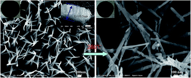
Sodium vanadate (Na5V12O32 or Na1.25V3O8) nanowire arrays were successfully prepared using a facile hydrothermal method with subsequent calcination. The length of the Na5V12O32 nanowire arrays on titanium foil were about 10.5 μm. The unique architecture renders a high-rate transportation of lithium ions that is attributed to their nanosized structure, active materials connected to the current collector and the high specific surface area. The Na5V12O32 nanowire arrays on titanium foil annealed at 250 °C as electrodes for lithium-ion batteries exhibit a significant capacity stability with a capacity from 339.3 to 289.7 mA h g−1 in 50 cycles at 50 mA g−1. The superior electrochemical performance demonstrated that the Na5V12O32 nanowire arrays are promising electrodes for secondary organic lithium-ion batteries.


One-dimensional (1D) nanostructures, such as nanorods, nanowires, and nanotubes, have attracted extensive attention due to their reduced size and morphology-dependent properties.1–3 These 1D nanostructured materials have exhibited novel optical, electrical, magnetic, and mechanical properties that are anomalous from those of bulk or nanoparticle materials. It has been noted that some 1D nanostructured materials have been used as active components or interconnects in fabricating nanoscaled electronic, optical, optoelectronic, electrochemical, and electromechanical devices.2,4 1D nanostructures used as lithium-ion battery (LIB) materials have exhibited appealing properties, such as reduced lithium ion (Li+) diffusion and electron transportation distance, enlarged contact area between the electrode and electrolyte, and tolerance to the stress upon Li+ ion take-up and removal processes.5–9

LIBs have drawn overwhelming research attention due to the environmental pollution caused by fossil fuels and the gradual depletion of oil resources. As an electrode material of LIBs, vanadium-oxide (V-O) compounds have been attracting much attention as they are known to exhibit a range of oxidation states from +2 to +5, allowing more than one Li+ ion to be inserted into a host vanadium unit, and thus they have immense potential to provide higher specific capacities.10,11 Reversible electrochemical Li+ intercalation into V2O5 at room temperature was first reported in 1975.12 The electrode performances of pure vanadium oxides were greatly improved by addition of Li+ ions into these host vanadium oxides, such as for LiV3O8, Li1+xV1−xO2,13,14 etc. These additional Li+ ions were arranged to form pillars between the vanadium oxide layers and thus stabilized the structure during Li+ insertion/extraction.15 In addition, these pillars increased not only the interlayer space but also the ion diffusion rate in the materials. All of these characteristics made them promising electrode materials for rechargeable lithium batteries. Xiong et al. fabricated LiV3O8 using an improved spray-drying method, which exhibited a discharge capacity as high as 340.2 mA h g−1 in the first cycle at a current density of 25 mA g−1.16 Song et al.17 obtained Li1+xVO2 without any impurities and the x value was 0.2 with a discharge capacity of 294 mA h g−1 and a cycle retention of >90%, after 25 cycles. Pan et al.18 produced LiV3O8 nanorods using a template-free, low-temperature method which delivered specific discharge capacities of 320 mA h g−1 and 239 mA h g−1 at current densities of 100 mA g−1 and 1 A g−1, respectively. Other cations with an even larger ionic radius than Li+, such as NH4+, Na+ and K+, have attracted much attention due to the larger interlayer distance compared to Li+, which potentially can improve the mobility of the Li+ ions during the Li+ ion intercalation/deintercalation process.19–25 Liu et al.26 reported that single crystalline NaV6O15 nanorods had a discharge capacity as high as 328 mA h g−1 at 20 mA g−1 in organic electrolyte. Zhou et al.27 obtained single crystalline Na2V6O16·0.14H2O (NaV3O8·0.07H2O) nanowires which delivered an initial specific discharge capacity of 122.7 mA h g−1 for an aqueous lithium-ion battery (based on the mass of anode material) at 60 mA g−1. As is known, Na5V12O32 (Na1.25V3O8) possesses a typical layered structure which consists of V3O8 layers and interstitial hydrated Na ions. The V3O8 layer is composed of VO6 octahedra and V2O8 units of edge-sharing square pyramids. The hydrated Na ions are located between the layers. The infinite chain structure formed by the atomically anisotropic arrangement in the crystal lattice has the potential to grow into 1D nanostructures with unique growth directions. Regardless of its fascinating structure and physicochemical properties, little work has been done on the synthesis of Na5V12O32.28

It is well known that fabrication methods, morphology, and preparation of the electrode methods affect the electrochemical properties of electrode materials significantly. The traditional preparation of electrodes for lithium-ion batteries includes mixing active materials with binders and conductive carbon additives. The presence of binders causes issues such as decreasing electrical conductivity and resistance which lowers the performance. Hence, the elimination of binders could decrease the weight of batteries and significantly improve the electrochemical performance of lithium-ion batteries.29,30 In this paper, Na5V12O32 nanowire arrays were facilely synthesized via a hydrothermal route with subsequent calcination and a growth mechanism of the unique nanowires on titanium foil is proposed. As a member of the aforementioned vanadium-based compounds, the electrochemical properties for lithium-ion batteries were tested in organic electrolyte and the host material stability was also analyzed.

The freshly prepared products were heat-treated at various temperatures in air. Fig. 1(a) displays the X-ray diffraction (XRD) patterns of these samples. The as-prepared sample and the 200 °C calcinated sample have a similar crystal structure with a low crystallinity. The diffraction peaks from the samples annealed at 250 °C and 300 °C are attributed to a monoclinic crystalline phase [space group: P21/m (no. 11)] of Na5V12O32 (JCPDS no. 24-1156) with lattice parameters of a = 12.14 Å, b = 3.61 Å, c = 7.32 Å and β = 106.73°. X-ray photoelectron spectroscopy (XPS) is an effective method that can provide an elemental analysis of the surface with specific information about the oxidation states of different elements in the material. A wide survey scan XPS (ranging from 100 to 1300 eV) of the as-prepared sample is shown in Fig. 2(a). The C 1s peak at 284.6 eV is used as a reference binding energy for calibration. A series of peaks from Cl 2p, C 1s, N 1s, V 2p, O 1s and Na 1s are clearly observed. The carbon peak of the sample is from surface contamination by CO2.9 As shown in Fig. 2(b), a full range XPS spectral analysis of the Na5V12O32 revealed five main peaks located at about 284.37, 516.91, 529.98 and 1070.86 eV, corresponding to C 1s, V 2p, O 1s and Na 1s, respectively. No other element peaks are detected, indicating that Na5V12O32 is highly pure. A high resolution XPS of V 2p is displayed in Fig. 2(c), which shows the enlarged spectrum between 514 eV and 528 eV of the sample. The peaks at 516.49 eV with the full width at half maximum (FWHM) of 1.49 eV and at 524.05 eV with the FWHM of 1.89 are attributed to the V 2p3/2 and V 2p1/2, respectively, which are centered at the V4+ position. The other peaks at 517.67 eV with the FWHM of 1.32 eV and at 525.28 eV with the FWHM of 1.42 eV correspond to the binding energy of the V 2p3/2 and V 2p1/2 electrons, respectively, for vanadium in the +5 oxidation state.9,30 The molar ratio of the V4+ to V5+ ions in the as-prepared sample is about 1/11. The XPS spectra of the as-prepared sample and the sample annealed at 250 °C clearly reveal that the annealing treatment causes the elimination of nitrogen and chlorine species. This is likely due to evaporation of NH3 or Cl2 gas species during the heat treatment. Another plausible reason could be the flexibility of the low-crystalline state. Incorporation of rather large amounts of additional impurities in a low crystallinity sample is allowed by reduced structural constraints in the amorphous network. During crystallization processes foreign atoms simply pull off from the volume of growing crystallites.


image file: c5ra01102g-f1.tif
Fig. 1 XRD patterns of the as-prepared sample and the samples annealed at different temperatures.

image file: c5ra01102g-f2.tif
Fig. 2 XPS survey spectrum of the as-prepared sample (a) and that annealed at 250 °C (b), and a high resolution XPS of V2p (c).

The morphology of the as-prepared dark-green Ti foil obtained after 1 h of reaction was investigated using scanning electron microscopy (SEM) (Fig. 3(a) and (b)), which revealed nanowires grown on the foil surfaces vertically. The diameter of the nanowires is about 50 nm and the length is about several microns, from the lateral view in the inset of Fig. 3(a). When annealed at 250 °C, Na5V12O32 maintains the wire-like structures assembled with particles. Moreover, the size of the nanowires is larger than that of the as-prepared nanowires, which is caused by growth from the merging of adjacent nanowires during annealing. It is apparent that the post-treatment temperature has an important effect on the morphological features of the samples. The main reason is that vanadate possesses a low melting point. Further annealed at 300 °C, the nanowire structures collapse to big rods as shown in Fig. S1(a and b) (ESI).


image file: c5ra01102g-f3.tif
Fig. 3 SEM images of the as-prepared sample on Ti foil (a and b) and that annealed at 250 °C (c and d). Inset in (a) and (c) are digital photos of the samples.

The detailed structure and the growth direction of the as-prepared nanowires were further examined by transmission electron microscopy (TEM). Fig. 4(a) shows an image in a lower magnification of several nanowires, which have a diameter of approximately 50 nm with smooth surfaces. The high resolution TEM (HR-TEM) image in Fig. 4(b) was obtained from the area marked with a rectangular frame in Fig. 4(a). The nanowire exhibits low crystallinity with limited areas of lattice fringes. Fig. 4(c) and (e) are the low-magnification TEM images of Na5V12O32 nanowires annealed at 250 °C. Consistent with the SEM images, the nanowires display uniform dimensions, 100 nm wide and several micrometers long. A selected-area electron diffraction (SAED) pattern was obtained from the area marked by a circle in Fig. 4(c). The SAED pattern further confirms that the Na5V12O32 structure is monoclinic crystalline in nature, which coincides to the XRD analysis. Each nanowire has a single crystal structure with a similar orientation in this local region. A closer observation of a typical nanowire is depicted by the HR-TEM image. As shown in Fig. 4(f), the lattice fringe with an inter-planar distance of 0.351 nm can be clearly observed, which is a good match with the d-spacing of (002).


image file: c5ra01102g-f4.tif
Fig. 4 (a) TEM and (b) HR-TEM images of the as-prepared nanowires, (c) & (e) TEM images and (d) SAED pattern of several nanowires, and (f) HR-TEM image of a Na5V12O32 nanowire. Inset in (a) is the SAED pattern of the as-prepared nanowires.

In order to investigate the formation mechanism of the nanowires, time-dependent experiments were carried out. The materials with reaction times of 10 min, 30 min, 1 h, 3 h and 5 h were prepared with a similar approach. Fig. S2 (ESI) shows the morphologies of the as-prepared products with various reaction times. There exists a compact film on the titanium foil after 10 min. In the sample with an intermediate reaction time of 30 min, tiny and short nanowires are well dispersed on the film. After a sufficient hydrothermal reaction time of 1 h, uniform nanowires are obtained. SEM images demonstrate that the intermediate sample with a reaction time of 3 h consists of longer nanowires with a larger diameter. After 5 h, the diameter of the wires is about 200 nm, without having connected to form sheets among the adjacent nanowires, which is different from our previously reported ammonium vanadium bronze (NH4V4O10).9 According to the morphology variations of the intermediate state after different reaction times, a growth mechanism of the unique nanowires is proposed, as shown in Fig. 5. Firstly, a thin film is coated on the titanium foil; secondly, tiny and short nanowires appear; then, uniform nanowires are obtained and after further extending the reaction time, the nanowires are larger and longer. Without the titanium foil in the autoclave, an as-prepared powder consisting of uniform nanowire flowers is obtained as shown in Fig. S3 (ESI).


image file: c5ra01102g-f5.tif
Fig. 5 The growth mechanism of the unique nanowires on titanium foil.

The electrochemical properties of the Na5V12O32 sample as an electrode in Li-ion storage were tested via discharge–charge measurements in organic electrolyte. Fig. 6(a) depicts the cycles of discharge–charge profiles of the Na5V12O32 nanowire arrays on Ti foil treated at 250 °C with a current density of 50 mA g−1. This sample gives the highest discharge capacity of 339.3 mA h g−1. There are three patent plateaus at 3.25 V, 2.8 V and 2.5 V, indicating multi-step intercalation of Li+ ions. The voltage profiles of the first Li+ ion intercalation plateau are lower than that in the subsequent intercalation processes, demonstrating that the samples experience successive and reversible phase transformations during the insertion/extraction and stress/strain, due to a large volume expansion in the first insertion which introduces a large strain over-potential. The large strain over-potential is significantly reduced in the subsequent cycles due to the introduction of defects in the first insertion, shifting the potential back to a higher value after the first insertion. The obvious flat voltage plateaus are almost stable at 2.69 V for charge and 2.53 V for discharge, with a polarization of 0.16 V. The resistance of the electrolyte, the electrode, the charge transfer at the interface and the membranes are responsible for the polarization.31 Surprisingly, the profiles show that the charge capacity is higher than the discharge capacity. In vanadium oxides, the additional alkaline ions are just arranged to form pillars between the vanadium oxide layers.19,20 The working mechanism of the cell is an electrochemical process. At the beginning, the device is at a discharge state. Under the driving of the potential with a direction from the anode to the cathode, the Li+ ions in the electrolyte will migrate along this direction through the separator and finally reach the cathode, as shown in Fig. 6(b). In the meanwhile electrons will pass through the contact point between the Li foil and the titanium substrate, and then up through the bottom of the titanium substrate.32 During the charge process, vanadium has V4+ and V5+ ions, and a valance state of +49/12, which could be oxidized to +5 with the Na+ ion extraction. Herein, we speculate that a great part of the extra charge capacity could be attributed to the extraction of Na+ ions from the host material as shown in Fig. 6(c). According to the above argument, the reversible insertion and extraction behavior of Li+ ions in Na5V12O32 nanowires may be amended as:

 
Na5V12O32 + xLi+ + xe → LixNa5V12O32 (1)
 
LixNa5V12O32xLi+yNa+ − (x + y)e → Na5−yV12O32 (y < 5) (2)
 
Na5−yV12O32 (y < 5) + zLi+ + ze → LizNa5−yV12O32 (y < 5) (3)


image file: c5ra01102g-f6.tif
Fig. 6 (a) Galvanostatic discharge–charge curves of the Na5V12O32 nanowires at a current density of 50 mA g−1. The working mechanism of the cell. (b) Schematic illustration of the cell in discharge state of Na5V12O32 on titanium substrate. (c) The extract mechanism of the Li+ ions and Na+ ions of the cell in charge state.

The cyclic voltammetry (CV) curves of Na5V12O32 at the rate of 0.5 mV s−1 for 3 cycles between 2 V and 4 V are shown in Fig. 7(a). There exists three anodic/cathodic peaks situated at 2.46/2.73, 2.87/2.99 and 3.27/3.32 for Na5V12O32, which correspond to the reversible insertion/extraction of Li+ ions into and from Na5V12O32 in the electrolyte. This illustrates that the Na5V12O32 material could present a good reversibility of the Li+ ion insertion/extraction. Fig. 7(b) presents the capacity retentions of the Na5V12O32 nanowires on titanium foil annealed at 250 °C, the Na5V12O32 powder annealed at 250 °C and the as-prepared sample. The electrode of Na5V12O32 nanowires on titanium foil annealed at 250 °C exhibits a light capacity decay from 339.3 to 289.7 mA h g−1 in 50 cycles at 50 mA g−1. The capacity loss mainly occurs in the first few cycles and the main reason may be that the small sized nanowires have a large surface area, and some Li+ ions are stored on the surface of the nanowires, which can contribute to the discharge capacity as an irreversible part. In comparison, the as-prepared nanowire arrays on titanium foil exhibit a gradual decreasing trend in the initial stage, and then become stabilized at lower values in successive cycles. The capacity of the Na5V12O32 powder annealed at 250 °C is lower than that for the former annealed nanowires, which indicates that active materials connected to the current collector can enhance electrochemical performance. The electrochemical impedance spectroscopy (EIS) measurements of the fresh electrodes were carried out by applying 100 kHz to 0.001 Hz frequency ranges with an ac oscillation amplitude of 5 mV at open potential. The impedance plots of the Na5V12O32 nanowires on Ti foil and Na5V12O32 nanowire powder electrodes are shown in Fig. S4 (ESI); all the plots show a semicircle at the high-to-medium frequencies and a straight slopping line at low frequency. The depressed semicircle in the high frequency range is related to the Li+ ion migration resistance (Rf) through the SEI film formed on the electrode or another coating layer. The inclined line in the lower frequency represents the Warburg impedance, which is associated with Li+ ion diffusion in the Na5V12O32 particles. The value of Rf of the Na5V12O32 nanowires on Ti foil electrode (64 Ω) is much smaller than that of the Na5V12O32 nanowire powder electrode (7179 Ω). This result reveals that the transfer rate of Li+ in the Na5V12O32 nanowires on Ti foil electrode is higher than in the powder electrode. The nanowires on the substrate (Ti) improve the electronic conductivity, which will improve the electrochemical performance of the Na5V12O32 nanowire arrays on Ti foil electrode. To further understand the high-rate performance of the Na5V12O32 annealed at 250 °C electrode, rate preferment was tested at different current densities ranging from 100 mA g−1 to 1000 mA g−1 in the potential range of 2.0–4.0 V. The results are shown in Fig. 7(c). The fifth specific discharge capacity of the electrode at 100 mA g−1, 200 mA g−1, 400 mA g−1, 600 mA g−1 and 1000 mA g−1 current densities is 283.3 mA h g−1, 213.1 mA h g−1, 166.4 mA h g−1, 142.1 mA h g−1 and 118.3 mA h g−1, respectively. When cycling at 100 mA g−1 again, the discharge capacity recovers to 254.7 mA h g−1.


image file: c5ra01102g-f7.tif
Fig. 7 (a) CV curves of Na5V12O32 nanowires on Ti foil. The scan rate is 0.5 mV s−1. (b) Cycling performances at a current density of 50 mA g−1. (c) Capacity retention for Na5V12O32 nanowires on Ti foil at different charge and discharge current densities from 100 to 1000 mA g−1.

Fig. 8 indicates that the preparation process affects the electrochemical properties of the electrode materials significantly. The capacity of the electrode prepared at 250 °C is much higher than the other electrodes in Fig. 8(a) and (b). The nanowires annealed at 200 °C performed better than the as-prepared nanowires, while the capacity and cycle stability were still low. A possible reason is that the decrease in annealing temperature will result in a poor crystallinity. On the other hand, the nanowires annealed at 250 °C retained a higher capacity of 289.72 mA h g−1 after 50 cycles. When the nanowires were annealed at an even higher temperature (300 °C), they did not perform well. Fig. S1 (a and b) demonstrates some extensive aggregation and collapse to big rods after annealing at 300 °C. The reaction time influence on the electrochemical performance was also investigated. Fig. 8(c) and (d) show the capacity and cycling performance of the Na5V12O32 electrodes prepared at different reaction times, respectively. The electrode fabricated through hydrothermal reaction for 1 h has the maximum capacity. The electrode material prepared in 30 min is a mixture of nanoparticles and nanowires. On the other hand, the nanowires combine into big rods or nanoribbons during a longer reaction time (more than 1 h), which also have a poor electrochemical performance.


image file: c5ra01102g-f8.tif
Fig. 8 The second cycle charge/discharge profiles (a) and cycling performances (b) of Na5V12O32 nanowire array electrodes prepared at different annealed temperatures; the second charge/discharge profiles (c) and cycling performances (d) of Na5V12O32 nanowire array electrodes prepared at different reaction times.

Conclusions

In summary, a hydrothermal method with subsequent calcination has been developed to fabricate Na5V12O32 nanowire arrays. From the growth mechanism study of the unique nanowire arrays, it was found that a thin film was coated on the titanium foil after 10 min, tiny and short nanowires appeared after 30 min, and uniform nanowires were obtained after 1 h. Further extension of the reaction time yielded larger and longer nanowires. As an electrode material of organic Li-ion batteries, the Na5V12O32 nanowire arrays exhibited excellent capacity and cycling stability. The Na5V12O32 nanowire arrays synthesized at 250 °C exhibit a light capacity decay in the potential range of 2.0 to 4.0 V, and after rate preferment at different current densities ranging from 100 mA h g−1 to 1000 mA h g−1, the discharge capacity recovers to 254.7 mA h g−1. The superior electrochemical performance of the Na5V12O32 nanowire arrays indicates their potential application in organic Li-ion batteries.

Acknowledgements

This work was supported by the Natural Science Foundation of China (no. 51201117), the Major State Basic Research Development Program (973 Program) (no. 2012CB722701), the Scientific Research Fund of Wuhan Textile University, and the Scholarship Award for Excellent Doctoral Student granted by Ministry of Education of China (no. 1343-71134001002). Dr. Guangzhong Li, Northwest Institute for Non-ferrous Metal Research, is acknowledged for help in the XPS testing.

References

  1. B. Su, Y. C. Wu and L. Jiag, Chem. Soc. Rev., 2012, 41, 7832–7856 RSC.
  2. Y. Xia, P. Yang, Y. Sun, Y. Wu, B. Mayers, B. Gates, Y. Yin, F. Kim and H. Yan, Adv. Mater., 2003, 15, 353–389 CrossRef CAS PubMed.
  3. D. Fang, K. L. Huang, S. Q. Liu and Z. J. Li, J. Alloys Compd., 2008, 464(1–2), L5–L9 CrossRef CAS PubMed.
  4. B. X. Li, Y. Xu, G. X. Rong, M. Jing and Y. Xie, Nanotechnology, 2006, 17, 2560–2566 CrossRef CAS PubMed.
  5. A. S. Arico, P. Bruce, B. Scrosati, J. M. Tarascon and W. V. Schalkwijk, Nat. Mater., 2005, 4, 366–377 CrossRef CAS PubMed.
  6. A. Q. Pan, H. B. Wu, L. Yu and X. W. Lou, Angew. Chem., Int. Ed., 2013, 52, 2282–2286 CrossRef PubMed.
  7. Y. Wang and G. Z. Cao, Adv. Mater., 2008, 20, 2251–2269 CrossRef CAS PubMed.
  8. D. Fang, L. C. Li, W. L. Xu, G. Z. Li, G. Li, N. F. Wang, Z. P. Luo, J. Xu, L. Liu, C. L. Huang, C. W. Liang and Y. S. Ji, J. Mater. Chem. A., 2013, 1, 13203–13208 CAS.
  9. D. Fang, L. C. Li, W. L. Xu, G. Z. Li, Z. P. Luo, C. W. Liang, Y. S. Ji, J. Xu and C. X. Xiong, RSC Adv., 2014, 4, 25205–25209 RSC.
  10. Y. Wang, K. Takahashi, K. Lee and G. Z. Cao, Adv. Funct. Mater., 2006, 16, 1133–1144 CrossRef CAS PubMed.
  11. L. Q. Mai, L. Xu, C. H. Han, X. Xu, Y. Z. Luo, S. Y. Zhao and Y. L. Zhao, Nano Lett., 2010, 10(11), 4750–4755 CrossRef CAS PubMed.
  12. M. S. Whittingham, J. Electrochem. Soc., 1975, 123, 315–320 CrossRef PubMed.
  13. G. Pistoia, M. Pasquali, G. Wang and L. Li, J. Electrochem. Soc., 1990, 137, 2365–2370 CrossRef CAS PubMed.
  14. A. R. Armstrong, C. Lyness, P. M. Panchmatia, M. S. Islam and P. G. Bruce, Nat. Mater., 2011, 10, 223–229 CrossRef CAS PubMed.
  15. M. E. Spahr, P. Novak, W. Scheifele, O. Haas and R. Nesper, J. Electrochem. Soc., 1998, 145, 1113–1121 CrossRef CAS PubMed.
  16. X. H. Xiong, Z. X. Wang, X. H. Li and H. J. Guo, Mater. Lett., 2012, 76, 8–10 CrossRef CAS PubMed.
  17. J. H. Song, H. J. Park, K. J. Kim, Y. N. Jo, J. S. Kim, Y. U. Jeong and Y. J. Kim, J. Power Sources, 2010, 6157–6161 CrossRef CAS PubMed.
  18. Q. Pan, J. Liu, J. G. Zhang, G. Z. Cao, W. Xu, Z. M. Nie, X. Jie, D. Choi, B. W. Arey, C. M. Wang and S. Q. Liang, J. Mater. Chem., 2011, 21, 1153–1161 RSC.
  19. J. Kawakita, T. Miura and T. Kishi, Solid State Ionics, 1999, 124(1–2), 21–28 CrossRef CAS.
  20. J. Kawakita, T. Miura and T. Kishi, Solid State Ionics, 1999, 124(1–2), 29–35 CrossRef CAS.
  21. H. Y. Wang, J. Wen, Y. Ren, K. L. Huang and S. Q. Liu, J. Power Sources, 2012, 199, 263–269 CrossRef CAS PubMed.
  22. A. J. Waldir, C. Ribeiro, E. R. Leite and V. R. Mastelaro, Mater. Chem. Phys., 2011, 127(1–2), 56–61 Search PubMed.
  23. Y. Xue, J. Wu, H. Y. Zhang, Y. Luo, X. D. Zhang, Z. L. Du and Y. Xie, RSC Adv., 2012, 2(18), 7290–7295 RSC.
  24. X. B. Huang, B. Dai, G. Q. Xu, X. Y. He, S. B. Zhou, Y. D. Chen and B. P. Liu, Int. J. Electrochem. Sci., 2014, 9, 6640–6647 CAS.
  25. H. L. Fei, X. M. Wu, H. Li and M. D. Wei, J. Colloid Interface Sci., 2014, 415, 85–88 CrossRef CAS PubMed.
  26. Y. G. Tang, D. Sun, H. Y. Wang, X. B. Huang, H. Zhang, S. Q. Liu and Y. N. Liu, RSC Adv., 2014, 4, 8328–8334 RSC.
  27. D. H. Zhou, S. Q. Liu, H. Y. Wang and G. Q. Yan, J. Power Sources, 2013, 227, 111–117 CrossRef CAS PubMed.
  28. S. Q. Liang, T. Chen, A. Q. Pan, D. W. Liu, Q. Y. Zhu and G. Z. Cao, ACS Appl. Mater. Interfaces, 2013, 5(22), 11913–11917 CAS.
  29. B. Liu, J. Zhang, X. F. Wang, G. Chen, D. Chen, C. W. Zhou and G. Z. Shen, Nano Lett., 2012, 2, 3005–3011 CrossRef PubMed.
  30. C. W. Liang, D. Fang, Y. H. Cao, G. Z. Li, Z. P. Luo, Q. H. Zhou, C. X. Xiong and W. L. Xu, J. Colloid Interface Sci., 2015, 439, 69–75 CrossRef CAS PubMed.
  31. A. Hauch and A. Georg, Electrochim. Acta, 2001, 8, 3457–3466 CrossRef.
  32. X. Y. Xue, S. H. Wang, W. X. Guo, Y. Zhang and Z. L. Wang, Nano Lett., 2012, 2, 5048 CrossRef PubMed.

Footnote

Electronic supplementary information (ESI) available: Experimental details, SEM images of the as-prepared samples annealed at 300 °C. SEM images of five hydrothermally prepared samples, prepared in (a) 10 min, (b) 30 min, (c) 1 h, (d) 3 h and (e) 5 h. The growth mechanism of the unique nanowires. SEM images of the as-prepared powders obtained in 3 h. Impedance measurements of the Na5V12O32 nanowires on Ti foil and the Na5V12O32 nanowire powder electrodes. See DOI: 10.1039/c5ra01102g

This journal is © The Royal Society of Chemistry 2015
Click here to see how this site uses Cookies. View our privacy policy here.