Multichannel detection of Cu2+ based on a rhodamine–ethynylferrocene conjugate

XingWen Chenga, Yi Zhou*a, Yuan Fanga, QingQing Ruia and Cheng Yao*ab
aCollege of Science, Nanjing Tech University, Nanjing 211816, China
bState Key Laboratory of Coordination Chemistry, Nanjing University, Jiangsu 210093, China. E-mail: zhouyi624@126.com; yaocheng@njtech.edu.cn; Fax: +86-25-5813-9482; Tel: +86-25-5813-9482

Received 5th January 2015 , Accepted 6th February 2015

First published on 9th February 2015


Abstract

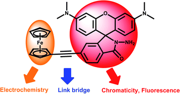
A novel multichannel chemosensor DR3 juxtaposed with a rhodamine chromophore and the electrochemical characterization of an ethynylferrocene group was developed. This chemosensor could selectively recognized Cu2+ in the presence of other competing ions in a wide pH range, which exhibits multiple responses for UV/vis absorption, fluorescence emission, and electrochemical parameters.


Introduction

Recently, due to its innate advantages such as high sensitivity, selectivity and real-time monitoring, luminescent chemosensors for the detection of transition metals have attracted increasing attention. These usually contain reaction sites and display an obvious change in optical characteristics upon host–guest interactions.1 Among the transition metals, copper is the third most abundant essential heavy metal in the human body, after zinc and iron, and plays a crucial role in biological processes. It is an important catalytic cofactor in redox chemistry for proteins.2 However, an abnormal level of Cu2+ in living systems may lead to various neurodegenerative diseases including Menkes, Wilson’s, and Alzheimer’s diseases.3–5 Thus, it is of significant importance to develop a luminescent chemosensor for monitoring Cu2+ in biological processes. As far as we know, a large number of Cu2+-chemosensors based on fluorescence enhancement have been reported.6–19 Most of them are based on the changes of UV/vis absorption and fluorescence emission. However, few multichannel Cu2+-selective chemosensors have been developed so far through multiple responses such as chromaticity, fluorescence, electrochemistry etc. Compared with single signal detection, multichannel detection has a higher sensitivity, more excellent selectivity and anti-interference ability. More importantly, a multichannel detection system can make self-calibration measurements come true through different analytic methods.

Zade et al. reported a thiophene-based salphen-type chemosensor for the detection of Cu2+ and Zn2+ with electrochemical properties.20 It is expected to introduce an excellent group to multichannel chemosensors, which have electrochemical characteristics, to establish multichannel analytic systems. Ferrocene has a brilliant ability to store electrons, strengthening the coordination between the chemosensor and the metal ion.21 It is well known that ferrocene-containing chemosensors exhibit an electrochemical response upon complexation of a suitable guest ion. Zhang and Zapata et al. also observed a significant potential shift of FeIII/FeII upon coordination of an analyte, which exhibits multi-responsive signaling.22 A further study relying on rhodamine-based multichannel chemosensors for Cr3+, Hg2+ etc. linked with ferrocene has been reported.23 However, multichannel Cu2+-chemosensors have barely been mentioned to the best of our knowledge.24

Herein, we synthesized a novel multichannel chemosensor DR3 for the detection of Cu2+ (Scheme 1). It is expected to achieve a three-channel Cu2+-selective chemosensor through introducing a ferrocene group to the rhodamine chromophore. We designed an acetylene group linked with the electrochemical properties of a ferrocene group and chromatic, fluorescent rhodamine with a ring-opening process, increasing the multichannel output signaling upon interaction with Cu2+. A hydrazide functional group was introduced into the chemosensor to act as a potential reaction site for Cu2+. Upon reaction with the copper ion, the fluorescence and absorption intensity evidently increases due to a process of spirolactam ring-opening and hydrolysis. This multichannel chemosensor DR3 exhibits suitable variations of the absorption spectrum, fluorescence emission and electrochemical parameters.


image file: c5ra00152h-s1.tif
Scheme 1 Design of Cu2+ multichannel chemosensor DR3.

Experimental section

Materials and measurements

All reagents were purchased from commercial suppliers and used without further purification. The 1H and 13C NMR spectra were measured on a Bruker Avance 500 or Brucker Avance 400 spectrometer in CDCl3. Electrospray ionization mass spectra (ESI-MS) were measured on a Micromass LCTTM system. UV-visible spectra were recorded on a Perkin-Elmer 35 spectrometer and fluorescence measurements were performed on a Perkin-Elmer LS 50B fluorescence spectrophotometer. Electrochemical measurements were performed with an Eco Chemie Autolab. The pH measurements were made with a PHS-3C Precision Ph/mV meter. TLC analysis was conducted on silica gel plates and chromatography was performed over silica gel (mesh 300–400).

UV/vis and fluorescence experiments

A stock solution of 1.0 mM DR3 was prepared in acetonitrile. Cu(NO3)2·3H2O was dissolved in doubly distilled water to form a 5.0 mM stock solution. For competing metal ions, various metal ion solutions of NaNO3, Co(NO3)2, KNO3, Zn(NO3)2·6H2O, Ni(NO3)2·6H2O, Mg(NO3)2·6H2O, Ca(NO3)2·4H2O, MnCl2·4H2O, Pb(NO3)2, HgCl2, AgNO3, Ba(NO3)2 were used. Before fluorescence and UV/vis titration investigations were conducted, the stock solution of DR3 was mixed with the stock solutions of metal salts in a 10 mL volumetric flask and diluted with H2O and CH3CN to volume. Spectral data were recorded at 2 min after the addition. For fluorescence measurements, excitation was provided at 530 nm, and emission was collected from 546 to 700 nm. The wide pH was adjusted by HCl or NaOH solutions.

Electrochemical test

Electrochemical characteristics were tested as previously reported in the literature.23 All measurements for DPV were carried out in a one-compartment cell under N2 gas, equipped with a glassy-carbon working electrode, a platinum wire counter electrode, and a silver reference electrode. All measurements for CV were carried out in a one-compartment cell under N2 gas, equipped with a glassy-carbon working electrode, a platinum wire counter electrode, and a saturated calomel electrode reference electrode. The supported electrolyte was a 0.10 mol L−1 CH3CN solution of tetrabutylammonium hexafluorophosphate (Bu4NPF6). The scan rate was 100 mV s−1.

Synthesis of compound DR1

Compound DR1 was synthesized as previously reported in the literature.9 JR1: 1H NMR (400 MHz, CDCl3) δ: 7.85 (d, J = 8.1 Hz, 1H, Ar–H), 7.70 (dd, J = 8.1, 1.6 Hz, 1H, Ar–H), 7.32 (d, J = 1.5 Hz, H, Ar–H), 6.66–6.57 (m, 2H, Ar–H), 6.48 (d, J = 2.5 Hz, 2H, Ar–H), 6.41 (dd, J = 8.8, 2.6 Hz, 2H, Ar–H), 2.99 (s, 12H, –CH3); DR1: 1H NMR (400 MHz, CDCl3) δ: 8.12 (d, J = 1.7 Hz, 1H, Ar–H), 7.83–7.66 (m, 1H, Ar–H), 7.05 (t, J = 8.8 Hz, 1H, Ar–H), 6.67–6.55 (m, 2H, Ar–H), 6.48 (d, J = 2.5 Hz, 2H, Ar–H), 6.41 (dd, J = 8.9, 2.6 Hz, 2H, Ar–H), 2.99 (s, 12H, –CH3).

Synthesis of compound DR2

Compound DR2 was synthesized according to the literature with some modifications.9 A mixture of DR1 (0.5 mmol, 233 mg), ethynylferrocene (0.5 mmol, 157 mg), 35 mg (0.05 mmol) of PdCl2(PPh3)2, and PPh3 (26 mg, 0.1 mmol), 4.8 mg (0.025 mmol) of CuI, THF (20 mL), NEt3 (5 mL) under nitrogen, was heated to 95 °C and refluxed for 12 h after completion of the reaction by TLC. The solvent was evaporated and the crude product was purified by column chromatography with CH2Cl2/NEt3 (v/v = 200/4) to afford a purplish red target product DR2 (247 mg, 83%). 1H NMR (400 MHz, CDCl3) δ: 8.08 (d, J = 0.7 Hz, 1H, Ar–H), 7.75–7.66 (m, 1H, Ar–H), 7.13 (d, J = 7.9 Hz, 1H, Ar–H), 6.63 (d, J = 8.8 Hz, 2H, Ar–H), 6.48 (d, J = 2.5 Hz, 2H), 6.40 (dd, J = 8.9, 2.6 Hz, 2H, Ar–H), 4.55 (t, J = 1.8 Hz, 2H, ferrocene-H), 4.30–4.28 (m, 2H, ferrocene-H), 4.27 (d, J = 4.5 Hz, 5H, ferrocene-H), 2.99 (s, 12H, –CH3); 13C NMR (101 MHz, CDCl3): 169.19, 152.91, 152.09, 151.91, 137.38, 128.75, 127.93, 127.34, 125.55, 124.08, 108.66, 106.40, 98.53, 90.75, 84.31, 71.62, 70.08, 69.19, 64.30, 46.15, 40.26. ESI-MS m/z 595.2 [M]+.

Synthesis of compound DR3

A solution of DR2 (0.25 mmol, 160 mg), excess 98% N2H4·H2O (1 mL) was resolved in 10 mL of ethanol and refluxed for 6 h. The solvent was evaporated and the crude product was purified by column chromatography with EtOAc/PE (v/v = 3/2) to get the desired product DR3 (110 mg, 73%). 1H NMR (400 MHz, CDCl3) δ: 8.06 (s, 1H, Ar–H), 7.55 (dd, J = 7.9, 1.4 Hz, 1H, Ar–H), 7.03 (d, J = 7.9 Hz, 1H, Ar–H), 6.52 (d, J = 8.8 Hz, 2H, Ar–H), 6.48 (d, J = 2.3 Hz, 2H, Ar–H), 6.39–6.36 (m, 2H, Ar–H), 4.52 (t, J = 1.7 Hz, 2H, ferrocene-H), 4.26 (s, 2H, ferrocene-H), 4.25 (s, 5H, ferrocene-H), 3.63 (s, 2H, –NH2), 2.97 (s, 12H, –CH3); 13C NMR (101 MHz, CDCl3): 165.56, 153.48, 151.55, 150.33, 135.50, 130.21, 127.94, 125.94, 124.30, 123.79, 108.81, 105.22, 99.01, 89.69, 84.90, 71.55, 70.05, 69.35, 69.03, 65.89, 40.31. ESI-MS m/z 609.2 [M + H]+.

Results and discussion

DR3 was prepared based on a two-step route, shown in Scheme 2. Firstly, compound DR2 was achieved in 83% yield by a Sonogashira reaction catalysed by PdCl2(PPh3)2, PPh3, and CuI. Then compound DR2 was reacted with hydrazine hydrate to obtain DR3 in 73% yield by refluxing for 6 h. These compounds were characterized and confirmed by 1H NMR, 13C NMR and ESI-MS (ESI, Fig. S1–S6).
image file: c5ra00152h-s2.tif
Scheme 2 Synthesis of DR3.

The influence of pH on the fluorescence of DR3 was investigated first, as shown in Fig. 1a. Under acidic conditions (pH < 5), ring opening of rhodamine occurred due to the protonation of the chemosensor.25 When the pH ranged from 5.0 to 9.0, no obvious characteristic fluorescence emission of rhodamine was observed. However, the addition of Cu2+ led to a fluorescence enhancement over a comparative range (5.0–9.0), which is attributed to the ring-opening process of rhodamine and hydrolysis. The multichannel chemosensors DR3 and DR3–Cu2+ remained unaffected in terms of fluorescence intensity in pH 6.0–8.0, suggesting that it was insensitive to pH around 7.0 and could be suitable for physiological conditions. These results were also the same for the UV/vis spectra in the range of pH 5–9, as illustrated in Fig. 1b.


image file: c5ra00152h-f1.tif
Fig. 1 (a) The fluorescence response of DR3 (10 μm) at 585 nm before and after the addition of 10 equiv. Cu2+ at different pH conditions. (b) The absorbance response of DR3 (10 μm) at 555 nm before and after the addition of 10 equiv. Cu2+ at different pH conditions.

The UV/vis spectrum of DR3 in a solvent system of CH3CN/HEPES 80[thin space (1/6-em)]:[thin space (1/6-em)]20 with titration of Cu2+ was shown in Fig. 2a. It exhibits almost no absorption in the visible wavelength range, indicating that chemosensor DR3 is predominantly in the form of spirolactam. Upon addition of Cu2+, a new absorption peak at 555 nm was observed, and the intensity gradually increased with increasing Cu2+ ion concentration. This could be attributed to the formation of a ring-opened amide form of DR3 upon interaction with Cu2+. The inset in Fig. 2a shows the absorbance at 555 nm as a function of Cu2+ concentrations. The ring-opening mechanism was also confirmed by the color change of 10 μM DR3 upon adding different cations (Fig. 2b). Among the metal ions, only Cu2+ can induce an obvious color change from colorless to pink in a solution of DR3, allowing colorimetric detection of Cu2+ by the naked eye. Fig. 2c shows the absorption response of DR3 towards various metal ions in HEPES/CH3CN (2[thin space (1/6-em)]:[thin space (1/6-em)]8, v/v, pH 7.0) solutions. DR3 exhibits almost no absorbance enhancement at around 555 nm upon addition of Hg2+, Pb2+, Ba2+, Mn2+, Ca2+, Mg2+, Ni2+, Ag+, Zn2+, K+, Cr3+, Co2+, Na+, DR3. These results indicate that chemosensor DR3 has a high selectivity towards Cu2+.


image file: c5ra00152h-f2.tif
Fig. 2 (a) Absorption spectra of DR3 (10 μM) in HEPES/CH3CN (2[thin space (1/6-em)]:[thin space (1/6-em)]8, v/v, pH 7.0) solutions upon addition of increasing concentrations of 0–8 equiv. Cu(NO3)2·3H2O. Inset: the absorbance at 555 nm as a function of Cu2+ concentrations. (b) Color changes upon addition of different cations in HEPES/CH3CN (2[thin space (1/6-em)]:[thin space (1/6-em)]8, v/v, pH 7.0) solutions. From left to right: DR3, Cu2+, Hg2+, Pb2+, Ba2+, Mn2+, Ca2+, Mg2+, Ni2+, Ag+, Zn2+, K+, Cr3+, Co2+ and Na+. (c) Absorption spectra of DR3 in HEPES/CH3CN (2[thin space (1/6-em)]:[thin space (1/6-em)]8, v/v, pH 7.0) solutions with 8 equiv. of metal ions: DR3, Cu2+, Hg2+, Pb2+, Ba2+, Mn2+, Ca2+, Mg2+, Ni2+, Ag+, Zn2+, K+, Cr3+, Co2+ and Na+.

Fig. 3a shows the fluorescence spectra of DR3 in HEPES/CH3CN (2[thin space (1/6-em)]:[thin space (1/6-em)]8, v/v, pH 7.0) solutions with the addition of Cu2+. Chemosensor DR3 shows a very weak emission at around 585 nm upon excitation at 530 nm. When Cu2+ was added to the DR3 buffer solution, a fluorescence intensity at 585 nm was observed, attributed to the ring-opening process of rhodamine derivatives. The solution showed an approximately 274-fold enhancement in the fluorescence intensity. This fact means that DR3 could act as an off-on luminescence chemosensor for Cu2+. The fluorescence intensity finally levelled off until the amount of added Cu2+ was 2.2 × 10⁻⁴ M (Fig. 3a inset). As an excellent luminescence chemosensor, it is important to have high selectivity. As illustrated in Fig. 3c, additions of other metal ions including Hg2+, Pb2+, Ba2+, Mn2+, Ca2+, Mg2+, Ni2+, Ag+, Zn2+, K+, Cr3+, Co2+, Na+ and DR3 induced no obvious fluorescence enhancement under the same conditions. These observations indicated that chemosensor DR3 could selectively recognize Cu2+ in a HEPES/CH3CN (2[thin space (1/6-em)]:[thin space (1/6-em)]8, v/v, pH 7.0) solution. For practical applications, the detection limit was also calculated as 6.85 × 10⁻⁶ M for Cu2+ (3σ/slope), which is sufficiently low for the detection of many chemical systems for Cu2+ (Fig. 3b).26


image file: c5ra00152h-f3.tif
Fig. 3 (a) Fluorescence spectra of DR3 in HEPES/CH3CN (2[thin space (1/6-em)]:[thin space (1/6-em)]8, v/v, pH 7.0) solutions upon addition of increasing concentrations of 0–24 equiv. Cu2+. Inset: fluorescence intensity at 585 nm as a function of Cu2+ concentrations. (b) Fluorescence intensity at 585 nm of DR3 (10 μM) in HEPES/CH3CN (2[thin space (1/6-em)]:[thin space (1/6-em)]8, v/v, pH 7.0) solutions as a function of Cu2+ concentration (0–3.5 μM). (c) Fluorescence intensity of DR3 (10 μm) in HEPES/CH3CN (2[thin space (1/6-em)]:[thin space (1/6-em)]8, v/v, pH 7.0) solutions upon addition of 24 equiv. metal ions (blank, Cu2+, Hg2+, Pb2+, Ba2+, Mn2+, Ca2+, Mg2+, Ni2+, Ag+, Zn2+, K+, Cr3+, Co2+, Na+).

As designed, DR3 shows an evident change in its reversible ferrocene/ferricinium redox cycles upon complexation. Differential pulse voltammetry (DPV) curves of DR3 were recorded in a CH3CN solution containing 0.1 M n-tetrabutylammonium hexafluorophosphate (n-Bu4NPF6) as a supporting electrolyte in the absence and presence of Cu2+.23a As shown in Fig. 4a, a significant displacement was observed upon addition of Cu2+. The oxidation peak was shifted in CH3CN from 0.728 to 0.54 V (ΔE1/2 = 188 mV). For cyclic voltammetry (CV), a significant shift of the redox potential of the ferrocenyl group was also observed (Fig. 4b). The CV behavior of chemosensor DR3 was measured in CH3CN, suggesting a reversible one-electron redox process. The addition of Cu2+ induces a positive shift of the ferrocene/ferricinium couple, which is attributed to the redox process of Cu2+. This fact indicated that DR3 could be a multi-signal chemosensor for Cu2+ for electrochemical measurements.


image file: c5ra00152h-f4.tif
Fig. 4 (a) DPV of DR3 (100 μM) in a CH3CN solution in the absence and presence of 1.6 equiv. of Cu2+, with n-Bu4NPF6 as a supporting electrolyte. (b) CV of DR3 (100 μM) in a CH3CN solution in the absence and presence of Cu2+, with n-Bu4NPF6 as a supporting electrolyte.

In order to explore the mechanism, the reaction product of DR3 and Cu2+ was detected by ESI spectra analyses (ESI, Fig. S7). The ion peak was detected at m/z 609.2, which corresponded to [DR3 + H]+. In addition, the main ion peak at m/z 595.2, corresponding to intermediate DR2, was detected after the addition of Cu2+ to a DR3 aqueous solution. This suggested that Cu2+ induces the hydrolysis and spirolactam ring-opening of rhodamine. To further confirm Cu2+ mediated hydrolysis, we carried out a chemical reversibility experiment in a CH3CN–water solution. Upon addition of 500 μM chelating agent, EDTA, to the DR3–Cu2+ complex in HEPES/CH3CN (2[thin space (1/6-em)]:[thin space (1/6-em)]8, v/v, pH 7.0) solutions, the color and fluorescence intensity showed no obvious changes (Fig. 5). These findings indicated that DR3 is an irreversible luminescence chemosensor for Cu2+. According to the obtained results, we proposed that the reaction process may proceed as per the route depicted in Scheme 3.


image file: c5ra00152h-f5.tif
Fig. 5 Fluorescence spectra in HEPES/CH3CN (2[thin space (1/6-em)]:[thin space (1/6-em)]8, v/v, pH 7.0) solutions: (a) DR3 (10 μM); (b) DR3 (10 μM) with Cu2+ (240 μM); (c) DR3 (10 μM) with Cu2+ (240 μM) and EDTA (500 μM).

image file: c5ra00152h-s3.tif
Scheme 3 The proposed reaction mechanism for DR3 with Cu2+.

Conclusions

In summary, we have designed a multichannel chemosensor DR3 for Cu2+ with the chromatic, fluorescent rhodamine derivatives and the electrochemical characterization of a ferrocenyl group. The novel chemosensor exhibits a multi-responsive colorimetric, fluorescent and electrochemical detection of Cu2+. Multichannel chemosensor DR3 could detect micromole levels of Cu2+ in a wide pH range (5.0–9.0). From the results above, we believe that DR3 could be further used for monitoring intracellular Cu2+ ions in biological systems.

Acknowledgements

The authors are grateful for the financial support from the open fund of the State Key Laboratory of Materials-Oriented Chemical Engineering (KL13-07), the China Postdoctoral Science Foundation (2014M550287), Specialized Research Fund for the Doctoral Program of Higher Education (20123221110012), Jiangsu Planned Projects for Postdoctoral Research Funds, and the State Key Laboratory of Coordination Chemistry of Nanjing University.

References

  1. Y. Yang, Q. Zhao, W. Feng and F. Y. Li, Chem. Rev., 2013, 113, 192 CrossRef CAS PubMed.
  2. H. Tapiero, D. M. Townsend and K. D. Tew, Biomed. Pharmacother., 2003, 57, 386 CrossRef CAS.
  3. D. J. Waggoner, T. B. Bartnikas and J. D. Gitlin, Neurobiol. Dis., 1999, 6, 221 CrossRef CAS PubMed.
  4. C. Vulpe, B. Levinson, S. Whitney, S. Packman and J. Gitschier, Nat. Genet., 1993, 3, 7 CrossRef CAS PubMed.
  5. K. J. Barnham, C. L. Masters and A. I. Bush, Nat. Rev. Drug Discovery, 2004, 3, 205 CrossRef CAS PubMed.
  6. C. W. Yu, W. Ting, K. Xu, J. Zhao, M. H. Li, S. X. Weng and J. Zhang, Dyes Pigm., 2013, 96, 38 CrossRef CAS PubMed.
  7. A. F. Liu, L. Yang, Z. Y. Zhang, Z. L. Zhang and D. M. Xu, Dyes Pigm., 2013, 99, 472 CrossRef CAS PubMed.
  8. S. L. Hu, S. S. Zhang, Y. Hu, Q. Tao and A. X. Wu, Dyes Pigm., 2013, 96, 509 CrossRef CAS PubMed.
  9. J. L. Fan, P. Zhan, M. M. Hu, W. Sun, J. Z. Tang, J. Y. Wang, S. G. Sun, F. L. Song and X. J. Peng, Org. Lett., 2013, 15, 492 CrossRef CAS PubMed.
  10. M. Kumar, N. Kumar, V. Bhalla, P. R. Sharma and T. Kaur, Org. Lett., 2012, 14, 406 CrossRef CAS PubMed.
  11. Z. Q. Hua, X. M. Wang, Y. C. Feng, L. Ding and H. Y. Lu, Dyes Pigm., 2011, 88, 257 CrossRef PubMed.
  12. J. L. Liu, C. Y. Li and F. Y. Li, J. Mater. Chem., 2011, 21, 7175 RSC.
  13. P. H. Xie, F. Q. Guo, D. Li, X. Y. Liu and L. Liu, J. Lumin., 2011, 131, 104 CrossRef CAS PubMed.
  14. D. P. Wang, Y. Shiraishi and T. Hirai, Chem. Commun., 2011, 47, 2673 RSC.
  15. W. Y. Liu, H. Y. Li, B. X. Zhao and J. Y. Miao, Org. Biomol. Chem., 2011, 9, 4802 CAS.
  16. C. W. Yu, J. Zhang, R. Wang and L. G. Chen, Org. Biomol. Chem., 2010, 8, 5277 CAS.
  17. V. Dujols, F. Ford and A. W. Czarnik, J. Am. Chem. Soc., 1997, 119, 7386 CrossRef CAS.
  18. J. T. Yeh, W. C. Chen, S. R. Liu and S. P. Wu, New J. Chem., 2014, 38, 4434 RSC.
  19. X. M. Wu, Z. Q. Guo, Y. Z. Wu, S. Q. Zhu, T. D. James and W. H. Zhu, ACS Appl. Mater. Interfaces, 2013, 5, 12215 CAS.
  20. A. K. Asatkar, S. P. Senanayak, A. Bedi, S. Panda, K. S. Narayan and S. S. Zade, Chem. Commun., 2014, 50, 7036 RSC.
  21. (a) A. P. D. Silva, H. Q. N. Gunaratne, T. Gunnlaugsson, T. J. M. Huxley, C. P. Mccoy, J. T. Rademacher and T. E. Rice, Chem. Rev., 1997, 97, 1515 CrossRef PubMed; (b) W. H. Zhu, L. W. Song, Y. H. Yang and H. Tian, Chem. - Eur. J., 2012, 18, 13388 CrossRef CAS PubMed; (c) M. Li, Z. Q. Guo, W. H. Zhu, F. Marken and T. D. James, Chem. Commun., 2015, 51, 1293 RSC.
  22. (a) B. C. Zhu, C. C. Gao, Y. Z. Zhao, C. Y. Liu, Y. M. Li, Q. Wei, Z. M. Ma, B. Du and X. L. Zhang, Chem. Commun., 2011, 47, 8656 RSC; (b) F. Zapata, A. Caballero, A. Espinosa, A. Tarraga and P. Molina, Org. Lett., 2007, 9, 2385 CrossRef CAS PubMed.
  23. (a) K. W. Huang, H. Yang, Z. G. Zhou, M. X. Yu, F. Y. Li, X. Gao, T. Yi and C. H. Huang, Org. Lett., 2008, 10, 2557 CrossRef CAS PubMed; (b) X. Q. Chen, S. W. Nam, M. J. Jou, Y. M. Kim, S. J. Kim, S. S. Park and J. Y. Yoon, Org. Lett., 2008, 10, 5235 CrossRef CAS PubMed; (c) H. Yang, Z. G. Zhou, K. W. Huang, M. X. Yu, F. Y. Li, T. Yi and C. H. Huang, Org. Lett., 2007, 9, 4729 CrossRef CAS PubMed.
  24. Y. S. Mi, Z. Cao, Y. T. Chen, Q. F. Xie, Y. Y. Xu, Y. F. Luo, J. J. Shi and J. N. Xiang, Analyst, 2013, 138, 5274 RSC.
  25. Y. Xiang and A. J. Tong, Org. Lett., 2006, 8, 1549 CrossRef CAS PubMed.
  26. D. N. Beatrice, M. Jerome, L. Dominique and F. F. Suzanne, Inorg. Chem., 2006, 45, 5691 CrossRef PubMed.

Footnote

Electronic supplementary information (ESI) available. See DOI: 10.1039/c5ra00152h

This journal is © The Royal Society of Chemistry 2015
Click here to see how this site uses Cookies. View our privacy policy here.