Synthesis of cadmium oxide and carbon nanotube based nanocomposites and their use as a sensing interface for xanthine detection

U. Jaina, J. Narangb, K. Ranic, Burnab, Sunnyb and N. Chauhan*b
aDepartment of Microbiology and Cell Biology, Indian Institute of Science, Bangalore, India
bAmity Institute of Nanotechnology, Amity University, Noida-201303, Uttar Pradesh, India. E-mail: nidhichauhan2007@rediffmail.com; Tel: +91 8130615833
cAmity Institute of Biotechnology, Amity University, Noida-201303, Uttar Pradesh, India

Received 2nd January 2015 , Accepted 12th March 2015

First published on 12th March 2015


Abstract

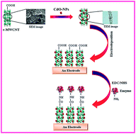
Xanthine oxidase (XOD) extracted from bovine milk was immobilized covalently via N-ethyl-N′-(3-dimethylaminopropyl)carbodiimide (EDC) and N-hydroxy succinimide (NHS) chemistry onto cadmium oxide nanoparticles (CdO)/carboxylated multiwalled carbon nanotube (c-MWCNT) composite film electrodeposited on the surface of an Au electrode. The nanocomposite modified Au electrode was characterized by Fourier transform infrared (FTIR), cyclic voltammetry (CV), scanning electron microscopy (SEM) and electrochemical impedance spectroscopy (EIS) before and after immobilization of XOD. Under optimal operation conditions (25 °C, +0.2 V vs. Ag/AgCl, sodium phosphate buffer, pH 7.5), the following characteristics are attributed to the biosensor: linearity of response up to xanthine concentrations of 120 μM, detection limit of 0.05 μM (S/N = 3) and a response time of at most 4 s. After being used 100 times over a period of 120 days, only 50% loss of the initial activity of the biosensor was evaluated when stored at 4 °C. The fabricated biosensor was successfully employed for the determination of xanthine in fish meat.


1. Introduction

Xanthine is a precursor of uric acid. Elevated levels of xanthine in blood and urine samples are indicative of pathological conditions such as xanthinuria, gout, or renal failure.1 A large number of mild xanthine derived stimulants, like caffeine and theobromine, are available in tea and coffee. Besides clinical diagnostics, xanthine is of great significance in the food industry. Fresh fish meat is required in food industries for the manufacturing of high quality products.2 ATP of dead fish is degraded into xanthine and increases during storage. Thus, xanthine attracts much attention as an indicator of fish freshness.3 Biochemical assays of xanthine are routinely performed, including enzymatic colorimetric,4 enzymatic fluorometric, fluorometric mass spectrometry fragmentography,5 HPLC6 and hair like segment gas chromatography.7 A common drawback of these methods is the painstaking sample preparation procedure, requiring special reagents and costly equipment.8 Amperometric biosensors may therefore afford a fast and cost-effective means of xanthine detection. In addition, the simplicity of operating with electrochemical signals makes them a promising alternative to the traditional analytical techniques.9,10

Integration of nanomaterials in biosensors can further enhance the properties of these biosensors. Nanomaterials, including nanoparticles, nanowires, nanotubes and nanochannels, have previously been incorporated in biosensing systems.11,12 Nanomaterials may generate novel interfaces that act as effective labels and amplify analysis. Furthermore, nanomaterials offer various advantages, including stability, durability, better sensitivity, accuracy, detection range and faster response times. Analysts have now focused on the use of cadmium oxide (CdO), particularly in the field of devices including solar cells, phototransistors and diodes, transparent terminals and gas sensors.13 CdO nanoparticles (CdO-NPs) of different morphologies were previously synthesized.14 CdO is known to possess high electrical conductivity, high carrier concentration and high transparency in the visible range of the electromagnetic spectrum. One important method is the formation of a thin CdO film by electrochemical deposition with specific composition, morphology and good adhesion between the deposited film and the substrate. CdO or oxyhydroxide layers on the electrode surface with excellent electrocatalytic activity are prepared by electrodeposition.15 The process makes it useful for a wide range of applications, such as biosensors and electrochemical devices.16,17

In the field of biosensing, multiwalled carbon nanotubes (MWCNTs) have received much attention because of their unique electronic, mechanical and structural characteristics. In addition, MWCNTs are promising for immobilization of proteins as they have a significant surface area, excellent electrical conductivity and good chemical stability.18,19 Together with metal nanoparticles, CNTs may serve as promising catalyst supports to form a composite exhibiting remarkable activity for small molecules.

The specific aim of this study is to detect xanthine by immobilizing commercial xanthine oxidase (XOD) on a CdO-NPs and MWCNT composite film electrodeposited onto an Au electrode, and its subsequent application. The electrochemical behavior of the CdO-NPs/MWCNT film was characterized by cyclic voltammetry and impedance spectroscopy. The analytical performance of the XOD/CdO-NPs/MWCNT modified electrode was then evaluated with respect to detection limit, linearity, stability and reproducibility. To our knowledge, it is the first demonstration of the combination of CdO-NPs and MWCNTs to achieve high sensitivity and selectivity for xanthine detection in fish.

2. Experimental methods

2.1. Chemicals and reagents

Xanthine oxidase (XOD) (E.C.1.1.3.2) from buttermilk (0.15 U mg−1), xanthine extrapure, N-ethyl-N′-(3-dimethylaminopropyl)carbodiimide (EDC), N-hydroxysuccinimide (NHS), Cd acetate and ammonia solution from SRL, Mumbai, India were used. Carboxylated multi-walled carbon nanotubes (functionalized MWCNT or c-MWCNT) (12 walls, length 15–30 μm, purity 90%, metal content: nil) from Intelligent Materials Pvt. Ltd., Panchkula (Haryana) India were used. Deionized water (DW) was used throughout the experiments. All other chemicals used were of analytical reagent grade.

2.2. Assay of XOD

XOD has previously been reported to catalyze the following reaction:
image file: c5ra00050e-t1.tif

The rate of formation of urate from xanthine is determined by measuring the increased absorbance at 290 nm due to uric acid. The assay of xanthine was carried out in a test tube as described by Bergmeyer et al., with modification and based on the oxidation of xanthine to uric acid by XOD.20 The assay mixture contained 2.8 mL of 50 mM sodium phosphate buffer (pH 7.0), 0.1 mL of xanthine (0.15 mM) and 0.1 mL of XOD (0.15 U mg−1). An increase in the absorbance at 290 nm was reported against a blank in a UV spectrophotometer. The activity was calculated as follows:

image file: c5ra00050e-t2.tif
where total vol. of the reaction mixture (mL) = 3 mL, extinction coeff. of uric acid = 1.22 × 104 cm−1, volume of enzyme used (mL) = 0.1 mL, and df = dilution factor. One unit will convert 1.0 nmol of xanthine to uric acid per min per mL at pH 7.5 at 25 °C.

2.3. Preparation of CdO-NPs

Chemical synthesis of CdO-NPs was performed in alcoholic media, such as ethanol, methanol or propanol. In alcoholic media, the growth of oxide particles is slow and controllable. Different solutions were prepared by dissolving 0.1 M of CdCl2 (20 mL), 0.1 M of NaOH (100 mL) and 10 mL of ethylene glycol with methanol (1[thin space (1/6-em)]:[thin space (1/6-em)]1). Ethylene glycol solution was added to the NaOH solution while it was continuously stirred. The resulting solution was stirred for one hour before adding CdCl2 solution to it. After three hours of constant stirring, a milky white solution was obtained. Size selective precipitation was carried out using acetone as a non-solvent. The precipitate was washed in methanol and was allowed to evaporate at room temperature to obtain cadmium hydroxide nanoparticles as a white powder. Cadmium hydroxide particles were then placed in the furnace and heated to 250 °C for five hours. After five hours, the CdO powder turns to a brown colour.21

2.4. Deposition of CdO-NPs onto c-MWCNTs

The deposition of CdO-NPs onto c-MWCNTs was carried out as described by Chauhan and Pundir in 2012 with little modification.22 First, 0.265 g of CdO-NPs was dispersed into 200 mL of DW by strong stirring. Then 1.0 mg of c-MWCNTs was added to the solution with ultrasonic treatment for about 10 min. Finally, 0.4 mol L−1 sodium hydroxide was added drop-wise into the solution until its pH reached 7.0. The CdO-NPs/c-MWCNTs solution was stirred for 30 min and desiccated at 50 °C for 2 h. Subsequently, this solution was centrifuged at 5000 × g for 15 min to separate the product from the mixture. Afterwards, the black coloured precipitates were washed with DW several times until the pH reached 7.0. The washed CdO-NPs/c-MWCNTs composites were treated with pure methanol and then desiccated at 50 °C for 6 h.

2.5. Construction of CdO-NPs/c-MWCNT modified Au electrode

A polycrystalline Au electrode (1.5 cm × 0.05 cm) was first polished with alumina slurry and then cleaned ultrasonically with ethanol and DW. It was then cleaned electrochemically by cycling the electrode potential between +1.6 and −0.4 V in 0.5 mol L−1 H2SO4 until a stable voltammogram was obtained. The electrodeposition of the CdO-NPs/c-MWCNTs nanocomposite onto the Au electrode was conducted in an electrochemical cell system using a potentiostat–galvanostat by immersing the Au electrode in 25 mL of solution containing 22 mL of electrolyte (2.5 mmol L−1 K3Fe(CN)6/K4Fe(CN)6 [1[thin space (1/6-em)]:[thin space (1/6-em)]1]) and 3 mL of nanomaterial, then applying 20 cycles of −0.2 to +0.6 V at a scan rate of 50 mV s−1. The resulting CdO-NPs/c-MWCNTs/Au electrode was washed thoroughly with DW to remove any unbound nanomaterial and stored in a dry Petri dish at 4 °C.

2.6. Immobilization of XOD enzyme onto CdO-NPs/c-MWCNT composite film coated Au electrode

The enzyme XOD was immobilized covalently onto the CdO-NPs/c-MWCNT composite film coated Au electrode using EDC and NHS chemistry, as described by Rahman et al. with modifications.23 First, free and unbound –COOH groups of the CdO-NPs/c-MWCNT composite film were activated by immersing them into 0.1 M sodium phosphate buffer, pH 7.5, containing EDC and NHS at the same concentration (10 mM) for 6 h, and then the excess EDC and NHS were removed by washing with 0.05 M sodium phosphate buffer (pH 7.4). Finally, the EDC–NHS-treated electrode was incubated in 5 mL of 0.05 M sodium phosphate buffer (pH 7.4) containing XOD (24 U) at 4 °C for 3 h and then washed with 0.05 M sodium phosphate buffer (pH 7.4). The resulting enzyme electrode was dried and stored in a refrigerator at 4 °C.

2.7. Cyclic voltammetric measurement and testing of xanthine biosensor

The cyclic voltammogram (CV) of XOD/CdO-NPs/c-MWCNT/Au electrode (working electrode) was recorded using a potentiostat–galvanostat from −0.4 to +0.4 V vs. Ag/AgCl as reference and Pt wire as the counter electrode in a 15 mL (0.05 M) sodium phosphate buffer (pH 7.5) containing 0.15 mM xanthine (0.5 mL). The maximum response was observed at +0.2 V and hence subsequent amperometric studies were carried out at this voltage. For further amperometric studies, the modified electrode (XOD/CdO-NPs/c-MWCNT/Au electrode) together with the reference electrode and the counter electrode were immersed in 25 mL of 0.05 M sodium phosphate buffer (pH 7.4) in a 50 mL beaker. The reaction was started by adding xanthine solutions of different concentrations to the reaction mixture and the current (mA) generated was recorded at +0.2 V.

2.8. Optimization of xanthine biosensor

The experimental conditions affecting the biosensor response were studied for effect of pH, incubation temperature, response time and substrate (xanthine) concentration. To determine the optimum pH, the pH was varied from 3.0 to 8.0 at intervals of 0.5. Similarly, the optimum temperature was studied by incubating the reaction mixture at different temperatures (20–50 °C) in increments of 5 °C and for different lengths of time (1–10 s) at an interval of 1 s. The effect of xanthine concentration on the biosensor response was determined by varying the concentration of xanthine from 0.05 μM to 120 μM at an interval of 20 μM.

2.9. Evaluation

The performance of this biosensor was evaluated for linearity, detection limit, analytical recovery, repeatability (precision), reproducibility and storage stability.

2.10. Amperometric determination of xanthine in fish meat

Labeo fish purchased from a local market was chopped and homogenized in 5 mL of 0.5 M HClO4 until a fine paste was obtained for the precipitation of proteins in the sample. The denatured samples were mechanically stirred for 10 min and then centrifuged at 4000 rpm for 5 min. The supernatant was collected and its pH was adjusted to 7.0 with NaOH. The supernatant (fish meat extract) was diluted 10 times. These denatured sample solutions were then divided into two parts. One part was used immediately and the other was stored at room temperature. To determine the xanthine content in the fish meat extract, the same procedure was used as that described for sensor response measurement under the optimal working conditions, except that xanthine was replaced by the sample. The xanthine content was interpolated from the calibration curve between xanthine concentrations vs. current (mA) prepared under optimal working conditions.

3. Results and discussion

A simple and efficient chemical method was introduced for the preparation of a CdO-NPs/c-MWCNT nanocomposite onto an Au electrode. Scheme 1 illustrates the CdO-NPs coated on the outer walls of the c-MWCNTs with other nanoparticles.22 Carboxyl groups present on the external surface of the MWCNTs are available to bind with –NH2 groups on the surface of XOD after the decoration of CdO-NPs on the surface of the c-MWCNT. XOD was covalently immobilized on the CdO-NPs/c-MWCNT composite film using EDC–NHS chemistry. EDC–NHS was used to activate the free –COOH groups present on the CdO-NPs/c-MWCNT composite film.
image file: c5ra00050e-s1.tif
Scheme 1 Schematic illustration of the stepwise amperometric xanthine biosensor fabrication process and immobilized XOD.

3.1. Characterization of CdO-NPs

The DLS of CdO-NPs is shown in Fig. 1. The results of the dynamic light scattering showed the size of the CdO-NPs (25 nm).
image file: c5ra00050e-f1.tif
Fig. 1 DLS graph; a typical intensity vs. size plot of CdO-NPs of size 25 nm in water.

The XRD patterns of the CdO nanostructure showed diffraction peaks absorbed at 2θ values (Fig. 2). The grain size of 25 nm was estimated by using the relative intensity peak (500) for CdO nanoparticles. The increase in the sharpness of the XRD peaks indicates that the particles are crystalline in nature. The reflections (111, 200, 220, 311 and 222) are clearly seen and closely matched the reference patterns for CdO (Joint Committee for Powder Diffraction Studies (JCPDS) File no. 05-0640). The sharp XRD peaks indicate that the particles were of a polycrystalline structure and that the nanostructure was grown with a random orientation.24


image file: c5ra00050e-f2.tif
Fig. 2 XRD pattern for CdO-NPs.

The TEM image of the CdO-NPs (Fig. 3A) corresponding to the same sample of DLS is shown in Fig. 1 and the XRD pattern is shown in Fig. 2. The particle size distribution is shown in Fig. 3B. These particles appeared as a single crystalline revealed by the high resolution electron microscope image (Fig. 3A). The particles are spherical or elliptical in shape. The histogram shows that the particle sizes range from 10 to 40 nm and possess an average size of 25 ± 1.5 nm (Fig. 3B).


image file: c5ra00050e-f3.tif
Fig. 3 TEM image of CdO-NPs of an average diameter of 25 nm (A). Particle size distribution histogram of CdO-NPs (B).

3.2. Surface characterization by SEM

The surface morphologies of the Au electrode, CdO-NPs/c-MWCNT/Au electrode and XOD/CdO-NPs/c-MWCNT/Au electrode were studied by SEM (Fig. 4). The bare Au electrode (Fig. 4a) showed a homogenous surface whereas the CdO-NPs/c-MWCNT/Au electrode has a uniform granular porous morphology, attributed to the homogeneous dispersion of CdO-NPs in the MWCNT network (Fig. 4b). After immobilization of XOD, the globular structural morphology of CdO-NPs/c-MWCNT/Au was changed into the regular form via covalent bonding between CdO-NPs/c-MWCNT and XOD (Fig. 4c).
image file: c5ra00050e-f4.tif
Fig. 4 Scanning electron micrographs of: (a) bare Au electrode, (b) CdO-NPs/c-MWCNT/Au electrode and (c) XOD/CdO-NPs/c-MWCNT/Au electrode.

3.3. FTIR spectra

The FTIR spectra of the c-MWCNT/Au electrode and the enzyme/CdO-NPs/c-MWCNT/Au electrode are shown in Fig. 5. In spectrum (i), the characteristic absorption peaks of c-MWCNTs were observed near 1518, 1156 and 2320 cm−1, which originated from the graphitic component of c-MWCNTs.25,26 A sharp absorption peak was observed at 1640 cm−1 because of the attachment of the carboxyl group onto the surface of the MWCNTs. The carboxyl groups attached to the MWCNTs were partly replaced by CdO-NPs (spectrum (i)). In the FTIR spectrum of the enzyme/CdO-NPs/c-MWCNT/Au electrode (spectrum (ii)), XOD binding is indicated by the appearance of additional absorption bands at 1641 and 1539 cm−1 assigned to the carbonyl stretch (amide I band) and –N–H bending (amide II band). As seen in Fig. 5 (spectrum (ii)), the presence of the 1637 cm−1 peak corresponding to the amide bands was confirmed with the attachment of XOD to CdO-NPs/c-MWCNT via carbodiimide chemistry.
image file: c5ra00050e-f5.tif
Fig. 5 FTIR spectra in KBr monitored in the region between 4000 cm−1 and 500 cm−1; (i) c-MWCNT/Au electrode and (ii) XOD/CdO-NPs/c-MWCNT/Au electrode.

3.4. Electrochemical impedance spectroscopic (EIS) studies

EIS studies provide useful information on impedance changes of the electrode surface during the fabrication process and were carried out to investigate immobilization of the enzyme onto the CdO-NPs/c-MWCNT/Au electrode. The diameter of the semicircle portion at higher frequencies in the Nyquist plot was equal to the charge transfer resistance (RCT), which controls the electron transfer kinetics of the redox probe at the electrode interface. The linear part at lower frequencies corresponding to the Warburg diffusion process is shown.27 The Nyquist plot (Fig. 6) displays the EIS studies of the bare Au electrode (curve a), the CdO-NPs/c-MWCNT/Au electrode (curve b) and the enzyme/CdO-NPs/c-MWCNT/Au electrode (curve c) in 0.05 M sodium phosphate buffer (pH 7.4) containing 5 mM K3Fe(CN)6/K4Fe(CN)6 (1[thin space (1/6-em)]:[thin space (1/6-em)]1) as a redox probe. The RCT values for the bare Au, CdO-NPs/c-MWCNT and XOD/CdO-NPs/c-MWCNT/Au electrodes were obtained as 196 Ω, 135 Ω and 180 Ω, respectively. The RCT of the CdO-NPs/c-MWCNT/Au electrode (curve b) was lower than the Au electrode (curve a), revealing its decreased resistance and high electron transfer efficiency. The CdO-NPs immobilized onto the c-MWCNTs were layered as a thin CdO film. This fact may suggest that the MWCNT has an obvious improvement effect, which makes the composites have more active sites for faradic reactions and a larger capacitance onto Au electrode. This results in enhanced conductivity, lowers the resistance and facilitates the charge-transfer of the composite. The RCT of the XOD/CdO-NPs/c-MWCNT/Au (curve c) bioelectrode was increased compared with the CdO-NPs/c-MWCNT/Au electrode. The increase in RCT is attributed to the fact that most biological molecules, including enzymes, are poor electrical conductors at low frequencies and hinder electron transfer. Furthermore, these results indicate the binding of XOD onto the CdO-NPs/c-MWCNT composite.
image file: c5ra00050e-f6.tif
Fig. 6 Nyquist plot of electrochemical impedance spectra for Au electrode (curve a), CdO-NPs/c-MWCNT/Au electrode (curve b) and XOD/CdO-NPs/c-MWCNT/Au electrode (curve c) in 0.05 M sodium phosphate buffer (pH 7.5) containing 5 mM K3Fe(CN)6/K4Fe(CN)6 (1[thin space (1/6-em)]:[thin space (1/6-em)]1) as a redox probe.

3.5. Cyclic voltammetry response of XOD/CdO-NPs/c-MWCNT modified Au electrode

A method is described for the construction of an electrochemical xanthine biosensor using XOD immobilized onto CdO-NPs/c-MWCNT composite film electrodeposited on the surface of an Au wire through electrodeposition. This construct was used as the working electrode along with Ag/AgCl as the reference electrode and Pt wire as the auxiliary electrode. The CdO-NPs/c-MWCNT/Au electrode potential peaks were not modified during cycling between −0.4 and +0.4 V. The structural properties of the deposited film were electrochemically stable. The same electrochemical behavior was observed for the composite material as for CdO-NPs and c-MWCNT.

In this improved amperometric biosensor, XOD was covalently immobilized on the CdO-NPs/c-MWCNT/Au electrode. During stepwise modification, the performance of the working Au electrode was evaluated by CV in 0.05 M phosphate buffer (pH 7.5). The CV of the Au electrode, the CdO-NPs/c-MWCNT/Au electrode and the XOD/CdO-NPs/c-MWCNT/Au electrode in the presence of 100 μL of xanthine (0.1 mM) in sodium phosphate buffer (pH 7.5) at a scan rate of 20 mV s−1 is shown in Fig. 7. No peak was observed for the Au electrode (curve a) in phosphate buffer after injecting 100 μL of xanthine into the reaction cell. The cyclic voltammogram of the CdO-NPs/c-MWCNT/Au electrode identified an oxidation peak at −290 mV (curve b). MWCNT is used as a good adsorbent for heavy metal ions and its solutions.28 This voltammogram also shows a single major reduction peak thereafter the current decreases according to diffused controlled process for cadmium reduction. Peaks are commonly associated with the reduction of Cd(II) to Cd(0), (Cd2+ + 2e → Cd0), as confirmed by other research.29,30 The current contribution of the cadmium dissolution peak was observed at a potential of about −0.290 mV (Fig. 7, curve b). This may be attributed to the dissolution ability of the deposited Cd atoms, which depends on the structure, grain size and porosity of the deposits. An oxidation peak at −0.200 mV (vs. Ag/AgCl) (curve c) was recorded by cyclic voltammetry of the XOD/CdO-NPs/c-MWCNT/Au electrode.


image file: c5ra00050e-f7.tif
Fig. 7 Cyclic voltammograms of: (a) bare Au electrode, (b) CdO-NPs/c-MWCNT/Au electrode, (c) XOD/CdO-NPs/c-MWCNT/Au electrode in pH 7.5 sodium phosphate containing 0.1 mM xanthine. Scan rate: 20 mV s−1.

The modification of the bare Au electrode with conductive CdO-NPs/MWCNTs resulted in a higher Ip and smaller ΔEp owing to an increase in the effective surface. The Ip of the XOD/CdO-NPs/MWCNT modified Au electrode was even higher, illustrating the oxidation of xanthine, which is catalyzed by the immobilized XOD on the CdO-NPs/MWCNT/Au surface.

3.6. Optimization of experimental conditions

Optimization studies were conducted using linear sweep voltammetry (LSV) on the effect of pH, incubation, temperature and response time. The effect of the pH value on the response current of the XOD/CdO-NPs/c-MWCNT/Au electrode was studied between 3.0 and 8.0 at an interval of 0.5. The response current was increased from 3.0 to 7.5 and thereafter decreased. The maximum current response was recorded at pH 7.5 (ESI Fig. 1A). Therefore, pH 7.5 was suitable for the maximum activity of immobilized XOD to soluble XOD.20 Additionally, in order to ensure optimization, the effect of temperature on the biosensor was studied. The response current reached a maximum at approximately 25 °C (ESI Fig. 1B) and then reduced with increasing temperature. The present biosensor showed the maximum response (comparatively faster) within 4 s (ESI Fig. 1C).

There was a linear relationship between biosensor response and xanthine concentration from 0.05 μM to 120 μM, although it was found to be constant after 120 μM (Fig. 8). There are many anodic and cathodic peaks associated with the figure due to the presence of electrolyte, i.e. K3Fe(CN)6/K4Fe(CN)6 (1[thin space (1/6-em)]:[thin space (1/6-em)]1), which acts as a redox probe. The current peaks are associated with desorption of ferri/ferro from the modified Au electrode surface. Some peaks are related to the reduction of oxygen and/or species. The inset in Fig. 8 displays the calibration curve (R2 = 0.9905) for the biosensor response to xanthine.


image file: c5ra00050e-f8.tif
Fig. 8 Effect of substrate concentrations on the response of the XOD/CdO-NPs/c-MWCNT/Au electrode in 0.05 M sodium phosphate buffer solution (pH 7.5) containing 5 mM K3Fe(CN)6/K4Fe(CN)6 (1[thin space (1/6-em)]:[thin space (1/6-em)]1) as a redox probe over a potential range of +0.4 to −0.4 V, scan rate 50 mV s−1 vs. Ag/AgCl reference electrode. The inset shows the calibration curve of xanthine concentration (μM) vs. current (mA).

3.7. Evaluation of xanthine biosensor

The following parameters were studied to evaluate the performance of this biosensor.
3.7.1. Linearity. There was a linear relationship between xanthine concentration ranging from 0.05 to 120 μM and current (A), which is better than the earlier biosensors based on a zinc oxide nanoparticle (ZnO-NPs)–polypyrrole (PPy) composite film modified Pt electrode (0.8 to 40 μM),2 a ZnO-NPs/chitosan/c-MWCNT/polyaniline modified Pt electrode (0.1 to 100 μM),31 an Au/polypyrrole (AuPPy) nanocomposites modified Pt electrode (0.4 to 100 μM),32 a double walled carbon nanotube (DWCNT) modified carbon paste electrode (2 to 50 μM),33 a CNT modified carbon paste electrode (1–100 μM),34 a chitosan bound gold coated iron nanoparticles (CHIT/Fe-NPs@Au)/PGE (0.1–300 μM)35 and a self-assembled biotinylated phospholipid membrane (20 to 100 μM).36
3.7.2. Detection limit. The detection limit of the biosensor was reported to be 0.05 μM (S/N = 3), which is lower than that of a ZnO-NPs–polypyrrole (PPy) composite film modified Pt electrode (0.8 μM),2 a ZnO-NPs/chitosan/c-MWCNT/polyaniline modified Pt electrode (0.1 μM),31 an Au/polypyrrole (AuPPy) nanocomposites modified Pt electrode (0.4 μM),32 an iron(III) mesotetraphenylporphyrin nanoparticles modified glassy carbon electrode (1 μM),37 a DWCNT modified carbon paste electrode (2 μM)33 and a CNT modified carbon paste electrode (1 μM).34
3.7.3. Analytical recovery. To study the analytical recovery of exogenously added xanthine, 0.1 mL of xanthine solutions (10 mg L−1, 20 mg L−l) in fish meat extract were added separately. The xanthine level in the fish meat extract was measured by the present method immediately before and after the addition of xanthine. The % recovery of the exogenously added xanthine was calculated by 95.6 ± 1.1% and 97.3 ± 1.7%, respectively (Table 1). The high analytical recovery shows the reliability of the method.
Table 1 Analytical recovery of exogenously added xanthine in fish meat samples, as measured by the XOD/CdO-NPs/c-MWCNT/Au electrode
Xanthine added (mg L−1) Xanthine found (mg L−1) %Recovery
22.21
10 31.77 95.6 ± 1.1
20 41.67 97.3 ± 1.7


3.7.4. Accuracy. The accuracy of present method was examined. The amounts of xanthine in fish samples (n = 12) were determined by enzymatic colorimetric method (x) and by the present method (y). The xanthine values obtained by the present biosensor were compared with enzymatic colorimetric method with good correlation (r = 0.98, significant at 1% level) (ESI Fig. 2). Comparatively, a higher detection limit was obtained by the present method than the colorimetric method. These results demonstrate the good accuracy of the described biosensor.
3.7.5. Precision study. In order to check the repeatability and reproducibility of the method, the xanthine content in the same fish sample was determined five times on a single day (within batch) and again after storage at −20 °C for one week (between batch). The results showed that the determinations were almost consistent. The coefficients of variation (CV) within and between the batch for fish sample determination were reported to be <4.3% and <5.16%, indicating the good reproducibility and reliability of the method.
3.7.6. Xanthine in fish meat. The xanthine concentration in fish meat was determined by the present biosensor at different storage times ranging from 1 to 18 days at room temperature. The xanthine level was increased from 1.2 to 37.1 mM g−1 during storage. The level was doubled after 4 days (Fig. 9).
image file: c5ra00050e-f9.tif
Fig. 9 Determination of xanthine in fish meat by xanthine biosensor based on XOD/CdO-NPs/c-MWCNT/Au electrode during storage at room temperature (30 ± 5 °C).
3.7.7. Long-term stability of the electrode. In order to examine the long-term storage stabilities, the activity of the XOD/CdO-NPs/c-MWCNT/Au electrode was tested with respect to storage time. The enzyme electrode was washed with reaction buffer 2–3 times before its reuse to avoid the possibility of plugging or masking of the electrode by soluble proteins in the fish meat extract. The electrode lost only 50% of its initial activity after 100 regular uses over a period of 120 days (Fig. 10), which is higher than earlier reported XOD biosensors: ZnO-NPs–polypyrrole (PPy) composite film modified Pt electrode (100 days),2 ZnO-NPs/chitosan/c-MWCNT/polyaniline modified Pt electrode (30 days)31 and Au/polypyrrole (AuPPy) nanocomposites modified Pt electrode (100 days).32 Thus, the covalent coupling between the enzyme and the nanocomposite material was reported to be a very efficient method for retaining the activity of immobilized XOD and preventing it from leaking out of the Au electrode.
image file: c5ra00050e-f10.tif
Fig. 10 Effect of storage stability on response of XOD/CdO-NPs/c-MWCNT/Au electrode.

A comparison of the analytical characteristics of the present biosensor with earlier nanomaterial-based amperometric xanthine biosensors is summarized in Table 2.

Table 2 Comparison of analytical properties of nanomaterials based amperometric xanthine biosensors
Sr. no. Support for immobilization Method of immobilization Detection limit (μM) Linear range (μM) Response time (s) Potential applied (V) Storage stability (days) Reference
1 XOD/ZnONPs/polypyrrole/Pt electrode Physisorption 0.8 0.8 to 40 5 0.38 100 2
2 XOD/ZnO-NP/CHIT/c-MWCNT/PANI/Pt electrode Covalent 0.1 0.1 to 100 4 0.5 30 24
3 XOD/Au/polypyrrole/Pt electrode Cross-linking 0.4 0.4 to 100 4 0.4 100 25
4 XOD/chitosan bound gold coated iron nanoparticles (CHIT/Fe-NPs@Au)/PGE Covalent 0.1 0.1 to 300 4 0.5 100 35
5 XOD/self-assembled biotinylated phospholipid membrane Streptavidin coupling 20 20 to 100 60 0.67 5 36
6 XOD/CdO-NPs/c-MWCNT/Au electrode Covalent 0.05 0.05 to 120 4 0.2 120 Present report


4. Conclusion

In summary, we have successfully designed a signal amplified electrochemical enzyme sensor for xanthine based on the synergistic catalysis of XOD and CdO-NPs/c-MWCNT nanostructures. The fabricated XOD/CdO-NPs/c-MWCNT/Au electrode exhibited a low detection limit (0.05 μM), wide linear range (0.05 to 120 μM), high stability (120 days) and rapid response (within 4 s) at a potential of +0.2 V vs. Ag/AgCl. We believe that the excellent electrocatalytic activity, biocompatibility and stability of the CdO-NPs/c-MWCNT can provide a new platform for biosensors.

Acknowledgements

Financial assistance by the Science and Engineering Research Board (SERB), New Delhi, in the form of the Young Scientist Scheme (Fast Track; file no. SB/YS/LS-106/2013) to Dr Nidhi Chauhan is greatly acknowledged.

References

  1. H. H. Shahbazian, H. Mombini, A. Moghaddam and M. Jasemi, J. Urol., 2006, 3, 225 Search PubMed.
  2. R. R. Devi, M. Thakur and C. S. Pundir, Biosens. Bioelectron., 2011, 26, 3420 CrossRef CAS PubMed.
  3. D. Shan, Y. Wang, H. Xue and S. Cosnier, Sens. Actuators, 2009, 136, 510 CrossRef CAS PubMed.
  4. W. Pu, H. Zhao, L. Wu and X. Zhao, Microchim. Acta, 2014, 182, 395 CrossRef.
  5. R. O. Olojoa, R. H. Xiab and J. J. Abramsona, Anal. Biochem., 2005, 339, 338 CrossRef PubMed.
  6. Z. Y. Cai, X. Zhao, X. Cui, L. Di and W. Li, J. Chin. Med. Mater., 2002, 25, 716 Search PubMed.
  7. S. Renata, L. Pagliarussi, A. P. Luis, L. Freitas and J. K. Bastos, J. Sep. Sci., 2002, 25, 371 CrossRef.
  8. T. Dodevska, E. Horozova and N. Dimcheva, Cent. Eur. J. Chem., 2010, 8, 19 CrossRef CAS PubMed.
  9. S. Mu and Q. Shi, Biosens. Bioelectron., 2013, 47, 429 CrossRef CAS PubMed.
  10. P. Kalimuthu, S. Leimkühler and P. V. Bernhardt, Anal. Chem., 2012, 84, 10359 CrossRef CAS PubMed.
  11. N. Chauhan and C. S. Pundir, Biosens. Bioelectron., 2014, 61, 1 CrossRef CAS PubMed.
  12. N. Chauhan, J. Narang, Sunny and C. S. Pundir, Enzyme Microb. Technol., 2013, 52, 265 CrossRef CAS PubMed.
  13. M. Negahdary, S. Rad, M. T. Noughabi, A. Sarzaeem, S. P. Noughabi, F. Mirzaeinasab, M. Mazdapour and S. Amraeifard, Adv. Stud. Biol., 2012, 3, 103 Search PubMed.
  14. G. Mazaheri, M. Fazilati, S. Rezaei-zarchi, M. Negahdary, A. Kalantar-Dehnavy and M. R. Hadi, Electron. J. Biol., 2012, 8, 1 Search PubMed.
  15. B. Khalilzadeh, M. Hasanzadeh, S. Sanati, L. Saghatforoush, N. Shadjou, J. E. N. Dolatabadi and P. Sheikhzadeh, Int. J. Electrochem. Sci., 2011, 6, 4164 CAS.
  16. P. A. Radi, G. Brito-Madurro, J. M. Madurro and N. O. Dantas, Braz. J. Phys., 2006, 36, 2A CrossRef PubMed.
  17. N. Mohammadi, Y. Ghanbari-kakavandi, A. M. Ali, S. Nasereslami, G. Mohseni, A. Hajiosseinlo, L. Sofimaryo, R. Soleimani, H. A. Dastjerdi and M. Negahdary, Int. J. Electrochem. Sci., 2013, 8, 7345 CAS.
  18. N. Chauhan and C. S. Pundir, Anal. Biochem., 2011, 413, 97 CrossRef CAS PubMed.
  19. N. Chauhan and C. S. Pundir, Anal. Chim. Acta, 2011, 701, 66 CrossRef CAS PubMed.
  20. H. U. Bergmeyer, K. Gawehn, M. Grass and H. U. Bergmeyer, Methods of enzymatic analysis, Academic Press Inc., 2nd edn, 1974, p. 443 Search PubMed.
  21. K. Manickathai, S. K. Viswanathan and M. Alagar, Indian J. Pure Appl. Phys., 2007, 46, 561 Search PubMed.
  22. N. Chauhan and C. S. Pundir, Electrochim. Acta, 2012, 67, 79 CrossRef CAS PubMed.
  23. M. M. Rahman, M. J. A. Shiddiky, M. A. Rahman and Y. B. Shim, Anal. Biochem., 2009, 384, 159 CrossRef CAS PubMed.
  24. H. P. Klug and L. E. Alexander, X-ray diffraction procedures for polycrystalline and amorphous materials, Wiley, 1954 Search PubMed.
  25. D. Cun-ku, L. Xin, Z. Yan, Q. Jing-yao and Y. Yun-Fang, Chem. Res. Chin. Univ., 2009, 25, 936 Search PubMed.
  26. N. Chauhan and C. S. Pundir, Anal. Chim. Acta, 2011, 701, 66 CrossRef CAS PubMed.
  27. J. J. Feng, G. Zhao, J. J. Xu and H. Y. Chen, Anal. Biochem., 2005, 342, 280 CrossRef CAS PubMed.
  28. C.-Y. Kuo and H.-Y. Lin, Desalination, 2009, 249, 792 CrossRef CAS PubMed.
  29. A. Dolati, A. Afshar and H. Ghasemi, Mater. Chem. Phys., 2005, 94, 23 CrossRef CAS PubMed.
  30. T. S. N. Sankaranarayanan, Met. Finish., 1999, 97, 94 CrossRef.
  31. R. Devi, S. Yadav and C. S. Pundir, Analyst, 2012, 137, 754 RSC.
  32. R. Devi, S. Yadav and C. S. Pundir, Colloids Surf., A, 2012, 394, 38 CrossRef CAS PubMed.
  33. U. Anik and S. Çevik, Microchim. Acta, 2009, 166, 209 CrossRef CAS.
  34. U. Anik and M. Cubukcu, Turk. J. Chem., 2008, 32, 711 CAS.
  35. R. Devi, S. Yadav, R. Nehra, S. Yadav and C. S. Pundir, J. Food Eng., 2013, 115, 207 CrossRef CAS PubMed.
  36. M. Rehak, M. Snejdarkova and M. Otto, Biosens. Bioelectron., 1994, 9, 337 CrossRef CAS.
  37. X. H. Li, Z. H. Xie, H. Min, Y. Z. Xian and L. Tongjin, Anal. Lett., 2008, 41, 456 CrossRef CAS.

Footnote

Electronic supplementary information (ESI) available. See DOI: 10.1039/c5ra00050e

This journal is © The Royal Society of Chemistry 2015