
Remotely triggered micro-shock wave responsive drug delivery system for resolving diabetic wound infection and controlling blood sugar levels†
Divya Prakash Gnanadhasab,
Monalisha Elangoa,
Midhun Ben Thomasac,
Jagadeesh Gopalanb and
Dipshikha Chakravortty*a
aDepartment of Microbiology and Cell Biology, Indian Institute of Science, Bangalore, India. E-mail: dipa@mcbl.iisc.ernet.in; Fax: +91-80-23602697; Tel: +91-80-22932842
bDepartment of Aerospace Engineering, Indian Institute of Science, Bangalore, India
cDepartment of Materials Engineering, Indian Institute of Science, Bangalore, India
First published on 16th January 2015
Abstract
A novel, micro-shock wave responsive spermidine and dextran sulfate microparticle was developed. Almost 90% of the drug release was observed when the particles were exposed to micro-shock waves 5 times. Micro-shock waves served two purposes; of releasing the antibiotic from the system and perhaps disrupting the S. aureus biofilm in the skin infection model. A combination of shock waves with ciprofloxacin loaded microparticles could completely cure the S. aureus infection lesion in a diabetic mouse model. As a proof of concept insulin release was triggered using micro-shock waves in diabetic mice to reduce the blood glucose level. Insulin release could be triggered for at least 3 days by exposing subcutaneously injected insulin loaded particles.
Biomaterials have been defined as “materials of synthetic as well as of natural origin in contact with tissues, blood, and biological fluids and intended for use for prosthetic, diagnostic, therapeutic, and storage applications without adversely affecting the living organism and its components”.1 They can be classified into different types such as metallic, ceramic, polymeric and composite biomaterials.2 Polymeric biomaterials are the topic of interest here and they have been used for a host of applications such as acrylic bone cements, orthopaedic implants, contraceptive reservoirs, tissue engineering and drug delivery systems.3 Over the past 20 years, significant advancements have been made in the field of “biomaterials” with the development of delivery systems produced from biocompatible and biodegradable materials for medical applications.4–6 Delivery systems which are triggered by pH,7 temperature,8 enzymes,9 light,10 ultrasound,11 electrical12 or redox stimuli13 and magnetic field14 are referred to as responsive drug delivery systems15,16 since they are stimuli dependent. These systems also generated interest as they release drug at a specific site and time. In this study we have used, micro-shock waves as an external stimulus to release drug from a delivery system. These shock waves are generated whenever there is a sudden release of energy in a confined area. Previously we have demonstrated bacterial transformation and needle free vaccination using micro-shock waves.17–19 Several biomaterials, such as positively and negatively charged polymers, based on their chemical properties have been used for developing different delivery systems for various purposes.20,21 One such positively charged polymer is spermidine (C7H19N3) which belongs to the class of polyamines. They are present in living cells in small quantity and are required for normal growth and various functions of the cells such as nucleic acid and protein synthesis. In this work, we have designed a novel spermidine–dextran sulfate (Sper–DS) micro particle aggregate system, loaded with either ciprofloxacin or insulin that responds to external exposure to micro-shock waves.
These drugs are released by exposing the delivery system externally by a hand held micro-shock wave generator. We have used two different model systems such viz. S. aureus skin infection and insulin delivery system as a proof of concept for our novel micro-shock wave responsive delivery system.
Sper–DS capsules were fabricated by the layer by layer (LbL) technique.22,23 The alternatively charged polyelectrolytes, spermidine and dextran sulfate were coated onto a negatively charged sacrificial template, calcium carbonate (Fig. S1a and b†). The 1 μm sized capsules were aggregated to form the Sper–DS microparticle aggregates (Fig. 1a and b). The size of the particles were observed from 1 μm to 100 μm under SEM. Factors such as pH and salt concentration were used to alter the Sper–DS capsule properties, to produce efficient ciprofloxacin loaded capsules.24 Here, 1 M NaCl was used as it ensured the screening of charges without change in the thickness of the walls. The pKa of the polyelectrolytes plays a crucial role in the LbL process. In this case, the pKa of spermidine and dextran sulfate are more than 8 and 2 respectively, which plays a significant role in ensuring that there is a strong electrostatic interaction between the layers.25 The fabrication process was carried out at a pH of 5.6 as there is a high concentration of protonated NH3+, the functional group of spermidine. Similarly, there is a high concentration of SO42− of dextran sulfate at the same pH. After the fabrication of the desired number of layers, the template dissolution (calcium carbonate) was carried out with 0.2 M EDTA to obtain hollow capsules. The SEM analysis revealed that the capsular system formed as aggregates. The initial concentration of drug (ciprofloxacin – 1 mg mL−1 and insulin 40 IU mL−1) and capsules was 0.4 mg mL−1. The loading was done at a ratio of 2![[thin space (1/6-em)]](https://www.rsc.org/images/entities/char_2009.gif) :1 as 400 μL of drug was incubated with 200 μL of hollow capsules. The drug encapsulation efficiency determined by measuring the fluorescence with excitation at 280 nm and emission at 450 nm for ciprofloxacin or by Bradford method for insulin indicated a loading efficiency of 77.08% for ciprofloxacin and 77.38% for insulin. At lower pH (pH 4.8) and higher pH (pH 9.0), faster release profile was observed and at 32 h almost 100% of the ciprofloxacin was released (Fig. S2a†). In case of insulin, only 4 mIU mL−1 was released in 20 h and 6 mIU mL−1 was released in 80 h at pH 7.4 (Fig. S5a†). It was observed that the release profile of the drug was slow at pH 7.4, as around 10% of ciprofloxacin was released over a period of 10 h and around 20% was released by 20 h Sustained release of ciprofloxacin for 40 h was observed in this capsular system at pH 7.4. An exponential release was observed after 40 h. SEM images of Sper–DS particles placed in PBS for 2 h, 24 h and 40 h showed that the particles were disintegrated around 40 h (Fig. S2b†). Once the particles are disintegrated, the surrounding media might regulate the release in an exponential manner after 40 h. Almost 100% of the ciprofloxacin was released in 80 h. Sustained release of antibiotics, antimicrobials or drugs from wound healing bandages are preferred for accelerating the wound healing and to avoid any further infection at the site.26 In view of the characteristic properties of the delivery system, we have used our microparticle system for treating S. aureus skin infection.
:1 as 400 μL of drug was incubated with 200 μL of hollow capsules. The drug encapsulation efficiency determined by measuring the fluorescence with excitation at 280 nm and emission at 450 nm for ciprofloxacin or by Bradford method for insulin indicated a loading efficiency of 77.08% for ciprofloxacin and 77.38% for insulin. At lower pH (pH 4.8) and higher pH (pH 9.0), faster release profile was observed and at 32 h almost 100% of the ciprofloxacin was released (Fig. S2a†). In case of insulin, only 4 mIU mL−1 was released in 20 h and 6 mIU mL−1 was released in 80 h at pH 7.4 (Fig. S5a†). It was observed that the release profile of the drug was slow at pH 7.4, as around 10% of ciprofloxacin was released over a period of 10 h and around 20% was released by 20 h Sustained release of ciprofloxacin for 40 h was observed in this capsular system at pH 7.4. An exponential release was observed after 40 h. SEM images of Sper–DS particles placed in PBS for 2 h, 24 h and 40 h showed that the particles were disintegrated around 40 h (Fig. S2b†). Once the particles are disintegrated, the surrounding media might regulate the release in an exponential manner after 40 h. Almost 100% of the ciprofloxacin was released in 80 h. Sustained release of antibiotics, antimicrobials or drugs from wound healing bandages are preferred for accelerating the wound healing and to avoid any further infection at the site.26 In view of the characteristic properties of the delivery system, we have used our microparticle system for treating S. aureus skin infection.
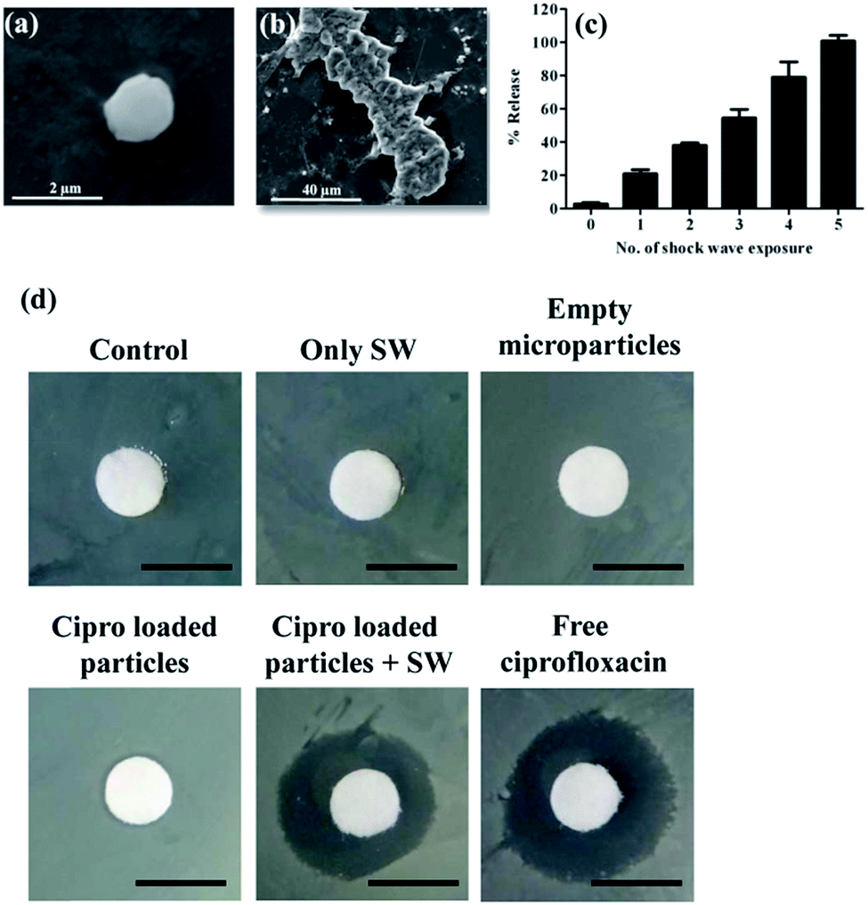 | ||
| Fig. 1 SEM images of Sper–DS capsular form (a) and aggregated form (b). (c) % Ciprofloxacin release upon exposure to different number of micro-shock waves. (d) A sterile filter disc was placed in S. aureus spread plate and the clearing zone was measured after 12 h of different treatments. Scale bar – 5 mm. SW – micro-shock wave. | ||
The biocompatibility of the capsules was checked in HeLa and Intestine 407 epithelial cell lines.27 The results show that the prepared capsule did not have cytotoxic effects, as viability of the cells was not affected up to 100 μg mL−1 concentration (Fig. S3†). From previous reports, it is known that spermidine is not cytotoxic up to 1 mg mL−1.28,29 Ciprofloxacin/insulin loaded micro particles were exposed to micro-shock waves19 as described in the method section and the release of ciprofloxacin/insulin was measured. A single time exposure of micro-shock wave could release around 20% of ciprofloxacin or insulin from the capsules (Fig. 1c). When the loaded micro particles were exposed to micro-shock waves 5 times, almost 100% release was observed. It clearly indicates that the Sper–DS capsular aggregates respond to micro-shock waves. To evaluate the killing efficiency of micro-shock wave exposed ciprofloxacin microparticles, disc diffusion assay was performed. When the discs were exposed to micro-shock waves in a plate containing S. aureus, the clearance zone appeared whereas no such clearance zone was observed without micro-shock wave exposure (Fig. 1d). These results show that ciprofloxacin is released from the microparticles only upon exposure to micro-shock waves and the released ciprofloxacin is active in killing S. aureus. When the capsules were exposed to micro-shock wave multiple times, an increase in the clearance zone was observed (Fig. S4†).
One of the most prominent human pathogens belongs to the genus Staphylococcus and is the most frequent cause of biofilm-associated infections. Biofilms are formed when it attaches to the human matrix protein or polymer surface of the indwelling medical devices through direct interaction. It is a major health issue as it is resistant to mechanical interference, host defense system and antibiotic treatment.30 The infection is even more lethal in diabetic individuals due to S. aureus foot infections. The ability of shock waves to disrupt the biofilm plays a major role in treating S. aureus infection (unpublished data).
Superficial S. aureus skin infection was created by tape striping method in BALB/c mice.31 After 3 days of infection, S. aureus colonization was observed in the wound site (Fig. 2a). BALB/c mice and alloxan induced diabetic BALB/c mice were taken for the studies. Blood glucose levels of the mice before and after the alloxan treatment was measured by taking blood from the tail vein. Mice with blood glucose levels >250 mg dL−1 were considered as diabetic and cohered in the diabetic group. After 3 days of S. aureus infection, the mice were treated with 10 μL of free ciprofloxacin (4 μg mL−1) or equivalent encapsulated ciprofloxacin with or without micro-shock wave exposure. The bacterial infection was assessed after 3 days of treatment. It was observed that only free ciprofloxacin or ciprofloxacin loaded capsules could not reduce S. aureus infection in skin, whereas micro-shock wave exposure reduced the skin infection significantly (Fig. 2b and c). A similar reduction was observed in diabetic mouse model (Fig. S5†). Around 10-fold higher S. aureus infection was observed in case of diabetic mice, which shows that infections can be life-threatening in diabetic condition. However, antibiotic along with shock wave therapy could rescue the diabetic mice from infection. It is known that shock waves can be used for inducing angiogenesis32–35 and to treat wound healing.36–38 All these properties of shock waves could further alleviate S. aureus wound infection in the skin. Our results (Fig. 2b and c and S5†) clearly indicate that along with micro-shock waves the Sper–DS microparticle system can be used to treat difficult S. aureus infection.
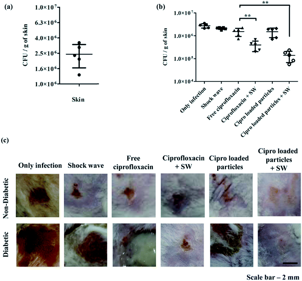 | ||
| Fig. 2 (a) The fur of BALB/c mice was stripped with adhesive tape and 5 μL of S. aureus (1 × 10⁶ bacteria) was placed in the skin. The bacterial burden was enumerated 3 days post infection at the infected site. (b) Mice were provided with different treatments after S. aureus skin infection. The bacterial burden was assessed after 3 days of treatment. (c) Photographs of mice skin with different treatments. | ||
We have used insulin delivery as a proof of concept for stimuli responsive release having distant targets using micro-shock waves. The release of insulin at physiological pH is very minimal (Fig. S6a†) and upon exposure to micro-shock waves the release of insulin was observed (Fig. S6b†). Alloxan induced diabetic mice were administered with 100 μL of insulin loaded micro particles subcutaneously and exposed to micro-shock waves. Diabetic mice without insulin treatment or treated with empty micro particles or insulin loaded micro particles without micro-shock wave exposure did not show any decrease in the blood glucose level (Fig. 3a).
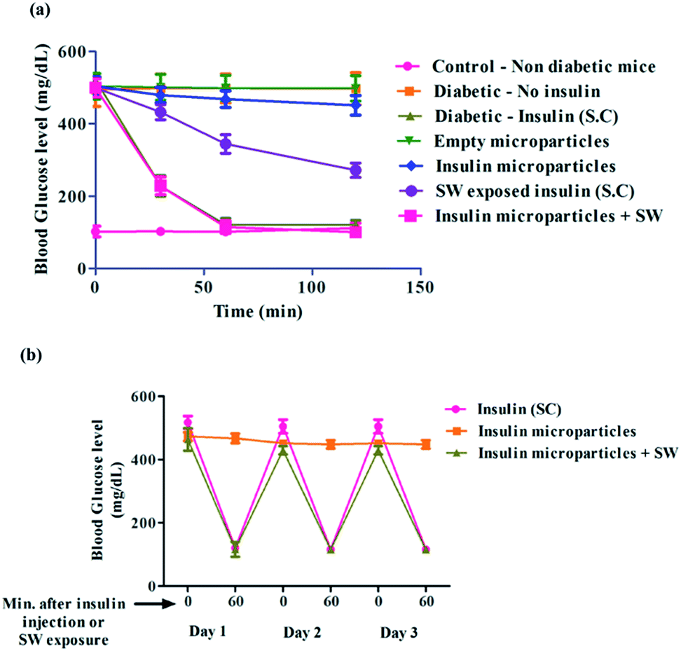 | ||
| Fig. 3 (a) Alloxan treated mice having blood glucose level >250 mg dL−1 were considered as diabetic and treated subcutaneously with insulin (1.0 mIU kg−1), empty micro particles, insulin loaded micro particles with or without micro-shock wave (SW) exposure. The blood glucose level was measured at different time points. (b) Insulin loaded micro particles were administered subcutaneously once and at different days the mice were exposed to micro-shock waves at the injection site. | ||
When the diabetic mice were treated with insulin (1.0 mIU kg−1) subcutaneously or exposed to micro-shock waves at the site of administration of insulin loaded micro particles, it showed a decrease in blood glucose level within 60 min. These results clearly indicate that micro-shock waves can be used as a trigger to release insulin in vivo. When diabetic mice were administered with 200 μL of insulin loaded micro particles subcutaneously and exposed to micro-shock waves at different days, a similar reduction in the blood glucose level was observed (Fig. 3b). This result shows that once the insulin loaded micro particles were deposited subcutaneously, micro-shock waves could be used as a trigger for insulin release over a period of at least 3 days. Thus, the number of insulin injections could be reduced by at least 33%. The delivery system could be improved by loading more insulin and injecting more number of particles into the body. Multiple deposition sites can be designed so that only remote trigger is needed to release insulin whenever required (Fig. S7†).
Conclusions
In conclusion, from these results it is clear that the micro-shock waves can be used as a stimulus to release the drug from Sper–DS delivery systems. To our knowledge, this is the first instance of use of micro-shock waves as a stimulus to release drug from a delivery system at topical or subcutaneous sites (Fig. 4). Apart from using shock wave as a stimulus for drug delivery; biofilm disruption, wound healing and angiogenesis properties of shock waves can be used to treat biofilm wound infection effectively in normal and diabetic individuals. Curing diabetic wound can lead to improved DALY scores and can be of tremendous help to the patients. A micro-shock wave trigger can be of potential use in the future and will open up further avenues for challenging discoveries in health and medicine. | ||
| Fig. 4 Micro-shock wave assisted treatment for S. aureus skin infection and for diabetes in mice. | ||
Acknowledgements
We thank the Electron microscopy facility, IISc for the help. We thank the Central Animal Facility, IISc for providing the mice. This work was supported by the grant Provision (2A) Tenth Plan (191/MCB) from the director of the Indian Institute of Science, Bangalore, India, and the Department of Biotechnology (DBT 311, NBA, sanctioned by the President), Life Science Research Board (LSRB0008) and DBT-IISc partnership program for advanced research in biological sciences and bioengineering to DC. Infrastructure support from ICMR (Center for Advanced Study in Molecular Medicine), DST (FIST), and UGC (special assistance) is acknowledged.Notes and references
- B. D. Ratner, A. S. Hoffman, F. J. Schoen and J. E. Lemons, in Biomaterials Science, ed. B. D. R. S. H. J. S. E. Lemons, Academic Press, 3rd edn, 2013, pp. xxv–xxxix Search PubMed.
- W. D. Callister and D. G. Rethwisch, Materials science and engineering: an introduction, Wiley, New York, 2007 Search PubMed.
- V. Mouriño and A. R. Boccaccini, J. R. Soc., Interface, 2010, 7, 209–227 CrossRef PubMed.
- B. D. Ratner and S. J. Bryant, Annu. Rev. Biomed. Eng., 2004, 6, 41–75 CrossRef CAS PubMed.
- Z. Yu, R. M. Schmaltz, T. C. Bozeman, R. Paul, M. J. Rishel, K. S. Tsosie and S. M. Hecht, J. Am. Chem. Soc., 2013, 135, 2883–2886 CrossRef CAS PubMed.
- B. R. Schroeder, M. I. Ghare, C. Bhattacharya, R. Paul, Z. Yu, P. A. Zaleski, T. C. Bozeman, M. J. Rishel and S. M. Hecht, J. Am. Chem. Soc., 2014, 136, 13641–13656 CrossRef CAS PubMed.
- X. Gu, J. Wang, Y. Wang, Y. Wang, H. Gao and G. Wu, Colloids Surf., B, 2013, 108, 205–211 CrossRef CAS PubMed.
- P. Du, H. Yang, J. Zeng and P. Liu, J. Mater. Chem. B, 2013, 1, 5298–5308 RSC.
- J. Hu, G. Zhang and S. Liu, Chem. Soc. Rev., 2012, 41, 5933–5949 RSC.
- F. D. Jochum and P. Theato, Chem. Soc. Rev., 2013, 42, 7468–7483 RSC.
- R. Tong, X. Lu and H. Xia, Chem. Commun., 2014, 50, 3575–3578 RSC.
- L. Peng, A. Feng, H. Zhang, H. Wang, C. Jian, B. Liu, W. Gao and J. Yuan, Polym. Chem., 2014, 5, 1751–1759 RSC.
- M. Zhao, A. Biswas, B. Hu, K.-I. Joo, P. Wang, Z. Gu and Y. Tang, Biomaterials, 2011, 32, 5223–5230 CrossRef CAS PubMed.
- S.-Y. Chen, S.-H. Hu and T.-Y. Liu, in Smart Materials for Drug Delivery: Volume 2, The Royal Society of Chemistry, 2013, vol. 2, pp. 32–62 Search PubMed.
- S. Mura, J. Nicolas and P. Couvreur, Nat. Mater., 2013, 12, 991–1003 CrossRef CAS PubMed.
- K. Radhakrishnan, J. Tripathy, D. P. Gnanadhas, D. Chakravortty and A. M. Raichur, RSC Adv., 2014, 4, 45961–45968 RSC.
- S. G. Rakesh, D. P. Gnanadhas, U. S. Allam, K. N. Nataraja, P. K. Barhai, G. Jagadeesh and D. Chakravortty, Appl. Microbiol. Biotechnol., 2012, 96, 647–662 CrossRef CAS PubMed.
- G. Jagadeesh, G. D. Prakash, S. G. Rakesh, U. S. Allam, M. G. Krishna, S. M. Eswarappa and D. Chakravortty, Clin. Vaccine Immunol., 2011, 18, 539–545 CrossRef CAS PubMed.
- G. Divya Prakash, R. V. Anish, G. Jagadeesh and D. Chakravortty, Anal. Biochem., 2011, 419, 292–301 CrossRef CAS PubMed.
- D. P. Gnanadhas, M. Ben Thomas, M. Elango, A. M. Raichur and D. Chakravortty, J. Antimicrob. Chemother., 2013, 68, 2576–2586 CrossRef CAS PubMed.
- M. B. Thomas, K. Radhakrishnan, D. P. Gnanadhas, D. Chakravortty and A. M. Raichur, Int. J. Nanomed., 2013, 8, 267–273 Search PubMed.
- G. B. Sukhorukov, in Studies in Interface Science, ed. D. Möbius and R. Miller, Elsevier, 2001, vol. 11, pp. 383–414 Search PubMed.
- G. B. Sukhorukov, E. Donath, S. Davis, H. Lichtenfeld, F. Caruso, V. I. Popov and H. Möhwald, Polym. Adv. Technol., 1998, 9, 759–767 CrossRef CAS.
- V. V. Lulevich and O. I. Vinogradova, Langmuir, 2004, 20, 2874–2878 CrossRef CAS.
- J.-P. Bégué and D. Bonnet-Delpon, in Bioorganic and Medicinal Chemistry of Fluorine, John Wiley & Sons, Inc., 2007, pp. 223–278 Search PubMed.
- Z. Değim, J. Drug Targeting, 2008, 16, 437–448 CrossRef PubMed.
- D. P. Gnanadhas, M. Ben Thomas, M. Elango, A. M. Raichur and D. Chakravortty, J. Antimicrob. Chemother., 2013, 68, 2576–2586 CrossRef CAS PubMed.
- S. Kaneko, M. Ueda-Yamada, A. Ando, S. Matsumura, E. Okuda-Ashitaka, M. Matsumura, M. Uyama and S. Ito, Invest. Ophthalmol. Visual Sci., 2007, 48, 455–463 Search PubMed.
- N. Seiler, B. Duranton, F. Gossé and F. Raul, Cell Biol. Toxicol., 2000, 16, 117–130 CrossRef CAS.
- J. W. Costerton, P. S. Stewart and E. P. Greenberg, Science, 1999, 284, 1318–1322 CrossRef CAS.
- E. Kugelberg, T. Norström, T. K. Petersen, T. Duvold, D. I. Andersson and D. Hughes, Antimicrob. Agents Chemother., 2005, 49, 3435–3441 CrossRef CAS PubMed.
- C. H. Ha, S. Kim, J. Chung, S. H. An and K. Kwon, Int. J. Cardiol., 2013, 168, 4168–4177 CrossRef PubMed.
- J. Holfeld, D. Zimpfer, K. Albrecht-Schgoer, A. Stojadinovic, P. Paulus, A. Thomas, W. Schaden, R. Kirchmair, S. Aharinejad and M. Grimm, Eur. Heart J., 2013, 34 Search PubMed.
- Y. Ping, G. Tao, P. Yun-zhu, W. Yu and C. Hong-yan, Heart, 2013, 99, A155–A156 CrossRef.
- C.-J. Wang, F.-S. Wang, J.-Y. Ko, H.-Y. Huang, C.-J. Chen, Y.-C. Sun and Y.-J. Yang, Rheumatology, 2008, 47, 542–546 CrossRef PubMed.
- T. A. Davis, A. Stojadinovic, K. Anam, M. Amare, S. Naik, G. E. Peoples, D. Tadaki and E. A. Elster, Int. Wound J., 2009, 6, 11–21 CrossRef PubMed.
- Y.-R. Kuo, C.-T. Wang, F.-S. Wang, Y.-C. Chiang and C.-J. Wang, Wound Repair Regen., 2009, 17, 522–530 CrossRef PubMed.
- B. Moretti, A. Notarnicola, G. Maggio, L. Moretti, M. Pascone, S. Tafuri and V. Patella, BMC Musculoskeletal Disord., 2009, 10, 54 CrossRef PubMed.
Footnote |
| † Electronic supplementary information (ESI) available: Materials and methods, figures. See DOI: 10.1039/c4ra15270k |
| This journal is © The Royal Society of Chemistry 2015 |