Ting Sua,
Xinyu Penga,
Jun Caoa,
Jing Changb,
Rong Liuc,
Zhongwei Gua and
Bin He*a
aNational Engineering Research Center for Biomaterials, Sichuan University, Chengdu 610064, China. E-mail: bhe@scu.edu.cn; Fax: +86-28-85412923; Tel: +86-28-85412923
bCollege of Marine Life Science, Ocean University of China, Qingdao 266003, China. E-mail: jingjing_ch@yahoo.com.cn; Fax: +86-532-82032105; Tel: +86-532-82032109
cCollege of Medical and Nursing, Chengdu University, Chengdu, 610106, China. E-mail: liurongscu@126.com; Fax: +86-86-85616523; Tel: +86-28-85616523
First published on 15th January 2015
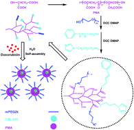
Multiple functionalization of nanoparticles has attracted great interest in drug delivery. In this paper, biodegradable poly(α,β-malic acid) with a hyperbranched architecture was synthesized via the polycondensation of L-malic acid, the functionalized poly(α,β-malic acid) was used as a nanocarrier platform with the immobilization of poly(ethylene glycol) (PEG) for long circulation, cinnamyl alcohol (CIN) for introducing π–π stacking interactions and 1-(3-aminopropyl)imidazole (API) for pH-sensitivity. The conjugates self-assembled into nanoparticles to load anticancer drug doxorubicin (DOX). The morphology, mean size and size distribution, drug release profile and in vitro anticancer activity of DOX loaded nanoparticles were studied. The results showed that the mean size of the nanoparticles was below 200 nm, the drug loading content was higher than 10 wt% and it increased with increasing CIN content because of the π–π stacking interaction between DOX and the carriers. The drug release of the nanoparticles was faster in the medium with pH 6.0 compared to pH 7.4. The nanoparticles exhibited an endosomal escape function to accelerate the release of DOX in cancer cells, which resulted in low IC50s to kill 4T1 breast cancer cells and HepG2 liver cancer cells in vitro.
Intelligent drug delivery are expected to release drugs in a controllable manner upon arrival at the target site in response to external or internal stimuli.14,18 pH-dependent drug release is one of the most successful strategies in tumor drug delivery systems.19–21 Taking the advantages of the weak acidic microenvironment of tumor tissues,22 many pH-sensitive nanoparticles were fabricated to improve therapeutic efficacy and reduce side effects.23–25 Poly(L-histidine) based nanoparticles exhibited excellent pH-sensitivity due to the protonation of side imidazole groups in weak acidic medium,26,27 however, the complicated synthesis and low yield of poly(L-histidine) limited its wide applications. With the inspiration of pH-sensitivity originated from the protonation of imidazole groups in poly(L-histidine), other nanoparticles with imidazole groups as pH-sensitive moieties were achieved.28–32
Poly(malic acid) (PMA) is a water-soluble, biodegradable, and bioabsorbable polymer,33,34 the degradation product malic acid is an intermediate product in tricarboxyl acid cycle in the metabolism of carbohydrates, which is non-toxic to cells and tissues. Poly(malic acid) has been reported as hydrogel,35 cell scaffold36 and drug carriers.37,38 The remarkable advantage of poly(malic acid) for biomedical applications is the large number of carboxyl groups on the backbones, which could be used for multiple functionalization. The synthesis of poly(malic acid) was focused on poly(β-malic acid) via ring-opening polymerization of malolactonate.39,40 The polycondensation of L-malic acid to receive poly(α,β-malic acid) was rarely reported. Different from the linear architecture of poly(β-malic acid), the polycondensation generated poly(α,β-malic acid) with hyperbranched architecture, the carboxyl groups were in the peripheral sites, which were more convenient and efficient for modification.
The goal of this study was to fabricate poly(α,β-malic acid) based nanoparticles for anticancer drug delivery. Poly(α,β-malic acid) was used as backbone to provide carboxyl groups for the immobilization of hydrophilic poly(ethylene glycol), hydrophobic cinnamyl alcohol and pH-sensitive 1-(3-aminopropyl)imidazole. The functionalized conjugates self-assembled into nanoparticles to trap anticancer drug doxorubicin. The nanoparticle was expected to own the integrated functions of long circulation, high drug loading content and pH-sensitive drug release.
Entry | Compositions | Mean sizea (nm) | PDI | ζ potentiala (mV) | |||||
---|---|---|---|---|---|---|---|---|---|
PEG | CIN | API | Blank | With DOX | Blank | With DOX | Blank | With DOX | |
a Measured by DLS (C = 1 mg mL−1), the average size of the three measurements was recorded. The results were expressed as mean ± SD (n = 3). | |||||||||
P1 | 10 | 90 | 0 | 9 ± 1.4 | 29 ± 1 | 0.88 | 0.24 | −25 ± 2 | −7.4 ± 0.3 |
P2 | 10 | 80 | 10 | 67 ± 12 | 94 ± 6 | 0.17 | 0.15 | −7.7 ± 0.2 | 9.8 ± 0.2 |
P3 | 10 | 70 | 20 | 58 ± 12 | 60 ± 12 | 0.14 | 0.18 | −5.6 ± 0.4 | 4.3 ± 0.4 |
P4 | 10 | 50 | 40 | 92 ± 11 | 168 ± 8 | 0.10 | 0.10 | −4.1 ± 0.2 | 15 ± 0.3 |
The cytotoxicity of blank nanoparticles was tested by Cell Counting Kit-8 assay (CCK-8, Dojindo, Japan) against 4T1 breast cancer cells, HepG2 liver cancer cells and C2C12 cells. 4T1 and C2C12 cells were seeded in 96-well plates with the cell density of 4 × 10³ mL−1, HepG2 cells were seeded in 96-well plates with a cell density of 6 × 10³ mL−1. Each well was cultured with 100 μL of medium. After 24 h incubation, the culture medium was removed and replaced with 100 μL of medium containing blank nanoparticles. The cells were incubated for another 48 h. The culture medium was removed and the wells were rinsed with PBS (pH = 7.4). 100 μL of CCK-8 (volume fraction 10%) solution was added to each well. After incubated for 2 h, the absorbance was measured at a Thermo Scientific MK3 (Thermo fisher, US) at the wavelength of 450 nm.
In order to explore the effect of API functionalized DOX loaded nanoparticles on endosomal escape for efficient intracellular trafficking, LysoTracker green was used to observe the cytoplasmic distribution of DOX loaded nanoparticles. 4T1 cells at a logarithm phase were seeded on glass dishes (diameter = 35 mm) at a cell density of 1 × 10⁴ mL−1. After incubated for 24 h, DOX loaded nanoparticles were dissolved in RPMI 1640 medium till the final DOX concentration was 10 μg mL−1, the culture medium was removed and 200 μL of the mixture was added into each dish. After incubated for 1 and 4 h, the culture medium was removed and the dishes were rinsed with PBS (pH = 7.4), and then stained with 50 nM LysoTracker green (Invitrogen, USA) for 60 min at 37 °C. The cells were washed by PBS (pH = 7.4) twice and observed by CLSM.
For the flow cytometry tests, 4T1 cells were seeded in 6-well plates at a density of 1 × 10⁶ cells per well and incubated for 24 h. The cells were treated with DOX loaded nanoparticles at the same DOX concentration (10 μg mL−1) for 1 and 4 h, respectively. The culture medium was eliminated, the cells were washed with PBS for three times and harvested by trypsinization. The cells were resuspended in PBS after centrifugation (1000 rpm, 5 min) and the fluorescence intensity was measured (excitation: 480 nm; emission: 590 nm) on a BD FACS Calibur flow cytometer (Beckton Dickinson).
![]() | ||
Scheme 1 The synthetic route (A) and the concept for a proposed behavior of polymeric nanoparticles for anticancer drug doxorubicin delivery (B). |
The molecular weight and molecular weight distribution of PMA was tested by GPC. The GPC spectrum of PMA was presented in Fig. S1 in the ESI.† A main peak was observed at the eluent time about 12.5 minutes and a weak shoulder peak was at 14.5 minutes in the spectrum, the intensity of the main peak was much stronger than that of shoulder peak. The calculated molecular weight was Mn = 3780 and the polydispersity was 1.14. The polydispersity of PMA was much narrower than that of theoretical calculated value in polycondensation, which was due to the hyperbranched architecture of PMA.
The 1H NMR spectra of PMA and copolymer P4 were shown in Fig. 1. The multi-peaks at δ = 3.0–3.2 ppm were assigned to the protons of CH2 in both α and β type units in poly(α,β-malic acid). The protons signals split into multiple peaks due to the random aggregation of α and β type of L-malic acid units in the main chains and the similar chemical environment.35 The doublets at δ = 5.5 and 5.6 ppm were attributed to the protons of CH in poly(α,β-malic acid). The peaks at δ = 3.4 and 3.6–3.8 ppm were assigned to the protons of OCH3 and OCH2CH2 in mPEG2k. The graft degree was calculated from the intensity ratio between CH3 in mPEG2k and CH in PMA. 10% of carboxyl groups were grafted on mPEG2k. The characteristic peaks of CIN were assigned to the protons of CHCH and CH2OCO at δ = 6.6, 6.2 and 4.7 ppm, respectively. The protons in the benzene ring (C6H5) in CIN were detected at δ = 7.2–7.5 ppm. The peaks at δ = 6.9–7.1 ppm were attributed to protons of imidazole ring in API, and the other three protons of NCH2CH2, CH2CH2CH2 and CH2CH2NH in API were appeared at δ = 3.2, 1.8 and 4.1 ppm, respectively. The calculated compositions of the four amphiphiles from 1H NMR spectra were presented in Table 1, they were nearly in agreement with the compositions in feedings. The 1H NMR spectra of copolymers P1, P2 and P3 were presented in Fig. S2 in ESI.†
The successful conjugation of each amphiphile was further confirmed by FTIR as shown in Fig. 2. It was obvious that the vibrations attributed to CH2 at around 2850 cm−1 and ether bond CH2OCH2 at around 1100 cm−1 were strengthened greatly after mPEG2k was grafted on PMA, and the vibrations of benzene ring at about 690 and 750 cm−1 were clear in the FTIR spectrum of P1. The characteristic peak at 1745 cm−1 in PMA, P1, P2, P3 and P4 was the stretching vibration absorbance of CO in ester bond. A new vibration band at 1645 cm−1 appeared in P2, P3 and P4, it was the stretching vibration absorbance of C
O in amide bond. At the same time, the peak at 1645 cm−1 in the amide bond became stronger comparing to the peak at 1745 cm−1 in ester bond with increasing the ratio of API from P2 to P4, suggesting that more API molecules were successfully immobilized on PMA backbones.31
The conjugates self-assembled into nanoparticles in aqueous solution. The size distribution and morphology of the nanoparticles were tested by DLS and TEM. The mean diameters and zeta potentials of blank and DOX loaded nanoparticles were summarized in Table 1. All the four conjugates self-assembled into monodisperse nanoparticles (Fig. 3A) and the mean size of P1, P2, P3 and P4 were 9, 67, 58 and 92 nanometers. It was interesting that the mean size of P1 was much smaller than that of the other three nanoparticles, the PDI of P1 was the largest. However, when DOX was loaded in the nanoparticles, the mean sizes of all the four nanoparticles were enlarged. The zeta potentials of the four blank nanoparticles increased with increasing the API compositions. The zeta potential of DOX loaded nanoparticles was higher than that of corresponding blank nanoparticles because of the amino group in DOX. The drug loaded nanoparticles were also monodisperse (Fig. 3B). The mean size of the drug loaded nanoparticles was smaller than 200 nanometers, which was in suitable size for passive targeting via EPR effect.46 The morphologies of the blank and DOX loaded nanoparticles were observed by TEM (Fig. 3C and D), the nanoparticles were well dispersed and the size was consistent with DLS results.47
![]() | ||
Fig. 3 DLS results of blank (A) and DOX loaded nanoparticles (B), TEM images of P4 bank nanoparticles (C) and DOX loaded nanoparticles (D). |
The drug loading content (DLC) and drug encapsulation efficiency (DEE) of the four nanoparticles were measured and the results were summarized in Table 2. P1-DOX nanoparticles exhibited the best DLC and DEE. The DLC and DEE of P1-DOX nanoparticles were 15 and 70.6 wt%, respectively, which were much higher than those of the other three nanoparticles. Both DLC and DEE of nanoparticles decreased when the API in the nanoparticles increased, it was probably attributed to the interaction between DOX and nanoparticles.
In our previous work, we reported that the formation of π–π stacking interaction was helpful to enhance the DLC of nanoparticles.16,17 In order to verify the π–π interaction and explain the DLC variation in the four nanoparticles, the π–π interaction between nanoparticles and DOX was investigated. The UV-Vis absorption and fluorescence spectra of DOX loaded nanoparticles were tested. The maximum UV absorbance (λmax) of free DOX·HCl was at 483 nm and the blank nanoparticle showed no evident absorption in the wavelength from 350 to 650 nm(Fig. 4A). After DOX was encapsulated into the nanoparticles, the absorbance λmax showed a red shift to 497, 498, 498 and 500 nm for P1-DOX, P2-DOX, P3-DOX and P4-DOX, respectively. It implied that π–π stacking interaction within the drug loaded nanoparticles was evoked.48 The π–π stacking interaction was further investigated via fluorescence measurement as showed in Fig. 4B. When the exciting wavelength was set at 483 nm, free DOX performed wide band from 600 to 700 nm. However, DOX loaded nanoparticles exhibited remarkable decrease in the fluorescence intensity of emission band comparing to free DOX at the same concentration. The significant intensity decrease indicated the quenching of fluorescence by energy transfer among π–π interaction overlapped systems.49 The higher quenching degree of DOX loaded nanoparticles likely demonstrated the stronger π–π interaction. It revealed that the π–π stacking interaction between nanoparticles and DOX was weakened with the composition increase of API in the nanoparticles. That was the intrinsic nature in nanoparticles to affect the drug loading content.
As we knew that the high buffering capacity enable nanoparticles to facilitate endosomal escape,50,51 which contributed to efficient drug release. The presence of imidazole units in PMA based nanoparticles was expected to achieve pH-responsive drug release via the protonation of imidazole groups in endosomes. Acid–base titration of the copolymers was carried out to exhibit the buffering capacity of the four conjugates (Fig. 5A). The results showed that all copolymers had a buffer platform, indicating all of them exhibited buffering capacity. With the graft degree of API increased, the P4 had the widest buffer platform comparing to the other three, it revealed that the P4 conjugate had better capacity for proton acceptance.
![]() | ||
Fig. 5 Acid–base titration curves of blank nanoparticles (A) and the release profiles of DOX loaded nanoparticles (B), means ± SD (n = 3). |
As the pH value in endosomes was about 6.0 and the nanoparticles were encapsulated in endosomes once they were internalized in cells. The drug release profiles of DOX loaded nanoparticles were tested in PBS solutions with pH = 7.4 and 6.0 (ionic strength = 0.01 M) at 37 °C (Fig. 5B). The amounts of released DOX at different predetermined time points were measured by fluorescence detector with excitation wavelength at 480 nm and emission wavelength at 550 nm. The release performed an early weak burst release in the first few hours and a sustained release in the followed stage for prolonged time. The four drug loaded nanoparticles practically showed no difference in DOX release in the medium with pH 7.4. The cumulated release was less than 20% even the release time was as long as 48 h. However, in the medium with pH = 6.0, the drug was released faster from nanoparticles, P4-DOX nanoparticles showed the fastest release within all the four nanoparticles, and the release rates of P2-DOX and P3-DOX nanoparticles showed nearly the same release rates. All the three nanoparticles with API in P2, P3 and P4 exhibited fast DOX release comparing to P1 without API. The release profiles revealed the pH-sensitivity of API.
The cytotoxicity of the polymeric nanoparticles was investigated via CCK-8 assay. The blank nanoparticles were incubated with 4T1 breast cancer cells, C2C12 cells and HepG2 liver cancer cells for 48 h with different concentrations. Fig. 6 showed that all the cell viabilities were higher than 90% after incubated with blank nanoparticle for 48 h even the concentration of nanoparticles was as high as 600 μg mL−1. It revealed that the four blank nanoparticles were nontoxic to cells.52
![]() | ||
Fig. 6 The cytotoxicity of blank nanoparticles incubated with 4T1 cells (A), C2C12 cells (B) and HepG2 cells (C) for 48 h. |
The delivery efficiency and intracellular localization of DOX loaded nanoparticles in 4T1 cells were investigated using confocal laser scanning microscopy (CLSM). In Fig. 7, red fluorescence of DOX was observed cytoplasm in both 4T1 and HepG2 cells in 1 h, which implied that most of the drug loaded nanoparticles were in cytoplasm. The cells treated with P4-DOX nanoparticles exhibited stronger red fluorescence for 1 h incubation, which demonstrated that more P4-DOX nanoparticles were internalized into the cells. The cells showed stronger red fluorescence for 4 h incubation compared with 1 h incubation, it implied that more drug loaded nanoparticles were internalized into cells and a sustained release of DOX from the DOX loaded nanoparticles was happened. The DOX in nanoparticles was more easily diffused in nuclei of HepG2 cells.
To identify the role of imidazole group in pH-responsive nanoparticles of P2, P3 and P4, the intracellular tracking of DOX loaded nanoparticles were studied via CLSM. Lysosomes in 4T1 cells were observed in green fluorescence after they were stained with specific LysoTracker green. DOX loaded nanoparticles were shown in red fluorescence. Co-localization of the DOX loaded nanoparticles overlapped with green-dyed lysosomes appeared yellow. As shown in Fig. 8, in the first hour, the red fluorescence was highly overlaid with the green fluorescence and all the four nanoparticles showed yellow fluorescence in the overlay images. Obviously, there was no difference among the four nanoparticles, illustrating that all the DOX loaded nanoparticles were located in lysosomes. In the 4 h images, the P1-DOX nanoparticles showed yellow, demonstrating that it was hard for P1-DOX nanoparticles to escape from endolysosomes.53 In contrast, although the P2-DOX, P3-DOX and P4-DOX nanoparticles were located in the endolysosomes for the first hour in yellow in the overlay images, however, the green fluorescence had a significant decline and the red fluorescence became stronger, rare yellow fluorescence was observed in the overlay of the images of P2-DOX, P3-DOX and P4-DOX nanoparticles. It clearly indicated that the efficient endolysosomal escape was happened in the imidazole modified DOX loaded nanoparticles (P2-DOX, P3-DOX and P4-DOX). These results revealed the pH-sensitive drug release from API modified PMA based nanoparticles.
The cellular internalization of drug loaded nanoparticles was further illustrated in flow cytometry. The intracellular delivery efficiency of DOX loaded nanoparticles in 4T1 cells was given in quantitative fluorescence intensity. Fig. 9A and B showed the results of 4T1 breast cancer cells treated with DOX loaded nanoparticles for 1 (Fig. 9A) and 4 h (Fig. 9B), respectively. The concentration of DOX was the same as 10 μg mL−1. The mean red fluorescence intensities of 4T1 cells for different incubation times were presented in Fig. 9C. The cells incubated with P4-DOX nanoparticles showed the highest red fluorescence intensity. There was no significant difference in the mean fluorescence intensity among the four nanoparticles for 1 h incubation as shown in the quantitative results in Fig. 9C. When the incubation time was extended to 4 h, all the nanoparticles showed a stronger red fluorescence in the cells, and the API modified nanoparticles (P2-DOX, P3-DOX and P4-DOX) exhibited higher mean fluorescence intensity comparing to P1-DOX nanoparticles. However, the strongest fluorescence was observed in P4-DOX nanoparticles. It also revealed the pH-sensitivity of API modified nanoparticles.
The in vitro anticancer activity of the four DOX loaded nanoparticles was evaluated in 4T1 breast cancer cells and HepG2 liver cancer cells via CCK-8 assay. As shown in Fig. 10, the IC50s (half maximal inhibitory concentration) values of the four DOX loaded nanoparticles of 4T1 cells for P1-DOX, P2-DOX, P3-DOX, P4-DOX and DOX·HCl were 7.89, 7.61, 4.53, 4.78 and 0.79 μg mL−1, and the IC50s for HepG2 cells were 3.27, 2.58, 1.95, 1.43 and 0.17 μg mL−1 as shown in Table 2. Once the drug loaded nanoparticles were internalized into cytoplasm via endocytosis, the protonation of imidazole groups began to take effect in the weak acidic environment of endosomes, and the resulting proton sponge effect accelerated the release of DOX.53 The imidazole group in API was beneficial to help drug loaded nanoparticles to escape from endosomes and release DOX to facilitate the diffusion into nucleus to inhibit the proliferation of cells. P3-DOX and P4-DOX nanoparticles showed the lower IC50s with more efficient in vitro anticancer activity due to the higher composition of API in the nanoparticles. DOX·HCl was a water-soluble molecule, which diffused much faster into cells to kill cells efficiently and resulted in lowest IC50s.
![]() | ||
Fig. 10 The IC50 of the DOX loaded nanoparticles incubated with 4T1 breast cancer cells (A) and HepG2 liver cancer cells (B). |
Footnote |
† Electronic supplementary information (ESI) available. See DOI: 10.1039/c4ra13686a |
This journal is © The Royal Society of Chemistry 2015 |